 Concordance: clinical impressions vs. pediatric oral histology
ABSTRACT
Several pathologies often lead to disagreements regarding their identification in clinical practice, particularly in the context of oral pathologies. The data obtained during anamnesis and physical examination frequently require revision to reach a diagnosis.
Objective: The present study aimed to evaluate the agreement between clinical and histopathological diagnoses and report the frequency of completing forms for specimens submitted to histopathological examination, examined retrospectively.
Methods: Data on 93,950 specimens submitted for histopathological examination were retrieved from medical records.
Results: A total of 5,052 reports from patients aged 0 to 14 years were included. Agreement was defined based on categorizing the nature of the injury according to its diagnostic category. The highest agreement rate was observed for mucocele and papilloma (86%), followed by pyogenic granuloma (60%). Low rates of histopathological confirmation of clinical impressions were observed for hemangioma (17.5%) and peripheral giant cell lesion (35%). Among lesions related to dental tissues, odontoma showed the highest agreement (84%), followed by keratocyst (74%), while radicular cyst showed the most discordance (21%).
Conclusion: This study found an acceptable level of agreement for the most frequent injuries. However, understanding the characteristics of oral lesions in children and their clinicopathological correlations is crucial for providing optimal treatment in each case.
Indexing terms
Child; Diagnosis, oral; Pathology, oral; Preschool
RESUMO
Diversas patologias conduzem frequentemente a divergências quanto à sua identificação na prática clínica, nomeadamente no contexto das patologias orais. Os dados obtidos durante a anamnese e o exame físico frequentemente necessitam de revisão para o diagnóstico.
Objetivo: O presente estudo teve como objetivo avaliar a concordância entre os diagnósticos clínicos e histopatológicos e relatar a frequência de preenchimento de formulários de espécimes submetidos ao exame histopatológico, examinados retrospectivamente.
Métodos: Dados de 93.950 espécimes submetidos para exame histopatológico foram obtidos de prontuários médicos.
Resultados: Foram incluídos 5.052 laudos de pacientes de 0 a 14 anos. A concordância foi definida a partir da categorização da natureza da lesão de acordo com sua categoria diagnóstica. A maior taxa de concordância foi observada para mucocele e papiloma (86%), seguido por granuloma piogênico (60%). Baixas taxas de confirmação histopatológica de impressões clínicas foram observadas para hemangioma (17,5%) e lesão periférica de células gigantes (35%). Entre as lesões relacionadas aos tecidos dentários, o odontoma apresentou maior concordância (84%), seguido pelo ceratocisto (74%), enquanto o cisto radicular apresentou maior discordância (21%).
Conclusão: Este estudo encontrou nível de concordância aceitável para as lesões mais frequentes. No entanto, compreender as características das lesões orais em crianças e suas correlações clinicopatológicas é crucial para proporcionar o tratamento ideal em cada caso.
Termos de indexação
Criança; Diagnóstico oral; Patologia bucal; Pré-escola
INTRODUCTION
Contrary to its simple appearance, the oral cavity of babies, children, and adolescents is complex. It can be affected by various cysts, benign and malignant tumors of the salivary glands, and odontogenic and non-odontogenic neoplasms [1]. Additionally, typical physiological processes, there are developmental changes specific to this age group [2]. Therefore, childhood and adolescent pathological conditions have characteristics and symptoms that are often different from pathologies in adults [3-5].
In the face of a pathological lesion, clinical diagnostic errors occur due to similarities in clinical presentations, lack of precise definitions for these characteristics, incompatibility of signs and symptoms in patients, and multiple manifestations for a lesion [6].
Although the histopathology report remains the gold standard, an excellent clinical impression is potentially a critical diagnostic tool in resource-limited rural settings. Thus, good agreement between clinical image and histopathological diagnosis is an essential tool in oral pathology diagnostics.
To this end, epidemiological studies constitute a fundamental instrument as they promote assessing the population’s health conditions in the recommended age group and favor the development of diagnostic hypotheses, assisting professionals with data on the prevalence of disease changes.
Therefore, the objective of this research was to analyze, through a retrospective study, the level of agreement between clinical and histopathological diagnoses of children aged 0 to 14 years obtained from archived reports from the Laboratory of the Discipline of Oral Pathology of the Faculty of Dentistry of the University of São Paulo.
METHODS
This project is part of the work “Oral and maxillofacial diseases affecting pediatric dentistry and pediatric dentistry patients in the last 20 years”, approved by the Research Ethics Committee of the Faculty of Dentistry of the University of São Paulo under number CAAE: 67845217.6.00000075.
For this retrospective study, reports of biopsies performed and received in the laboratory of the Oral Pathology Discipline of the Faculty of Dentistry of the University of São Paulo, a reference laboratory, were collected over 20 years. The age range of patients analyzed was 0 to 14 years old. To better organize the data, the age groups were divided into three groups, following the division proposed by IBGE: 0 to 4 years, 5 to 9 years, and 10 to 14 years.
Information on age, diagnostic hypothesis, and final diagnosis were tabulated, and the results were described. Reports with inappropriate material for analysis, inconclusive diagnoses, and missing data for appropriate agreement comparisons were excluded from the calculations.
RESULTS
In a database of 93,950 reports, 5,052 corresponded to the age group analyzed: 373 from 0 to 4 years old, 1,927 from 5 to 9 years old, and 2,752 from 10 to 14 years old. Reports with missing diagnostic data or no diagnostic hypothesis to verify agreement, biopsies unsuitable for analysis, and collections outside the oral region or from non-human subjects were discarded from the final analysis. Therefore, in the age group of 0 to 4 years, 64.9% of diagnostic agreement was obtained between the described hypotheses and the histopathological diagnosis (187/288); in the age group of 5 to 9 years, 63.1% (1,052/1,665); and in the age group of 10 to 14 years, 57.6% (1,406/2,438).
Age range from 0 to 4 years: The most prevalent lesions in this group were mucocele, papilloma, giant cell fibroma, pyogenic granuloma, and hemangioma, respectively—all soft tissue injuries.
All of these lesions had a higher agreement rate, except for giant cell fibroma (55% diagnostic disagreement) and hemangioma (80% disagreement) (table 1).
Age range: 5 to 9 years. In this group, the most prevalent injuries were separated into “injuries related to soft tissues” and “injuries related to dental tissues” for teaching purposes. The most common soft tissue lesion found was mucocele, while the dental tissue lesion was odontoma. The soft tissue lesions with the highest rate of diagnostic disagreement were peripheral giant cell lesion/CGPL (65%), hemangioma (60%), and inflammatory fibrous hyperplasia/HFI (54%). In comparison, the dental tissue lesion with the highest rate of diagnostic disagreement was odontoma (50%) [table 2].
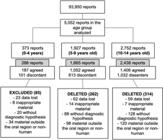
Infographic representing the distribution of agreement between the analyzed reports and exclusion criteria.
Age range: 10 to 14 years. In this age group, the most prevalent lesions were separated like the previous group, and the division “odontogenic tumors” was added for teaching purposes. The most pervasive soft tissue injury continued to be mucocele and dental tissue injury, odontoma. Ameloblastoma was also more present in this group. The soft tissue lesion with the highest rate of diagnostic disagreement was the hemangioma (80%). In comparison, the dental tissue lesion with the highest rate of diagnostic controversy was the radicular cyst (71%) [table 3].
DISCUSSION
This study represents a pioneering effort in assessing the agreement between clinical and histopathological diagnoses and the completion rate of biopsy forms for oral lesions in the pediatric age group. Despite methodologies already recognized [7-9] employed by other researchers, this is the first study to specifically evaluate the concordance rate of oral lesions solely in children.
Our study employed flexible criteria to compare clinical and histopathological diagnoses. If the referral report indicated a clinical impression of mucus retention and the microscopic evaluation revealed a diagnosis within the same diagnostic category as mucocele or ranula, the case was considered accordingly [10].
Of the data collected, 373 cases occurred in early childhood, 1,927 in the second childhood age group, and 2,752 patients were between 10 and 14 years old. This age difference can be attributed to several factors. Firstly, it may be due to the lack of awareness among family members about taking their baby to a pediatric dentist even before the eruption of the first tooth, which typically occurs around six months of age [11]. Alternatively, it could be due to the prevalence of oral diseases in this age group resulting from viruses or fungi, such as primary herpetic gingivostomatitis, hand-foot-and-mouth disease, or candidosis, which are diagnosed clinically and do not require biopsies [12-14].
All pathologies observed in the 0 to 4 years age group were restricted to soft tissue. Even though primary teeth are fully developed around three years of age, the pathologies observed in this group included mucocele, papilloma, pyogenic granuloma, giant cell fibroma, and one case of hemangioma [15-17]. Mucocele had the highest diagnostic certainty at 82%, likely due to its pathognomonic etiology and characteristic clinical features [18,19]. The other lesions observed in early childhood with high diagnostic agreement rates were papilloma, with 93%, and pyogenic granuloma, with 86% agreement [17,20-23].
In the 5 to 9 years age group, in addition to the soft tissue pathologies observed in babies, several cases related to dental tissue were observed. Pericoronal capping remained prevalent, mainly due to the eruption of the first permanent molars, during which pericoronitis is common [24]. Dentigerous cysts had a 91% correct diagnostic hypothesis rate, followed by keratocysts (86%) and odontomas (84%) [25]. These cysts are more common in adults and are typically painless, often diagnosed incidentally during routine panoramic x-rays [26,27].
In children between 10 and 14 years old, ameloblastoma was observed, consistent with its occurrence as the most common benign odontogenic tumor in this age group, although it is considered rare overall [28,29].
Our findings indicate moderate to high levels of agreement between clinical and histopathological diagnoses. However, there were discrepancies in the completion of biopsy forms, with relatively low completion rates for some pathological categories. It is challenging to directly compare these results with those from other studies due to marked methodological differences, especially concerning agreement criteria and the professionals who carried out the assessments.
These findings highlight the need to improve knowledge about the attitudes and behaviors of dentists, pediatric dentists, and dental students regarding the requirement for histopathological examination and to familiarize themselves with the most frequent pathologies in children.
CONCLUSION
The level of disagreement between clinical and histopathological diagnoses was higher than the level of agreement.
A thorough anamnesis, combined with a solid understanding of the most common oral lesions observed in pediatric patients, is essential for enhancing the accuracy of clinical diagnoses and aligning them with anatomopathological findings.
How to cite this article
-
Rezende KM, Gentile AC, Silva MB, Marinho GB, Haddad AE, Bönecker M. Concordance: clinical impressions vs. pediatric oral histology. RGO, Rev Gaúch Odontol. 2024;72:e20240027. http://dx.doi.org/10.1590/1981-86372024002720240010
REFERENCES
-
1 Tatli U, Erdogan O, Uguz A, Ustun Y, Sertdemir Y, Damlar I. Diagnostic concordance characteristics of oral cavity lesions. ScientificWorldJournal. 2013;2013:785929. http://dx.doi.org/10.1155/2013/785929
» https://doi.org/10.1155/2013/785929 -
2 Rezende KM, de Barros Gallo C, Nogueira GP, Corraza AC, Haddad AE, Gallottini M, et al. Retrospective study of oral lesions biopsied in babies and toddlers. ral Dis. 2024;30(3):1242-1244. http://dx.doi.org/10.1111/odi.14552
» https://doi.org/10.1111/odi.14552 -
3 Jones AV, Franklin CD. An analysis of oral and maxillofacial pathology found in adults over a 30-year period. J Oral Pathol Med. 2006;35(7):392-401. http://dx.doi.org/10.1111/j.1600-0714.2006.00451.x
» https://doi.org/10.1111/j.1600-0714.2006.00451.x -
4 DE Souza VG, Gomes BP, Cavalcanti TR, Câmara J, Libório-Kimura. Oral lesions in children/teenagers: a cross-sectional study of 225 reports in a pathology service in Amazon. Minerva Dent Oral Sci. 2021;70(6):263-8. http://dx.doi.org/10.1111/10.23736/S2724-6329.20.04431-3
» https://doi.org/10.1111/10.23736/S2724-6329.20.04431-3 -
5 Glickman A, Karlis V. Pediatric benign soft tissue oral and maxillofacial pathology. Oral Maxillofac Surg Clin North Am. 2016;28(1):1-10. http://dx.doi.org/10.1016/j.coms.2015.07.005
» https://doi.org/10.1016/j.coms.2015.07.005 - 6 Kondori I, Mottin RW, Laskin DM. Accuracy of dentists in the clinical diagnosis of oral lesions. Quintessence Int. 2011;42(7):575-7.
-
7 Logan RM, Goss AN. Biopsy of the oral mucosa and use of histopathology services. Aust Dent J. 2010;55 Suppl 1:9-13. http://dx.doi.org/10.1111/j.1834-7819.2010.01194.x
» https://doi.org/10.1111/j.1834-7819.2010.01194.x - 8 Melrose RJ, Handlers JP, Kerpel S, Summerlin DJ, Tomich CJ, American Academy of O, et al. The use of biopsy in dental practice. The position of the American Academy of Oral and Maxillofacial Pathology. Gen Dent. 2007;55(5):457-61; quiz 62-3, 88.
-
9 Patel KJ, De Silva HL, Tong DC, Love RM. Concordance between clinical and histopathologic diagnoses of oral mucosal lesions. J Oral Maxillofac Surg. 2011;69(1):125-33. http://dx.doi.org/10.1016/j.joms.2010.07.075
» https://doi.org/10.1016/j.joms.2010.07.075 - 10 Kuc I, Peters E, Pan J. Comparison of clinical and histologic diagnoses in periapical lesions. Oral Surg Oral Med Oral Pathol Oral Radiol Endod. 2000;89(3):333-7.
-
11 Djokic J, Bowen A, Dooa JS, Kahatab R, Kumagai T, McKee K, et al. Knowledge, attitudes and behaviour regarding the infant oral health visit: are dentists in Ireland aware of the recommendation for a first visit to the dentist by age 1 year? Eur Arch Paediatr Dent. 2019;20(2):65-72. http://dx.doi.org/10.1007/s40368-018-0386-0
» https://doi.org/10.1007/s40368-018-0386-0 -
12 Vainionpaa A, Tuomi J, Kantola S, Anttonen V. Neonatal thrush of newborns: Oral candidiasis? Clin Exp Dent Res. 2019;5(5):580-2. http://dx.doi.org/10.1002/cre2.213
» https://doi.org/10.1002/cre2.213 - 13 Saguil A, Kane SF, Lauters R, Mercado MG. Hand-foot-and-mouth disease: rapid evidence review. Am Fam Physician. 2019;100(7):408-14.
-
14 Esposito S, Principi N. Hand, foot and mouth disease: current knowledge on clinical manifestations, epidemiology, aetiology and prevention. Eur J Clin Microbiol Infect Dis. 2018;37(3):391-8. http://dx.doi.org/10.1007/s10096-018-3206-x
» https://doi.org/10.1007/s10096-018-3206-x -
15 Hoger PH, Hamm H. [Infantile hemangioma : Clinical manifestation, treatment, and differential diagnoses]. Dermatologie (Heidelb). 2023;74(5):331-9. http://dx.doi.org/10.1007/s00105-023-05145-2
» https://doi.org/10.1007/s00105-023-05145-2 -
16 Sharma S, Chandra S, Gupta S, Srivastava S. Heterogeneous conceptualization of etiopathogenesis: Oral pyogenic granuloma. Natl J Maxillofac Surg. 2019;10(1):3-7. http://dx.doi.org/10.4103/njms.NJMS_55_18
» https://doi.org/10.4103/njms.NJMS_55_18 - 17 Rezende KM, Moraes PC, Oliveira LB, Thomaz LA, Junqueira JL, Bonecker M. Cryosurgery as an effective alternative for treatment of oral lesions in children. Braz Dent J. 2014;25(4):352-6.
-
18 Guimaraes MS, Hebling J, Filho VA, Santos LL, Vita TM, Costa CA. Extravasation mucocele involving the ventral surface of the tongue (glands of Blandin-Nuhn). Int J Paediatr Dent. 2006;16(6):435-9. http://dx.doi.org/10.1111/j.1365-263X.2006.00759.x
» https://doi.org/10.1111/j.1365-263X.2006.00759.x -
19 Plantier DB, Neto DB, Pinna FR, Voegels RL. Mucocele: clinical characteristics and outcomes in 46 operated patients. Int Arch Otorhinolaryngol. 2019;23(1):88-91. http://dx.doi.org/10.1055/s-0038-1668126
» https://doi.org/10.1055/s-0038-1668126 - 20 Miranda GGB, Chaves-Junior SC, Lopes MP, Rocha TBD, Colares DF, Ito FA, et al. Oral mucoceles: A Brazillian Multicenter study of 1,901 cases. Braz Dent J. 2022;33(5):81-90.
-
21 Adachi P, Soubhia AM, Horikawa FK, Shinohara EH. Mucocele of the glands of Blandin-Nuhn--clinical, pathological, and therapeutical aspects. Oral Maxillofac Surg. 2011;15(1):11-3. http://dx.doi.org/10.1007/s10006-010-0221-1
» https://doi.org/10.1007/s10006-010-0221-1 -
22 Amaral MB, Freitas IZ, Pretel H, Abreu MH, Mesquita RA. Low level laser effect after micro-marsupialization technique in treating ranulas and mucoceles: a case series report. Lasers Med Sci. 2012;27(6):1251-5. http://dx.doi.org/10.1007/s10103-012-1176-2
» https://doi.org/10.1007/s10103-012-1176-2 -
23 Joshi SR, Pendyala GS, Choudhari S, Kalburge J. Mucocele of the glands of blandin-nuhn in children: a clinical, histopathologic, and retrospective study. N Am J Med Sci. 2012;4(9):379-83. http://dx.doi.org/10.4103/1947-2714.100977
» https://doi.org/10.4103/1947-2714.100977 - 24 Lautenschlager Gde A, Gallina MC, Ferreira Junior O, Lara VS. Primary failure of tooth eruption associated with secondarily inflamed dental follicle: inflammatory follicular cyst? Braz Dent J. 2007;18(2):144-7.
-
25 Vasiapphan H, Christopher PJ, Kengasubbiah S, Shenoy V, Kumar S, Paranthaman A. Bilateral dentigerous cyst in impacted mandibular third molars: a case report. Cureus. 2018;10(12):e3691. http://dx.doi.org/10.7759/cureus.3691
» https://doi.org/10.7759/cureus.3691 - 26 Counts AL, Kochis LA, Buschman J, Savant TD. An aggressive dentigerous cyst in a seven-year-old child. ASDC J Dent Child. 2001;68(4):268-71.
- 27 Alvarado Monterrubio G. [Panoramic radiograph in the pediatric radiographic examination]. Rev ADM. 1990;47(3):123-7.
-
28 Gyulai-Gaal S, Takacs D, Szabo G, Suba Z. Mixed odontogenic tumors in children and adolescents. J Craniofac Surg. 2007;18(6):1338-42. http://dx.doi.org/10.1097/scs.0b013e3180a7730f
» https://doi.org/10.1097/scs.0b013e3180a7730f -
29 Bansal S, Desai RS, Shirsat P, Prasad P, Karjodkar F, Andrade N. The occurrence and pattern of ameloblastoma in children and adolescents: an Indian institutional study of 41 years and review of the literature. Int J Oral Maxillofac Surg. 2015;44(6):725-31. http://dx.doi.org/10.1016/j.ijom.2015.01.002
» https://doi.org/10.1016/j.ijom.2015.01.002
Edited by
-
Assistant editor: Luciana Butini Oliveira