 In vitro evaluation of different techniques for aesthetic treatment of enamel white spot lesions after orthodontic treatment
ABSTRACT
Objective To evaluate two in vitro methods (microabrasion and resin infiltration) for the aesthetic treatment of white spot lesions in enamel after orthodontic treatment.
Methods The sample consisted of 40 human teeth, divided into two groups: Microabrasion and Resin Infiltration. Orthodontic brackets were bonded to the teeth, which were subjected to a dynamic DEM-REM cycle to induce lesions. After debonding, microabrasion (Whitness RM − FGM) and resin infiltration (ICON - DMG) were performed. The variables evaluated were: color (spectrophotometer), surface roughness (contact profiler − Ra), mineral content (micro-Raman spectroscopy and EDS), and surface micromorphology (scanning electron microscope − SEM). All tests were conducted at baseline, after the DEM-REM cycle, and after treatments. Color and roughness were analyzed by the Kruskal-Wallis test (= 5%); Micro-Raman data, SEM images, and EDS were described qualitatively.
Results Color (ΔE) and roughness were altered after white spot induction but returned to baseline values after treatments regardless of the group (p>0.05). Enamel surface mineral content (EDS) returned to baseline levels after microabrasion, which was corroborated by PO43- peaks (micro-Raman). In surface micromorphology, microabrasion exhibited a smooth and polished surface, and infiltration a dense surface with small irregularities.
Conclusion Both techniques were successful in masking white spot lesions, with final color and roughness similar to that of sound enamel. The mineral content of microabrasion-treated enamel resembled that of sound enamel, while infiltration showed a slight reduction. After both treatments, the enamel surface appeared smooth and mineralized; in the resin infiltration group, discreet protrusions were observed.
Indexing terms
Dental caries; Orthodontics; Tooth remineralization
RESUMO
Objetivo Avaliar dois métodos in vitro (microabrasão e infiltração de resina) para o tratamento estético de lesões de mancha branca no esmalte após tratamento ortodôntico.
Métodos A amostra consistiu em 40 dentes humanos, divididos em dois grupos: Microabrasão e Infiltração de Resina. Os braquetes ortodônticos foram colados aos dentes, que foram submetidos a um ciclo dinâmico de desmineralização-remineralização (DES-RE) para induzir as lesões. Após a remoção dos braquetes, foram realizados microabrasão (Whitness RM − FGM) e infiltração de resina (ICON − DMG). As variáveis avaliadas foram: cor (espectrofotômetro), rugosidade superficial (perfilômetro de contato − Ra), conteúdo mineral (microespectroscopia Raman e EDS) e micromorfologia superficial (microscopia eletrônica de varredura − MEV). Todos os testes foram realizados na linha de base, após o ciclo DES-RE e após os tratamentos. A cor e a rugosidade foram analisadas pelo teste de Kruskal-Wallis (p= 5 %); os dados de micro-Raman, as imagens de MEV e os dados de EDS foram descritos qualitativamente.
Resultados A cor (ΔE) e a rugosidade foram alteradas após a indução de manchas brancas, mas retornaram aos valores basais após os tratamentos, independentemente do grupo (p> 0 , 05 ). O conteúdo mineral da superfície do esmalte (CME) retornou aos níveis basais após a microabrasão, o que foi corroborado pelos picos de PO43- (micro-Raman). Na micromorfologia da superfície, a microabrasão exibiu uma superfície lisa e polida, enquanto a infiltração apresentou uma superfície densa com pequenas irregularidades.
Conclusão Ambas as técnicas foram eficazes no mascaramento das lesões de manchas brancas, com cor e rugosidade finais semelhantes às do esmalte hígido. O conteúdo mineral do esmalte tratado com microabrasão assemelhou-se ao do esmalte hígido, enquanto a infiltração apresentou uma leve redução. Após ambos os tratamentos, a superfície do esmalte apresentou-se lisa e mineralizada; no grupo de infiltração com resina, observaram-se discretas protrusões.
Termos de indexação
Cárie dentária; Ortodontia; Remineralização dentária
INTRODUCTION
Enamel demineralization, particularly in the form of non-cavitated white spot lesions, represents an undesirable side effect of fixed orthodontic treatment, with a prevalence around 50% of the treated patients [1].
When a fixed orthodontic appliance is installed, maintaining proper oral hygiene becomes more difficult, favoring the development of a mature and pathogenic biofilm. The significant increase of potentially cariogenic bacteria, such as Streptococcus mutans and Lactobacillus spp., along with a drop in the biofilm’s pH, promotes demineralization of the enamel surface, leading to white spot lesions around brackets [2].
Demineralized enamel presents greater opacity and reduced fluorescence compared to healthy enamel. This characteristic arises due to a decrease in the refractive index and an increase in light scattering caused by the porous structure of the lesion [3]. These optical effects are not corrected by conventional remineralization techniques. The main challenge with conventional remineralization techniques is that, when remineralizing agents are applied, they tend to affect only the outermost enamel layer, while the subsurface remains porous. This results in the persistence of the characteristic opacity of white spot lesions, which is particularly problematic aesthetically when located on the labial surfaces of incisors [4]. Consequently, microabrasion and resin infiltration techniques are used to restore the optical properties of the affected enamel.
Microabrasion is an invasive technique that removes the outermost enamel layer by combining erosion and abrasion processes. The technique involves applying an acid combined with abrasive particles, rubbed onto the enamel surface. The result is a denser, aprismatic, and smoother surface that reflects light differently, camouflaging visible color changes such as white spots [5]. Despite its high aesthetic success rate, the microabrasion procedure leads to significant enamel loss. Recent studies [6] have shown that manual application of acid with abrasive, although effective in approximately 90% of evaluated cases, results in pronounced enamel loss − on average 234 µm, reaching up to 450 µm. Even so, this loss was considered clinically acceptable, since values below 250 µm are generally well tolerated in conservative enamel treatments.
An alternative to this wear is the use of resin infiltration, which is a microinvasive technique that fills the microporosities of demineralized enamel with fluid resin and forms a barrier within the tissue. This prevents light scattering and visually masks white spot lesions. Moreover, it provides significant mechanical reinforcement, with some studies [7] showing an increase of about 68% in microhardness of white spot lesions and a 54% reduction in surface roughness. In healthy teeth, the infiltrant also reduces the depth of demineralization by half compared to untreated controls, offering greater resistance to future acid attacks. Additionally, a systematic review [8] showed that resin infiltration appears to provide the best results for masking white spots, even when compared to fluoride varnishes, which are cosidered the gold standard treatment for enamel remineralization.
Therefore, this study aims to investigate the differences between the described techniques in relation to enamel, factors such as color, surface roughness, mineral content, and superficial micromorphology.
METHODS
This study was submitted to the Research Ethics Committee of the State University of Ponta Grossa (Paraná, Brazil) and approved under protocol number 3.674.434. It followed the CRIS 17 guidelines (Checklist for Reporting In Vitro Studies) and addressed the following research question: What is the most suitable aesthetic treatment for white spot lesions after orthodontic treatment: resin infiltration (Icon) or microabrasion?
Sample size calculation
Based on a previous study [9], for a test power of 95% and a significance level of 5%, a sample size of 20 specimens per group was calculated. The calculation was performed using the website <www.openepi.com/SampleSize/SSCohort.htm>.
Specimen selection and initial preparation
Forty extracted human permanent central incisors comprised the sample for this study. To be included, the teeth had to be intact, without visible developmental enamel defects or clinical cracks. Teeth were obtained from the Human Tooth Bank from State University of Ponta Grossa.
Sample preparation consisted of dental prophylaxis and removal of the root portion. Then, each tooth crown was embedded in an acrylic resin block, exposing only the buccal surface for adhesion procedures.
Study group definition
Once all specimens were embedded in acrylic blocks, they were randomly assigned into two groups using a random number list generated by <sealaedenvelope.com>.
The specimens were divided according to the aesthetic treatment of white spot lesions: Resin Infiltration Group (Inf) and Microabrasion Group (Micro).
Surface roughness
Before the initial roughness evaluation, the area to be occupied by the brackets was marked on the enamel surface of each tooth. This was necessary because the initial analysis was conducted before bracket bonding, but the same regions were consistently analyzed throughout all stages. Surface roughness (Ra) was measured using a contact profilometer (Mitutoyo Surftest 301, Serial No. 15700438, Japan).
Four measurements were taken, outlining the area around the orthodontic bracket in a square pattern parallel to the bracket’s limits. The mean roughness of each specimen was calculated by averaging the Ra values of all readings.
Color analysis
For initial color analysis of the dental crowns, all specimens were evaluated using a spectrophotometer (VITA Easyshade® Compact, SN: 602649, Vita, Säckingen, Germany). The probe tip was positioned directly on the tooth surface, which was divided into three equal parts (cervical, middle, and incisal thirds). To prevent light interference, all measurements were taken in an room with low and controlled lighting, without direct natural light interference or shadows.
For this study, the measurement used to assess color changes was ΔE, representing the total color difference compared to sound enamel. Each final tooth value was the average of ΔE values from the three measured areas on the buccal surface.
Micro-Raman spectroscopy
Micro-Raman spectroscopy was used to investigate mineral changes in enamel around the brackets. All specimens were analyzed using a micro-Raman spectrometer (Senterra, Bruker Optik GmbH, Ettlingen, Baden-Württemberg, Germany). Prior to analysis, the Raman spectrometer was calibrated using an internal silicon reference for a static measurement at a single peak of 520 cm⁻¹.
The following parameters were used: 20 mW power with neon laser, 532 nm wavelength, ≈3 µm spatial resolution, ≈5 cm⁻¹ spectral resolution, 30-second accumulation time with 6 co-additions, and 20x magnification (Olympus, UK) with an approximate beam diameter of 1 µm [10]. Phosphate groups (PO₄³⁻), associated with apatite crystals, were detected in the ~960 cm⁻¹ band, while other mineral-related vibrations were found at ~431 cm⁻¹ and ~589 cm⁻¹ [11]. Three readings were taken from each region around the brackets (cervical, mesial, incisal, and distal) from each tooth. Data were processed using Opus Spectroscopy software (version 6.5, Bruker Optik GmbH, Ettlingen, Germany) to determine peak values.
Qualitative evaluation of enamel surface topography
To analyze qualitatively the surface topography, three samples from each group were randomly selected and examined under a Scanning Electron Microscope (SEM) (Mira 3 / Tescan) after each stage (initial, post-lesion induction, post-aesthetic treatment).
Sample preparation included ultrasonic cleaning in deionized water for 20 minutes, dehydration in silica for 24 hours, mounting on stubs, and gold coating (40–60 nm) for SEM observation at up to 2000× magnification.
Direct bonding of brackets
After all initial tests were performed on sound enamel, orthodontic brackets (Morelli) were bonded to the buccal surface of each tooth crown.
Bonding steps included: prophylaxis with oil-free paste, rinsing and drying, etching with 37% phosphoric acid for 15 seconds, rinsing and drying, application of cement (Orthocem – FGM) to the bracket base, placement on the enamel surface, and light curing for 20 seconds on each bracket margin.
Artificial induction of demineralization adjacent to the bracket
After bonding, the teeth were coated with nail varnish, leaving only the enamel surrounding the bracket exposed to the cariogenic challenge. The teeth were immersed in Buskes demineralizing solution [12]: 2.87 mL acetic acid, 0.441 g CaCl₂•2H₂O, 0.408 g KH₂PO₄, 2.06 g etidronic acid, and 4.7 mL KOH to maintain a pH of 5.0 at 37°C.
The dynamic demineralization and remineralization cycle model proposed by [13] was used, which consists of immersing the specimens in a demineralizing solution for 6 hours, followed by rinsing with deionized water, drying with air jets, and immersion in a remineralizing solution represented in this study by artificial saliva [14] for 18 hours at 37°C. These procedures were performed daily for 8 days.
The solutions were replaced at each immersion to maintain constant pH levels. After 8 days, the teeth were removed from the demineralizing solution, and the brackets were debonded from the enamel surface using a bracket removal plier. Cement residues were carefully removed to avoid damaging the dental enamel, using a high-speed multilaminated bur (24 blades) for orthodontic adhesive removal.
After cement removal, all specimens were subjected again to the previous tests: surface roughness, color analysis, and micro-Raman spectroscopy, Energy Dispersive Spectroscopy (EDS), and SEM/FEG.
Resin infiltration treatment
Specimens in the Resin Infiltration Group were treated with Icon resin infiltrant (DMG, Hamburg, Germany). The treatment sequence is detailed in chart 1.
Characteristics of the materials used in the group treated with resin infiltrant and microabrasion, including composition and description of the application method.
Microabrasion treatment
Specimens in the Microabrasion Group were treated with Whitness RM (FGM, Joinville, SC, Brazil). Composition and application procedures are provided in chart 1.
After treatment, all specimens from both groups were once again subjected to the following tests: surface roughness, color analysis, and micro-Raman spectroscopy, EDS, and SEM/FEG as previously described.
Statistical analysis
The data regarding color and roughness assessments were initially presented using descriptive statistics. For inferential analysis, the Shapiro-Wilk test was applied to assess data normality. As the data did not follow a normal distribution, they were analyzed usingKruskal-Wallis test. These analyses were performed using SigmaPlot 12.0 (Systat Software), with a significance level set at 0.05.
Data obtained from mineral content analysis (micro-Raman spectroscopy and EDS) and surface micromorphology analysis (SEM) were described qualitatively.
RESULTS
Color analysis
Mean values of spectrophotometric color evaluation at the three time points of the study are described in table 1; ΔE indicates the “magnitude” of the absolute differences in color coordinates between a standard and different samples, but it does not show how the colors are different. In this study, considering the ΔE value of sound enamel as the standard for each specimen, the differences were observed after the enamel surface treatments by comparing them to this standard.
Mean values of color spectrophotometric evaluation at the three time points of the study (ΔE) and mean roughness values at the three time points of the study.
At the beginning of the study, when sound enamel was evaluated, no color difference in teeth from the two study groups were detected (p>0.05), that is, both groups started from a common level in terms of color.
After the artificial induction of white spot lesions, there was a numerical change in the ΔE value, meaning there is some difference compared to the standard sample (sound enamel). However, this difference was not statistically significant (p<0.05).
After the treatment of white spot lesions with resin infiltrant or microabrasion, a numerical difference in ΔE was again observed compared to post-demineralization evaluation, but without statistical significance (p>0.05). It was also observed that the ΔE values are very close to those obtained in sound enamel (p>0.05).
Roughness
It was observed that all samples started with similar enamel surface roughness values (p>0.05). The induction of white spot lesions, despite showing a numerical increase in roughness, did not present a statistically significant difference (p>0.05). After treatment with microabrasion and resin infiltrant, the enamel roughness was similar to the initial roughness (p>0.05). All the values are displayed on table 1.
Micro-raman spectroscopy
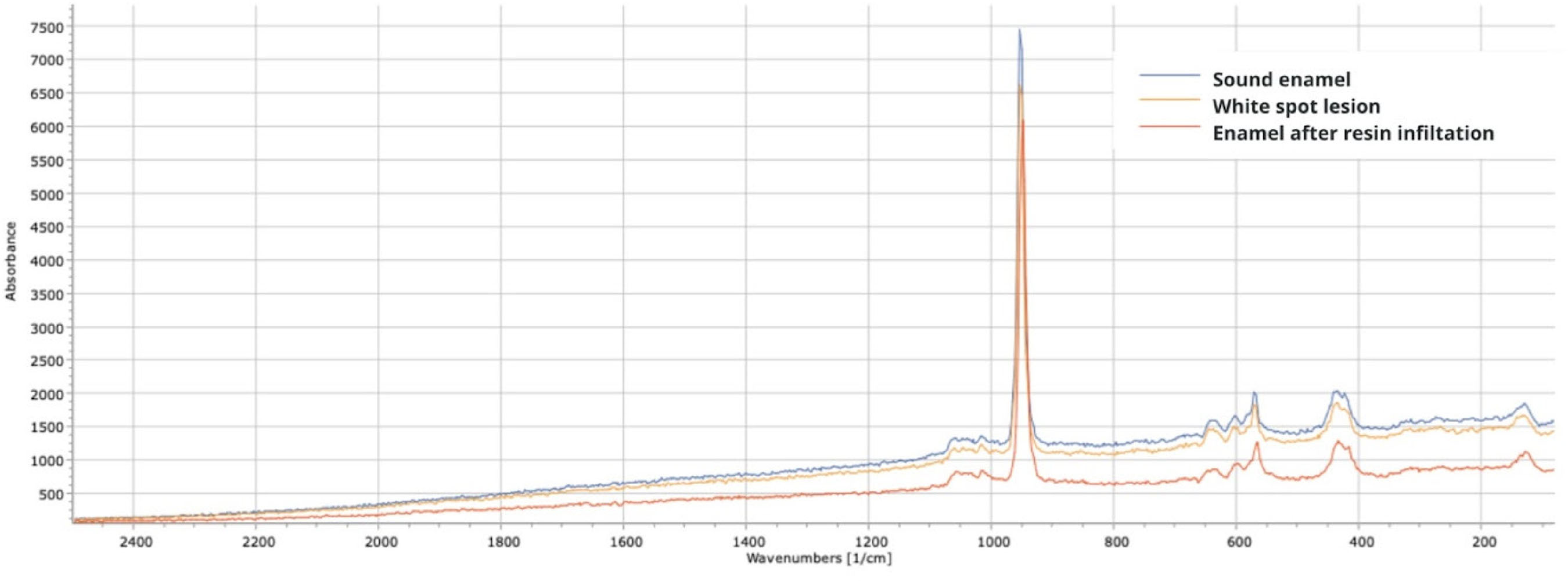
The data obtained through micro-raman spectroscopy show the detected spectrum after excitation, in the range of 150 to 1200 cm⁻¹, at the initial moment, after artificial caries induction, and after treatment with microabrasion and resin infiltrant. the data are presented in figures 1 and 2 described qualitatively.
The presence of phosphate groups (PO₄³⁻) is associated with apatite crystals and is detected in the ~960 cm⁻¹ band. other markers of mineral components linked to PO₄³⁻ vibrations are detected in the ~431 cm⁻¹ and ~589 cm⁻¹ bands. these phosphate group vibrations are characteristic indicators of apatite minerals [15].
Analyzing the study groups in this research, phosphate group (PO₄³⁻) vibrations are found in the 958 cm⁻¹ band in both the microabrasion-treated group (figure 1) and the group treated with resin infiltrant (figure 2), along with other markers showing vibrations at 427 cm⁻¹ and 586 cm⁻¹.

Micro-raman spectrum of sound permanent tooth enamel, after demineralization, and after treatment with microabrasion.

Micro-raman spectrum of sound permanent tooth enamel, after demineralization, and after treatment with resin infiltrant.
It is observed that the peaks identified after artificial induction of white spot lesions are lower than those observed in sound enamel samples, indicating mineral loss. after treatment, the distance between the peaks of sound enamel and post-treatment, observed around the ~960 cm⁻¹ wavelength, are closer especially in the group treated with microabrasion indicating a mineral content closer to that of sound enamel.
Energy-dispersive x-ray spectroscopy (sem/edx)
The images obtained by scanning electron microscopy are shown in figure 3 A to F. Figure 3 A and D show the surface of sound enamel. The surface appears smooth, uniform, and a layer of aprismatic enamel can be observed. No relevant irregularities are present. In B and E, after artificial induction of white spot lesions, a highly irregular surface is observed, with removal of the aprismatic layer. The presence of grooves and depressions of varying depths can be seen, which is representative of mineral loss characteristic of white spot lesions. “Gaps” can be observed in the enamel structure, which appears porous and uneven. In C, it is observed that the enamel, after the application of the resin infiltrant, acquires a more regular appearance; however, it does not reach the same appearance as sound enamel. Small elevations are observed, corresponding to the enamel prism pattern. In contrast, enamel treated with microabrasion (F) has an appearance equivalent to that of sound enamel, with a smooth surface and no irregularities.
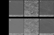
A to F: Morphological characteristics of sound enamel (A, D), after artificial induction of white spot lesions (B, E), and after treatment with resin infiltrant (C) and microabrasion (F). Magnification: 2000×.
The EDS data should be considered for qualitative analysis of the specimens, focusing on the superficial enamel. It is observed that sound enamel shows a higher content of calcium and phosphorus. After demineralization, the weight percentage of calcium and phosphorus is reduced, meaning the main mineral components of enamel were partially removed. With microabrasion, the mineral content returns to a level very similar to that of sound enamel. This is not observed after treatment with resin infiltrant, and the mineral content remains similar to that of enamel with white spot lesions (figure 4).

Representative images of the weight percentage of carbon, fluoride, calcium, and phosphorus in sound enamel, enamel with white spot lesions, and after treatment with resin infiltrant.
DISCUSSION
Both tested techniques (microabrasion and resin infiltration) have the ability to mask white spot lesions after orthodontic treatment. However, the mineral content and micromorphological characteristics of the enamel surface did not return to similar baseline levels after treatment with resin infiltrant. It is important to emphasize that neither of the two techniques is capable of “remineralizing” white spot lesions. Both techniques employ strategies to modify light reflection patterns, thereby restoring the aesthetic appearance of the teeth.
The white spot lesions treated in this study were artificially created in an in vitro model that aims to simulate the conditions of lesions that develop in vivo. In this scenario, the teeth were exposed to a demineralizing solution that is undersaturated with respect to hydroxyapatite, simulating the conditions of cariogenic dental biofilm fluid and enabling lesion formation [16]. By performing the procedure in a controlled laboratory environment, similar lesions were produced for both study groups in terms of depth and mineral content. This was confirmed by analyses such as micro-Raman and FEG. When performing the demineralization cycle using the Buskes solution combined with a remineralization period through immersion of the specimens in artificial saliva, a subsurface lesion is created, since the superficial region is able to remineralize during the process, mimicking what occurs in the oral environment [16].
The microabrasion technique promotes superficial enamel loss, and studies indicate an invasive nature of the procedure, especially if the treatment is repeated over the long term [6]. This level of wear is related to the application time, the concentration of the acid used, and the manual method of applying the abrasive agent. Furthermore, factors such as the type of enamel treated and the severity of the stains also influence the wear resulting from the procedure [6].
In this study, the material used for microabrasion was Whitness RM (FGM, Joinville, SC, Brazil), which contains 6% hydrochloric acid and silicon carbide in its composition. This combination, also present in other materials such as Opalustre (Ultradent Prod. Inc., Utah, USA), promotes significant substance loss, but a significant increase in microhardness is also reported [17]. In enamel treated with microabrasion, the formation of a highly polished surface is observed, resulting from the combination of chemical erosion and mechanical abrasion, which compacts the mineralized tissue in the interprismatic spaces during wear [18]. This explains the smooth and dense surface observed in the scanning electron microscopy images and the calcium and phosphorus levels after microabrasion similar to those of sound enamel identified by EDS.
Resin infiltration, in turn, is considered a minimally invasive treatment, with results obtained in a single session. The very fluid resin is capable of penetrating by capillarity into the microporosities of demineralized enamel; thus, it not only modifies the light reflection pattern but also occludes the acid diffusion pathways, which also favors lesion arrest [8]. It is stated that the surface resulting after infiltration can be considered a hybrid surface, with the spaces between apatite crystals filled by resin [19]. Enamel microhardness also increases significantly after the application of the resin infiltrant. However, although the structure treated with ICON shows greater mechanical resistance than demineralized enamel, it still proves less resistant to pH variations in the oral environment when compared to sound enamel. Furthermore, there is evidence that the microhardness of enamel treated with resin infiltrant increases significantly after the procedure.
The results obtained in this study show a lower level of mineral content after treatment with resin infiltrant, as observed in the qualitative EDS analysis. However, considering the characteristics already described, the fact that specimens treated with resin infiltrant showed lower mineral content may have a minimized significance, both from the standpoint of protection against future DEM-REM cycles and the mechanical resistance of the enamel.
The mineral variations observed in the different groups, although qualitative in nature, are confirmed by the two mineral analysis methods used − micro-Raman spectroscopy and EDS. These findings can be validated by future research, but certainly, the present study indicates a trend showing differences in mineral content resulting from treatment with microabrasion and resin infiltration techniques.
Color analysis was performed using a spectrophotometer to enable quantitative evaluation and eliminate subjective factors inherent to color assessment by the human eye. The literature recommends that the tooth surface to be analyzed be divided into sections for proper measurement [20], as done in this study. A difference of at least 3.7 points in ΔE is also considered necessary for a clinically perceptible color change in the tooth [20]. Thus, it can be considered in this study that both techniques were able to mask the effects of white spot lesions on the aesthetics of teeth after orthodontic treatment.
This result was also found in a clinical trial [9], which showed that the proposed aesthetic treatments were able to restore the appearance of enamel with white spots. However, another study reported that neither of the proposed methods − microabrasion nor resin infiltrant − was able to fully restore the tooth color [21].
One of the limitations of the present study is that color evaluation was performed only immediately after treatment; therefore, it is not possible to make any claims about the color stability or masking of the lesion over time. An in vitro study [5] evaluated the color change and stability of white spot lesions treated with microabrasion, resin infiltration, and remineralization with nano-hydroxyapatite (nHAP). After simulating pigment exposure (immersion in coffee for 7 days), all the treatments demonstrated good color stability, with the exception ofnHAP, which showed greater susceptibility to staining. Another clinical study [22] assessed the color durability of lesions treated with resin infiltrant and demonstrated that the aesthetics obtained after the procedure remained stable for up to four years. The long-term color stability of teeth treated with microabrasion is scarcely discussed in the literature. Therefore, this is a topic that still requires further investigation in future research.
Roughness was another criterion evaluated and showed minimal numerical variation in both study groups after aesthetic treatment, with no statistical differences. This is a beneficial finding, since a smooth surface minimizes niches for biofilm accumulation. The literature presents conflicting results regarding the influence of microabrasion on enamel roughness: while [23] report minimal changes − especially after polishing, which progressively smooths the surface by forming a superficial layer of approximately 15 µm composed of abrasive residues and smear layer, called “glazed enamel” − more recent studies demonstrate a significant increase in roughness when using Opalustre (a compound similar to Whiteness RM used in this study) without adequate polishing. Another study [18], for example, observed a significant increase in roughness in bovine incisors subjected to 15 applications of Opalustre (6.6% HCl + silicon carbide) or Whiteness RM, although homemade mixtures (10% HCl + pumice) resulted in lower roughness.
Regarding the roughness of enamel treated with resin infiltrant, the literature shows that the use of this technique allows the formation ofa very smooth surface [24]. This finding corroborates the data obtained in ourstudy. Despite showing slight elevations related to enamel prisms, the average roughness values and the surface micromorphology obsereved in the SEM images does not exhibit microporosities or irregularities capable of interfering with dental biofilm accumulation. It has also been demonstrated that this characteristic favors lower adhesion of Streptococcus mutans on the enamel surface [25].
One of the main limitations of this in vitro study is its inability to fully replicate the clinical conditions of the oral environment, which are essential for evaluating long-term aesthetic outcomes. Factors such as saliva, dietary habits, and variations in oral hygiene can significantly influence the appearance and stability of treated enamel surface. Additionally, mechanical stresses like brushing abrasion, thermal cycling, and staining from food and beverages are difficult to simulate precisely in vitro. Also, our study did not evaluate the color stability over time, which is a very important clinical variable.
Finally, it is essential to highlight that the techniques described here for masking white spot lesions have limitations, the main one being the lesion depth. Previous research has shown that microabrasion typically removes approximately 250 µm of the superficial enamel, and resin infiltration penetrates the enamel pores to about 100 µm [9]. Common sense and the clinician’s experience are essential tools to define the suitable treatment plan. Regarding white spot lesions, there is a correlation between the intensity of the lesion’s color and the depth of enamel demineralization [26]. In cases where lesions are deeper relative to the enamel thickness, it is likely that the desired aesthetic appearance will not be achieved with either technique [26], and the combination of techniques is probably desirable. In any case, considering proper diagnosis and indication, both procedures can be safely used with good immediate clinical results.
CONCLUSION
Based on the results of this study and within the limitations of the in vitro design, the following conclusions can be drawn:
-
● Both techniques (resin infiltration and microabrasion) were successful in masking white spot lesions.
-
● The final surface roughness of the treated enamel was comparable to that of sound enamel.
-
● The mineral content of enamel treated with microabrasion was similar to that of sound enamel, whereas enamel treated with resin infiltration showed a slightly reduced mineral content.
In both treatments, the post-treatment enamel surface appeared smooth and mineralized; however, the resin-infiltrated enamel presented slight, discrete elevations, likely corresponding to enamel prisms.
-
How to cite this article
Adimari LAW, Gaspar E, Rosa NVC, Wambier LM, Chibinski ACR. In vitro evaluation of different techniques for aesthetic treatment of enamel white spot lesions after orthodontic treatment. RGO, Rev Gaúch Odontol. 2026;74:e20260014. http://dx.doi.org/10.1590/1981-86372026001420250086
Data Availability
The research data are available from the corresponding author upon reasonable request.
REFERENCES
-
1 Hussain U, Wahab A, Kamran MA, Alnazeh AA, Almoammar S, Alshahrani SSM, et al. Prevalence, incidence and risk factors of white spot lesions associated with orthodontic treatment: a systematic review and meta-analysis. Orthod Craniofac Res. 2025;28(2):379-99. doi: https://doi.org/10.1111/ocr.12888
» https://doi.org/10.1111/ocr.12888 -
2 Thanetchaloempong W, Koontongkaew S, Utispan K. fixed orthodontic treatment increases cariogenicity and virulence gene expression in dental biofilm. J Clin Med. 2022;11(19):5860. doi: https://doi.org/10.3390/jcm11195860
» https://doi.org/10.3390/jcm11195860 -
3 Prada AM, Potra Cicalău GI, Ciavoi G. A Review of white spot lesions: development and treatment with resin infiltration. Dent J (Basel). 2024;12(12):375. doi: https://doi.org/10.3390/dj12120375
» https://doi.org/10.3390/dj12120375 -
4 Monjarás-Ávila AJ, Hardan L, Cuevas-Suárez CE, Alonso NVZ, Fernández-Barrera MÁ, Moussa C, et al. Systematic review and meta-analysis of remineralizing agents: outcomes on white spot lesions. Bioengineering (Basel). 2025;12(1):93. doi: https://doi.org/10.3390/bioengineering12010093
» https://doi.org/10.3390/bioengineering12010093 -
5 Novozhilova N, Mun A, Polyakova M, Mikheikina A, Zaytsev A, Babina K. Color change and color stability of white spot lesions treated with resin infiltration, microabrasion, or nano-hydroxyapatite remineralization: an in vitro study. Dent J (Basel). 2025;13(3):112. doi: https://doi.org/10.3390/dj13030112
» https://doi.org/10.3390/dj13030112 -
6 Nevárez-Rascón M, Molina-Frechero N, Adame E, Almeida E, Soto-Barreras U, Gaona E, et al. Effectiveness of a microabrasion technique using 16% HCL with manual application on fluorotic teeth: a series of studies. World J Clin Cases. 2020;8(4):743-56. doi: https://doi.org/10.12998/wjcc.v8.i4.743
» https://doi.org/10.12998/wjcc.v8.i4.743 -
7 Soveral M, Machado V, Botelho J, Mendes JJ, Manso C. Effect of resin infiltration on enamel: a systematic review and meta-analysis. J Funct Biomater. 2021;12(3):48. doi: https://doi.org/10.3390/jfb12030048
» https://doi.org/10.3390/jfb12030048 -
8 Bourouni S, Dritsas K, Kloukos D, Wierichs RJ. Efficacy of resin infiltration to mask post-orthodontic or non-post-orthodontic white spot lesions or fluorosis: a systematic review and meta-analysis. Clin Oral Investig. 2021;25(8):4711-9. doi: https://doi.org/10.1007/s00784-021-03931-7
» https://doi.org/10.1007/s00784-021-03931-7 -
9 Gu X, Yang L, Yang D, Gao Y, Duan X, Zhu X, et al. Esthetic improvements of postorthodontic white-spot lesions treated with resin infiltration and microabrasion: a split-mouth, randomized clinical trial. Angle Orthod. 2019;89(3):372-7. doi: https://doi.org/10.2319/041218-274.1
» https://doi.org/10.2319/041218-274.1 -
10 Gutiérrez MF, Malaquias P, Hass V, Matos TP, Lourenço L, Reis A, et al. The role of copper nanoparticles in an etch-and-rinse adhesive on antimicrobial activity, mechanical properties and the durability of resin-dentine interfaces. J Dent. 2017;61:12-20. doi: https://doi.org/10.1016/j.jdent.2017.04.007
» https://doi.org/10.1016/j.jdent.2017.04.007 -
11 Comodi P, Liu Y, Frezzotti ML. Structural and vibrational behaviour of fluorapatite with pressure. Part II: in situ micro-Raman spectroscopic investigation. Physics Chem Minerals. 2001;28(4):225-31. doi: https://doi.org/10.1007/s002690100155
» https://doi.org/10.1007/s002690100155 -
12 Buskes JA, Christoffersen J, Arends J. Lesion formation and lesion remineralization in enamel under constant composition conditions: a new technique with applications. Caries Res. 1985;19(6):490-6. doi: https://doi.org/10.1159/000260887
» https://doi.org/10.1159/000260887 -
13 Queiroz CS, Hara AT, Paes Leme AF, Cury JA. pH-cycling models to evaluate the effect of low fluoride dentifrice on enamel de- and remineralization. Braz Dent J. 2008;19(1):21-7. doi: https://doi.org/10.1590/s0103-64402008000100004
» https://doi.org/10.1590/s0103-64402008000100004 -
14 Göhring TN, Zehnder M, Sener B, Schmidlin PR. In vitro microleakage of adhesive-sealed dentin with lactic acid and saliva exposure: a radio-isotope analysis. J Dent. 2004 Mar;32(3):235-40. doi: https://doi.org/10.1016/j.jdent.2003.11.003
» https://doi.org/10.1016/j.jdent.2003.11.003 -
15 Buchwald T, Okulus Z, Szybowicz M. Raman spectroscopy as a tool of early dental caries detection–new insights. J Raman Spectrosc. 2017;48(8):1094-102. https://doi.org/10.1002/jrs.5175
» https://doi.org/10.1002/jrs.5175 -
16 Magalhães AC, Moron BM, Comar LP, Wiegand A, Buchalla W, Buzalaf MA. Comparison of cross-sectional hardness and transverse microradiography of artificial carious enamel lesions induced by different demineralising solutions and gels. Caries Res. 2009;43(6):474-83. doi: https://doi.org/10.1159/000264685
» https://doi.org/10.1159/000264685 -
17 Yazkan B, Ermis RB. Effect of resin infiltration and microabrasion on the microhardness, surface roughness and morphology of incipient carious lesions. Acta Odontol Scand. 2018;76(7):473-81. doi: https://doi.org/10.1080/00016357.2018.1437217
» https://doi.org/10.1080/00016357.2018.1437217 -
18 Silva PLPD, Maciel PP, Martins LBC, Carvalho FGD, Santos RLD, Medeiros ESD, et al. Weight-loss and surface roughness of enamel after microabrasion procedure with different agents. Rev Odontol Unesp. 2021;50:e20210020. https://doi.org/10.1590/1807-2577.03421
» https://doi.org/10.1590/1807-2577.03421 -
19 Chabuk MM, Al-Shamma AM. Surface roughness and microhardness of enamel white spot lesions treated with different treatment methods. Heliyon. 2023;9(7):e18283. doi: https://doi.org/10.1016/j.heliyon.2023.e18283
» https://doi.org/10.1016/j.heliyon.2023.e18283 -
20 Gençer MDG, Kirzioğlu Z. A comparison of the effectiveness of resin infiltration and microabrasion treatments applied to developmental enamel defects in color masking. Dent Mater J. 2019;38(2):295-302. doi: https://doi.org/10.4012/dmj.2018-074
» https://doi.org/10.4012/dmj.2018-074 - 21 Silva LO, Signori C, Peixoto AC, Cenci MS, Faria-E-Silva AL. Color restoration and stability in two treatments for white spot lesions. Int J Esthet Dent. 2018;13(3):394-403.
-
22 Puleio F, Di Spirito F, Lo Giudice G, Pantaleo G, Rizzo D, Lo Giudice R. Long-term chromatic durability of white spot lesions through employment of infiltration resin treatment. Medicina (Kaunas). 2023;59(4):749. doi: https://doi.org/10.3390/medicina59040749
» https://doi.org/10.3390/medicina59040749 -
23 Rodrigues MC, Mondelli RF, Oliveira GU, Franco EB, Baseggio W, Wang L. Minimal alterations on the enamel surface by micro-abrasion: in vitro roughness and wear assessments. J Appl Oral Sci. 2013;21(2):112-7. doi: https://doi.org/10.1590/1678-7757201302117
» https://doi.org/10.1590/1678-7757201302117 -
24 Ibrahim DFA, Hasmun NN, Liew YM, Venkiteswaran A. Repeated etching cycles of resin infiltration up to nine cycles on demineralized enamel: surface roughness and esthetic outcomes in vitro study. Children (Basel). 2023;10(7):1148. doi: https://doi.org/10.3390/children10071148
» https://doi.org/10.3390/children10071148 -
25 Arslan S, Zorba YO, Atalay MA, Özcan S, Demirbuga S, Pala K, et al. Effect of resin infiltration on enamel surface properties and Streptococcus mutans adhesion to artificial enamel lesions. Dent Mater J. 2015;34(1):25-30. doi: https://doi.org/10.4012/dmj.2014-078. Erratum in: Dent Mater J. 2016;35(2):333. doi: https://doi.org/10.4012/dmj.2014-078-e
» https://doi.org/10.4012/dmj.2014-078 -
26 Abbas BA, Marzouk ES, Zaher AR. Treatment of various degrees of white spot lesions using resin infiltration-in vitro study. Prog Orthod. 2018;19(1):27. doi: https://doi.org/10.1186/s40510-018-0223-3
» https://doi.org/10.1186/s40510-018-0223-3
Edited by
-
Assistant editor
Luciana Butini Oliveira