 Rehabilitation of a young patient with palate obturator prosthesis: 11 years of follow-up
ABSTRACT
The patient was referred to the Hospital das Clínicas de São Paulo for resection of the lesion by a head and neck surgeon. Resection was performed in January 2008 and the final diagnosis was low-grade osteoblastic osteosarcoma. He was referred to the prosthetic clinic at the Institute of Science and Technology-UNESP in São José dos Campos, presenting loss of the right hemi-maxilla. The patient was rehabilitated in 2012 with a removable partial palate obturator prosthesis and then distanced himself from treatment, which caused deleterious consequences due to the inherent wear of the material and the maladaptation of the prosthesis, causing him difficulties in speaking and swallowing. After a period of 11 years, the patient returned in 2023 for follow-up and new rehabilitation, being rehabilitated with a removable partial palate obturator prosthesis, where it was possible to obtain a satisfactory maxillofacial prosthesis that provides the patient with the possibility of speaking, eating and swallowing better comfort, despite the limitations that the case presented, in addition to providing psychosocial improvement to the patient.
Indexing terms
Dental prosthesis; Dentistry; Diagnosis; Maxillofacial Prosthesis; Osteosarcoma
RESUMO
O paciente foi encaminhado ao Hospital das Clínicas de São Paulo para ressecção da lesão por um cirurgião de cabeça e pescoço. A ressecção foi realizada em janeiro de 2008 e o diagnóstico final foi osteossarcoma osteoblástico de baixo grau. Ele foi encaminhado à clínica de próteses do Instituto de Ciência e Tecnologia-UNESP em São José dos Campos, apresentando perda da hemi-maxila direita. O paciente foi reabilitado em 2012 com uma prótese de obturador palatino parcial removível e, posteriormente, se distanciou do tratamento, o que causou consequências deletérias devido ao desgaste inerente do material e à má adaptação da prótese, causando-lhe dificuldades na fala e deglutição. Após um período de 11 anos, o paciente retornou em 2023 para acompanhamento e nova reabilitação, sendo reabilitado com uma prótese de obturador palatino parcial removível, onde foi possível obter uma prótese maxilofacial satisfatória que proporciona ao paciente a possibilidade de falar, comer e engolir com maior conforto, apesar das limitações que o caso apresentou, além de proporcionar melhoria psicossocial ao paciente.
Termos de indexação
Prótese dentária; Odontologia; Diagnóstico; Prótese maxilofacial; Osteossarcoma
INTRODUCTION
Deformities in the head and neck regions are defined as oral and maxillofacial deficiencies [1] and cause various social, psychological and even family problems for the patient. These deformities may originate from development, pathologies or consequences of accidents.
Deformities arising from pathologies are often associated with jaw tumors, such as sinus tumors, which are difficult to diagnose due to their frequent asymptomatic course until infiltration of adjacent structures occurs, negatively affecting treatment results, where patients are diagnosed already in advanced stage [2].
Despite being a different specialty in dentistry, and its knowledge being little publicized, oral and maxillofacial prosthesis is extremely old in society. According to Murphey and collaborators in 1945, the Romans and Greeks decorated their statues with artificial eyes made with precious stones such as gold, as early as 200 BC, in ancient China even statues had, in place of eyes, precious stones such as jade in the eyeball region [3], thus representing a search for improvement in the replication of humanoid forms for society [4].
The greatest difficulty encountered in oral and maxillofacial rehabilitation must be the fact that the human face being complex, consisting of several soft and hard tissues, each with the function of allowing the individual to bring expressions according to the movement of the muscles, providing the ability to express their feelings and even assist in your communication [1,5]. That said, the branch of oral and maxillofacial prostheses aims to carry out rehabilitative treatments, restoring quality of life and reintegration of the mutilated patient into society, bringing maximum comfort, function and aesthetics within the limitations of each case, extending both to intra-oral losses (teeth, palate and oral structures) in terms of major losses of the maxilla and mandible, also covering cleft lip and palate and extraoral losses (in the face region), such as extensive facial, nasal, palpebral and ocular losses [6,7]. This is extremely important, since there are already studies on the quality of life of patients who use maxillofacial prostheses and present high levels of emotional suffering, physical limitations, body image disorders and impairments in social relationships [8-10].
CASE REPORT
A 27-years-old, white male patient, born in São José dos Campos, Brazil was forwarded to the prosthesis clinic at the Institute of Science and Technology-UNESP (ICT-UNESP) in São José dos Campos, presenting loss of the right hemi-maxilla. This case report was reviewed and approved by the Research Ethics Committee of UNESP, CAAE 81815624.5.0000.0077
History of the disease: in October 2007 (at the age of 12) the patient noticed the appearance of a vesicle in the hard palate region. He was evaluated by a dentist who performed drainage of the lesion, which revealed serosanguineous secretion coming out and was advised on therapy with amoxicillin. The lesion grew again, this time accompanied by “softening” of the upper right molars. The patient underwent biopsy at the Faculty of Dentistry of São José dos Campos, currently the ICT-UNESP of São José dos Campos.
Anatomopathological result: it was observed a malignant mesenchymal neoplasm containing a group of pleomorphic polygonal cells in which there is deposition of osteoid in varying degrees of mineralization, forming irregular bone trabeculae and numerous irregulars or rounded mineralized masses, with aspects of dystrophic calcification. Neoplastic cells presented nuclei that were sometimes eccentric, vacuolated and with evident or hyperchromatic nucleoli. The anatomopathological test was carried out at ICT UNESP by the department of oral biopathology.
Diagnostic hypothesis: compatible with osteoid osteosarcoma, requiring immunohistochemical analysis to confirm the diagnosis.
Diagnosis: The immunohistochemical profile was compatible with an osteoblastic lesion with low proliferative activity. The immunohistochemistry test was carried out by the pediatric oncology group in São José dos Campos.
The patient was referred to the Hospital das Clínicas in São Paulo for resection of the lesion with a head and neck surgeon, as the lesion was already growing rapidly, compromising swallowing and airways. Tumor resection was performed in January 2008 and the material was sent for diagnostic confirmation.
Diagnosis of tumor excision in the right maxilla: was Low-grade osteoblastic osteosarcoma. The neoplasm measured 10 cm, affecting the bone of the hard palate and mucosa, with erosion.
After excision of the lesion, an immediate post-surgical removable partial prosthesis was performed, to which the patient did not adapt aesthetically at another institution in the city of São José dos Campos.
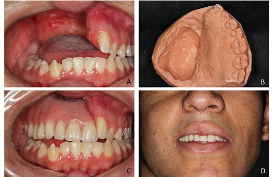
After authorization from the responsible doctor, an emergency temporary removable partial obturator prosthesis was performed in 2012, at the ICT-UNESP in São José dos Campos, so that the patient could eat and improve aesthetics. The initial state showing the loss of the hemi-maxilla, the plaster model obtained to create the prosthesis and the temporary prosthesis installed can be seen in figure 1A to D.
A) Situation of the initial case before starting the creation of the temporary prosthesis in the frontal photograph. B) Plaster model obtained for the manufacture of the provisional prosthesis. C) Temporary prosthesis installed. D) Final appearance of the provisional prosthesis.
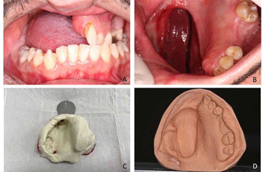
Patient went to live in Japan and after 10 years, he returned to the ICT-UNESP in São José dos Campos to have a new removable partial obturator prosthesis made due to the disfitting of the previous one. The loss of the right hemi-maxilla can be seen in figures 2A and 2B.

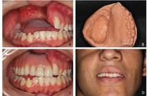
Images from the case report. A) Status of the initial case in frontal photography. B) Occlusal photograph of the maxilla showing the oral defect. C) Alginate molding carried out for the patient’s rehabilitation. D) Plaster model obtained from the impression.
On facial examination, a loss of lip support was observed due to the absence of teeth in the mid-arch, and the patient also mentioned breathing, chewing and speech problems. The treatment consisted of making a removable partial palate obturator prosthesis in thermally activated acrylic resin with a metal frame and clips.
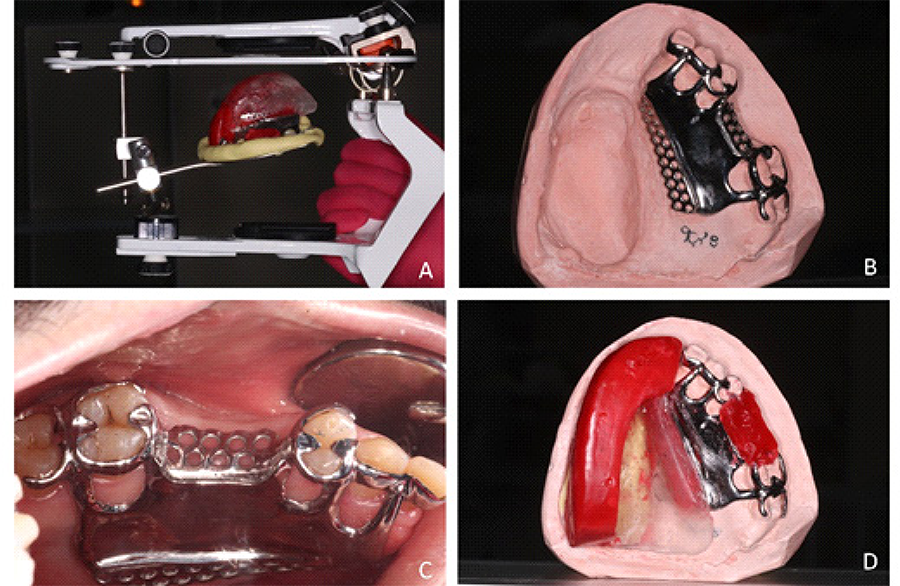
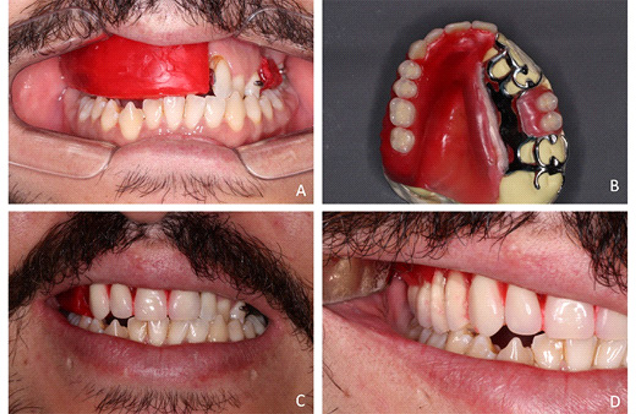
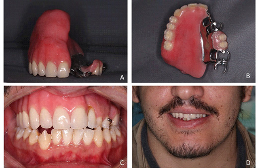
To manufacture the prosthesis, the entire maxilla was molded in alginate (figure 2C) to obtain the plaster model (figure 2D) and the antagonist arch was molded to mount the case in a semi-adjustable articulator (figure 3A). Frame planning and necessary preparations were carried out to receive the prosthesis frame on the remaining teeth, as well as preparations for supports and guide planes. After casting the frame, it was tested and adjusted (figure 3B and 3C), and then a test base was created next to the metal frame to create the wax plane to record the intermaxillary and aesthetic relationships and subsequently carry out the assembly of teeth (figure 3D and figure 4B to D). After the aesthetic test, the prosthesis was acrylicized and installed (figure 5A to D).

Images of the case report. A) Assembly of the upper model on a semi-adjustable articulator. B). Metal frame for removable partial obturator prosthesis. C). Test of the metal frame in the mouth. D) Mounting of orientation plane in wax.
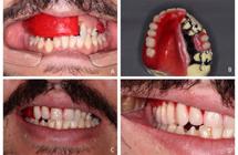
Images of the case report. A) Wax orientation plane test in mouth with distension of lips and cheeks. B) Assembly of artificial teeth. C) Testing of artificial teeth. D) Testing of artificial teeth with distension of lips and cheeks.
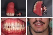
Images of the case report. A) Acrylic prosthesis in frontal view. B) Acrylic prosthesis in occlusal view. C) Installation of the acrylic prosthesis. D) Final appearance of the prosthesis.
DISCUSSION
Obturator, facial and maxillofacial prostheses are important not only in functional and aesthetic rehabilitation, but also in the patient’s resocialization. The use of prostheses to rehabilitate patients who have had extensive tissue loss is an alternative for aesthetic and functional facial reconstruction, since in these cases conventional reconstructive surgery cannot be applied [11].
After surgery to remove cancer in the head and neck region, the rehabilitation of the patient’s aesthetics and function (chewing, swallowing and speaking) can be reestablished through the use of maxillofacial prostheses [12], which can be planned and manufactured immediately after surgery to restore chewing and swallowing function in cases similar to those reported and final prostheses, with greater functional and aesthetic planning after the surgeon/doctor authorizes it.
The removal of tumors in the maxilla is the main reason for maxillary resection surgery, which can involve the maxillary sinus, nasal cavity and hard palate, structures that, when affected, can lead to communication between the oral and nasal cavities, causing the entry of a food bolus and liquids in the nasal cavity, which can lead to difficulty in eating, such as choking (with consequent weight loss and nutritional deficiency) and distortion of speech sounds due to the air exit from the nasal cavity to the oral cavity, which can lead to unintelligible speech, and in some cases aesthetic-social damaged [12]. The case presented shows an extensive loss of the maxilla, with buco-sinus communication where the patient had difficulty swallowing and speaking.
Maxillofacial obturator prostheses need to fulfill some functions, such as facilitating feeding, protecting the wound or keeping the defective area clean, increasing trauma healing, helping in the remodeling and reconstruction of the palatal contour, promoting speech and improving aesthetics [11,13-15], especially in cases as presented, as the patient is a young and active man where the aesthetic part has a strong participation in the psychosocial relationship. With the creation of the maxillofacial obturator prosthesis, the patient began to have better oral speech, easier drinking and eating, as oral-sinus communication was interrupted by the use of the prosthesis, in addition to aesthetic and psychosocial improvements.
In addition to these attributions, the retention of maxillofacial obturator prostheses depends on factors such as direct or indirect retention promoted by the remaining teeth, size of the defect, retention of available tissue around the cavity and development of muscular control [11], in addition to the size and location of the defect that can cause adaptation difficulties and instability of the prosthetic part. In this case, retention of the prosthesis was obtained by the remaining teeth 22, 23, 24, 27 and 28, which received metal clasps, by the left hemipalate and by the size of the defect. These requirements are important for the success of the treatment, as they increase the quality of life of patients after maxillectomy.
The use of a maxillofacial obturator prosthesis is necessary to perform the functional separation of the oral and nasal cavities and must be used for the rest of the patient’s life, requiring periodic evaluation due to the useful life of the material used in manufacturing the prostheses [12].
CONCLUSION
It was possible to obtain a satisfactory maxillofacial prosthesis that gives the patient the possibility of speaking, eating and swallowing with better comfort despite the limitations that the case presented, in addition to enabling the patient to achieve psychosocial improvement.
The prosthetic piece has limitations regarding the time of use and must be evaluated periodically as it may present maladaptations and cause speech and swallowing problems, in addition to the deterioration of the material over time.
The patient was informed of the need for periodic replacements, as he had been using the temporary prosthesis for 10 years.
With the possibility of following the case for 11 years, it was possible to observe that the previous use of the temporary prosthesis made the patient’s adaptation to the new prosthesis easier.
It was possible to observe that, as discussed in the discussion, the patient presented speech and swallowing impairments, as well as psychosocial impairments resulting from the maladaptation of the temporary prosthesis, which was already worn out due to use and in maladaptation due to tissue modification and tooth loss.
-
How to cite this article
Paes Júnior TA, Kukulka EC, Lima BSS, Miranda PRP, Furquim MA, Almeida JD, et al. Rehabilitation of a young patient with palate obturator prosthesis: 11 years of follow-up. RGO, Rev Gaúch Odontol. 2024;72:e20240046. http://dx.doi.org/10.1590/1981-86372024004620240016
REFERENCES
-
1 Lopes BCS, Bueno RB, Custodio WC, de Godoi APT. Color stability of silicones for use in maxillofacial prosthesis submitted to human sweat. Braz J Dev 2021;7:79879-79895. https://doi.org/10.34117/bjdv7n8-279
» https://doi.org/10.34117/bjdv7n8-279 -
2 Walshaw EG, Taylor R, Anderson J, Sexton P, Parmar JD, Carter LM. The psychological sequelae of maxillofacial trauma: a scoping review of the literature. Br J Oral Maxillofac Surg 2022;60:1303-1320. https://doi.org/10.1016/j.bjoms.2022.09.013
» https://doi.org/10.1016/j.bjoms.2022.09.013 -
3 Murphey PJ, Pitton RD, Schlossberg L, Harris LW. The development of acrylic eye prosthesis at the national naval medical center. IJD, Int J Dent. 1945;32:1227-1244. https://doi.org/10.14219/jada.archive.1945.0182
» https://doi.org/10.14219/jada.archive.1945.0182 - 4 Dias RB, Herrera LP, Reis RCD, Coto, NP. Contribuição da prótese bucomaxilofacial na internacionalização da Odontologia. Rev Assoc Paul Cir Dent 2016;70:122-125.
-
5 Swift A, Green JB, Hernandez CA, Aguilera SB, Fagien S, Gold MH, Casabona G, Frank K, Nikolis A, Cotofana S. Tips and tricks for facial toxin injections with illustrated anatomy. Plast. Reconst. Surg. 2022;149(2):303e-312e. https://doi.org/10.1097/PRS.0000000000008708
» https://doi.org/10.1097/PRS.0000000000008708 - 6 Simões FG, de Brito R. A especialidade de prótese bucomaxilofacial e sua atuação na Odontologia. RSBO 2009;6:327-331.
- 7 Rodrigues RGS, Rodrigues DS, de Oliveira DC. Reabilitação com prótese bucomaxilofacial: revisão de literatura. Rev Saúde Multid. 2019;5:20-27.
-
8 Schoen PJ, Raghoebar GM, van Oort RP, Reintsema H, van der Laan BF, Burlage FR, Roodenburg JL, Vissink A. Treatment outcome of bone-anchored craniofacial prostheses after tumor surgery. Cancer. 2001;92:3045-3050. https://doi.org/10.1002/1097-0142(20011215)92:12<3045::aid-cncr10147>3.0.co;2-k
» https://doi.org/10.1002/1097-0142(20011215)92:12<3045::aid-cncr10147>3.0.co;2-k -
9 Atay A, Peker K, Günay Y, Ebrinç S, Karayazgan B, Uysal Ö. Assessment of health-related quality of life in Turkish patients with facial prostheses. Health Qual Life Outcomes. 2013 Jan 26;11:11. https://doi.org/10.1186/1477-7525-11-11
» https://doi.org/10.1186/1477-7525-11-11 -
10 Irish J, Sandhu N, Simpson C, Wood R, Gilbert R, Gullane P, et al. Quality of life in patients with maxillectomy prostheses. Head Neck. 2009;31:813-821. https://doi.org/10.1002/hed.21042
» https://doi.org/10.1002/hed.21042 -
11 Goiato MC, Pesqueira AA, da Silva CR, Gennari Filho H, dos Santos DM. Patient satisfaction with maxillofacial prosthesis. Literature review. J Plast Reconstr Aesthet Surg. 2009;62:175-180. https://doi.org/10.1016/j.bjps.2008.06.084
» https://doi.org/10.1016/j.bjps.2008.06.084 -
12 Phasuk K, Haug SP. Maxillofacial prosthetics. Oral Maxillofac Surg Clin North Am. 2018;30:487-497. https://doi.org/10.1016/j.coms.2018.06.009
» https://doi.org/10.1016/j.coms.2018.06.009 -
13 Lang BR, Bruce RA. Presurgical maxillectomy prosthesis. J Prosthet Dent. 1967;17(6):613-619. https://doi.org/10.1016/0022-3913(67)90133-3
» https://doi.org/10.1016/0022-3913(67)90133-3 - 14 Chalian VA. Maxillofacial prosthetics: multidisciplinary practice. Preston Street Baltimor U.S.A: The Willians & Wilkins Company; 1971.
-
15 Keyf F. Obturator prostheses for hemimaxillectomy patients. J Oral Rehabil. 2001;28:821-829. https://doi.org/10.1046/j.1365-2842.2001.00754.x
» https://doi.org/10.1046/j.1365-2842.2001.00754.x
Edited by
-
Assistant editor:
Luciana Butini Oliveira