
 Click-evoked auditory brainstem response: The influence of age and risk factors for hearing loss
ABSTRACT
Purpose: to analyze the relationship between infants’ auditory brainstem response results and their corrected gestational age, considering prematurity and risk factors for hearing loss.
Methods: a cross-sectional, observational study with 62 infants, divided into G1 (27 to 36 weeks) and G2 (37 to 41 weeks). After normal NHS results, the click-evoked ABR investigated the integrity of the auditory pathways and the electrophysiological threshold. Data were analyzed using descriptive statistics, as well as the association with the Mann-Whitney test and the correlation between electrophysiological threshold and corrected gestational age using the Spearman’s test with a significance level of 5%.
Results: neither group had statistically significant different absolute latency values and interpeak intervals in the auditory pathway integrity study. The electrophysiological threshold study found a statistically significant difference between G1 and G2. Likewise, the difference between the electrophysiological threshold and the presence of risk factors for hearing loss and between the electrophysiological threshold and prematurity was statically significant. The electrophysiological threshold was weakly correlated with the corrected gestational age.
Conclusion: the corrected gestational age in the study population did not influence the absolute latency parameters and interpeak intervals. However, the electrophysiological threshold was better in the group whose corrected gestational age was 37 or more weeks. Moreover, the presence of risk factors for hearing loss helped increase the electrophysiological threshold.
Keywords:
Hearing; Infant, Newborn; Evoked Potentials, Auditory; Auditory Threshold; Risk Index
RESUMO
Objetivo: analisar a relação entre os resultados dos Potenciais Evocados Auditivos de Tronco Encefálico e a idade gestacional corrigida de lactentes, considerando prematuridade e Indicadores de Risco para Deficiência Auditiva (IRDA).
Métodos: estudo observacional transversal, composto por 62 lactentes, divididos em: G1 - 27 a 36 semanas e G2 - 37 a 41 semanas. Após resultado normal na TAN, foi realizado o PEATE por clique para pesquisa de integridade das vias auditivas e de limiar eletrofisiológico. Os dados foram analisados por meio de estatística descritiva, de associação amparada pelo teste Mann-Whitney e de correlação entre limiar eletrofisiológico e idade gestacional corrigida por meio do teste Spearman. O nível de significância adotado foi de 5% e intervalos de confiança de 95%.
Resultados: os dois grupos apresentaram valores de latências absolutas e intervalos interpicos sem diferença estatisticamente significante na pesquisa de integridade de vias auditivas. Na pesquisa de limiar eletrofisiológico, foi observada diferença com relevância estatística entre G1 e G2. Na relação entre limiar eletrofisiológico e presença de IRDA e limiar eletrofisiológico com prematuridade também foi observada diferença com relevância estatística. Houve correlação fraca entre limiar eletrofisiológico e idade gestacional corrigida.
Conclusão: conclui-se que, na população avaliada, os parâmetros de latência absoluta e os intervalos interpicos não foram influenciados pela idade gestacional corrigida. Entretanto, o limiar eletrofisiológico foi melhor no grupo com idade gestacional corrigida superior a 37 semanas. Pode-se também observar que a presença de indicadores de risco para a deficiência auditiva contribuiu para o aumento do limiar eletrofisiológico.
Descritores:
Audição; Recém-Nascido; Potenciais Evocados Auditivos; Limiar Auditivo; Indicador de Risco
INTRODUCTION
Hearing disorders can lead to deficits in oral language and cognitive, intellectual, cultural, and social development. Hence, measures to detect hearing disorders should be taken as early as possible, favoring the development of language and allowing the establishment of social function1.
According to the Pan American Health Organization (PAHO), an estimated 217 million people in the Americas have hearing loss, with a predictable increase to 322 million by 20502.
In developed countries, it is estimated that one in every thousand births has some degree of hearing loss, and this prevalence is higher in newborns with some risk factor for hearing loss (RFHL)3,4. It is known that RFHL may be associated with prematurity, due to the preterm infant’s health conditions5.
Prematurity is closely linked to auditory pathway maturation and may negatively impact the development of auditory and language skills5. This information is even more relevant when considering that the prevalence of prematurity in Brazil is around 11.1%, according to a study6 that researched this prevalence between 2011 and 2021.
Maternity hospitals must offer neonatal hearing screening (NHS) to identify hearing loss early. In Brazil, performing the NHS is a right guaranteed by Law no. 12,303, of August 2, 2010, which establishes the mandatory performance of the test called transient evoked otoacoustic emissions (TEOAE). However, infants with RFHL should be submitted to automated auditory brainstem response (A-ABR), which is efficient for detecting cochlear and retrocochlear hearing loss7. This is also an important test in the subsequent NHS stages - diagnosis and monitoring8.
The ABR assesses auditory pathway integrity and determines the electrophysiological threshold3. It is interpreted by analyzing the absolute latencies, interpeak intervals, morphology and amplitude of the waves generated, and the reproducibility of the tracing3,9. Several stimuli can be used to elicit neural responses, such as CE-chirp and speech stimuli. However, the click stimuli are still the most common in clinical practice1,3,10.
The interference of prematurity and RFHL in auditory pathway maturation is widely discussed9. However, studies seldom address their real impact on ABR regarding parameters that can be considered normal in these cases, and it is not correct to use normative data from full-term infants because they generate confounding factors in the audiological diagnosis.
Thus, this study aimed to analyze the relationship between ABR results and the corrected gestational age (CGA) between 27 and 41 weeks of infants in an NHS program, considering prematurity and RFHL.
METHODS
This research was approved by the Research Ethics Committee of the Universidade Federal de Minas Gerais, MG, Brazil, under evaluation report number 934.475. Data were collected at the rooming-in ward and the neonatology unit of the Clinics Hospital of the Universidade Federal de Minas Gerais (HC-UFMG). All parents/guardians of the research volunteers signed an informed consent form, as provided for in Resolution 466 of December 12, 2012.
This cross-sectional observational study was carried out in a reference NHS service.
The sample had 62 preterm and full-term infants born at the HC-UFMG maternity hospital, which provides care for high-risk pregnancies and deliveries from all over the state of Minas Gerais. The infants selected for the study had gestational ages (GA) between 24 and 41 weeks (gestational weeks on the day of birth). On the day of the examination, the chronological age (CA) in weeks was adjusted for the CGA - i.e., the GA was added to the CA in weeks, resulting in the CGA. The infants were assessed with a CGA between 27 and 41 weeks.
All infants, including extremely premature ones and with a few weeks of life, only participated in the study after ensuring the conditions for carrying out the exams, without compromising the care of the child, and authorized by the physician responsible for them, as the objective of this study was related to GA.
The sample was stratified into groups for the study. Infants ≤ 36 gestational weeks were considered preterm, and those ≥ 37 gestational weeks were considered full-term11.
Thus, groups were divided according to CGA on the day of the examination: Group 1 (G1) had infants whose CGA was between 27 and 36 weeks (n = 32), and Group 2 (G2) had infants whose CGA was between 37 and 41 weeks (n = 30).
The first stage of the study was carried out through NHS, with TEOAE and A-ABR, as recommended in the literature5,7. The research included the cases with a “pass” result.
This study performed the following procedures:
-
Medical history survey: To obtain identification, pregnancy, perinatal, infant health, and family history data.
-
Otoscopy: To inspect the external auditory meatus (EAM).
-
-NHS using TEOAE and A-ABR.
-
TEOAE: The equipment used to record TEOAEs was the Elios® from ECHODIA. The TEOAE recording protocol uses non-linear click stimuli at 80 dB SPL in 1, 2, 3, 4, and 5 KHz. TEOAEs are present when reproducibility is greater than or equal to 70% and the signal-to-noise ratio is greater than or equal to 3 dB in at least three of the five frequency bands.
-
A-ABR: This procedure was performed automatically by ECHODIA brand Elios® equipment, investigating the presence of wave V in two scans at 40 dB nHL. To be considered normal, the exam should present wave V in both scans, with a latency difference of up to 0.3 ms between them and reproducibility equal to or greater than 75%. The frequency investigated with click stimuli ranges from 100 Hz to 5 KHz. To perform the A-ABR, surface electrodes were placed in the Fp1, Fz, A1, and A2 positions, after cleaning the skin with abrasive gel, ensuring acceptable impedance ≤ 7 kOhms, and presenting stimuli through insert earphones.
-
The screening “pass” criteria included the bilateral presence of TEOAE and A-ABR. Infants with a “pass” result were then submitted to the diagnostic ABR test.
-
Diagnostic ABR: Auditory pathway integrity was assessed in two scans, with click stimuli at 80 dB nHL to analyze waves I, III, and V and interpeak intervals I-III, III-V, and I-V. Continuing the test, the research was performed at 60, 40, 35, 30, 25, 20, 15, and 10 dB nHL or until the electrophysiological threshold was found. Only wave V was researched after analyzing the waves at 80 dB nHL. Recordings were made in Elios® from ECHODIA with the following protocol: click stimuli; alternating polarity; stimulus presentation rate - 23 clicks/s; 1,000 mediations; low-pass filters of 3,000 Hz and high-pass filters of 50 Hz; acceptable impedance (artifacts) ≤ 7 kOhms. Infants were assessed in natural sleep in an acoustically treated room, using the same electrodes already positioned for the A-ABR, verifying the impedance, and presenting the stimuli through insert earphones.
The inclusion criterion was an NHS “pass” result (presence of TEOAE and normal A-ABR, bilaterally). The exclusion criteria were not completing the diagnostic ABR exam and the parent/guardian withdrawing from participating in the research.
The information collected was entered into a Google form and exported to an Excel spreadsheet, and the data were statistically analyzed using the SPSS program. Descriptive analysis of the data was performed using frequency distribution for categorical variables (sex, presence and type of RFHL, and prematurity) and measures of central tendency (mean, median), standard deviation, minimum, and maximum for continuous variables (GA, CGA, and birth weight). Then, sample distribution analysis was performed, observing an asymmetric distribution of continuous variables: absolute latencies of waves I, III, and V and interpeak intervals I-III, I-V, and III-V at 80 dB nHL, and electrophysiological thresholds. Thus, the Wilcoxon test compared the right and left ears, and the Mann-Whitney test assessed whether there was a difference between females and males. No variable had a statistically significant difference in these analyses. Therefore, it was decided to group them for analysis per CGA group (G1 and G2).
The Mann-Whitney test compared G1 and G2 regarding the absolute latency of waves I, III, and V and interpeak intervals I-III, I-V, and III-V at 80 dB nHL, and the electrophysiological thresholds. This test also compared electrophysiological thresholds considering the presence of RFHL and prematurity, regardless of the group. The Spearman’s test was applied to verify the correlation between CGA and electrophysiological threshold, considered weak when 0 < r < 0.4; moderate when 0.4 < r < 0.7; and strong when 0.7 < r < 1.0. Moderate and strong correlations with p < 0.05 were considered significant. The significance level was 5%, with 95% confidence intervals.
RESULTS
The sample had 62 infants - 31 males and 31 females; 87.09% (n = 54) of the sample had at least one RFHL, and 56.45% (n = 35) were premature. The distribution of the population characteristics (GA, CGA, birth weight, prematurity, and RFHL) is shown in Tables 1 and 2, stratified by groups according to GA on the day of the examination - G1 with infants whose CGA was between 27 and 36 weeks (n = 32), and G2 with infants whose CGA was between 37 and 41 weeks (n = 30).
The most frequent RFHL in NHS was a neonatal intensive care unit (NICU) stay of more than 5 days (50.79%, n = 32), followed by hyperbilirubinemia (26.98%, n = 17), low birth weight (≤ 1500 g) (23.80%, n = 15), mechanical ventilation (20.63%, n = 13), congenital infections (toxoplasmosis, rubella, cytomegalovirus, herpes, syphilis, HIV) (17.46%, n = 11), ototoxic medication (15.87%, n = 10), craniofacial anomaly (12.70%, n = 8), 1-minute APGAR score from 0 to 4 (11.29%, n = 7), and syndromes (4.76%, n = 3). The data are shown in Figure 1.
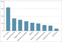
Table 3 presents the response pattern of the absolute latency of waves I, III, and V and interpeak intervals I-III, I-V, and III-V at 80 dB nHL and electrophysiological thresholds. The group comparison revealed a statistically significant difference between their thresholds (p < 0.001).
The presence of one or more RFHLs in the study population influenced the electrophysiological threshold. The analysis of examination results of infants with and without RFHL showed a statistically significant difference (p = 0.05). The same was true for prematurity (p = 0.02), as shown in Table 4.
The correlation analysis between the electrophysiological threshold and CGA demonstrated that the electrophysiological threshold improved with increasing age (p = 0.006; r = -0.236) (Figure 2).
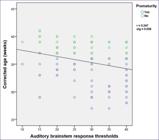
Chart with the correlation between electrophysiological thresholds and corrected gestational age
DISCUSSION
The sample had 62 infants, 35 (56.45%) of whom were born prematurely, 32 (51.61%) with CGA on the day of the examination less than or equal to 36 weeks, of which 18 (56.30%) were males, corroborating data from IBGE (2010) that indicate a higher premature birth rate in male infants3.
Previous research observed differences between females and males, with greater latencies in males10. The authors attributed this result to the anatomical differences between the sexes10. This study found no statistically significant differences in any variable regarding sex.
Prematurity can have consequences, not because of the condition itself, but because of the special care that preterm infants generally require in the NICU and the RFHLs that they may present, such as low birth weight, use of ototoxic medication, mechanical ventilation, and so forth8. GA and length of NICU stay at birth are important variables related to the probability of NHS “failure”, and there is a higher occurrence of hearing loss in preterm infants12.
This study had more preterm than full-term infants. However, the study population comprised infants born in a reference hospital for high-risk pregnancies, which justifies their greater number in the sample.
Regarding GA, another study found that most babies in the NICU were premature13,14. These findings allow us to infer that the NICU population is generally made up of preterm infants with low weight and other risk factors, requiring specialized care.
The results of this study also showed that some of the most common RFHLs were NICU stay of more than 5 days - 50.79% (32 infants), mechanical ventilation - 20.63% (13 infants), and ototoxic medication - 15.87% (10 infants). These coincide with the results of a study conducted by Silva et al.13, whose authors showed that 55% of the cases were of patients with a NICU stay of more than 5 days, 26% remained on mechanical ventilation, and 19% used ototoxic medication.
NHS is the gold standard strategy due to its efficacy in detecting newborns and infants with suspected HL in early childhood15. However, it is always necessary to include an ABR assessment to rule out retrocochlear hearing impairment.
Many parameters can be analyzed to determine whether ABR responses are within normal limits. Some are directly linked to specific parameters used and verified while acquiring the tracing and interpreting results. These parameters are characterized by analyses of absolute wave latency, interpeak interval latency, changes in absolute latencies as a function of decreased intensity, and morphology and reproducibility of tracings16-21.
The analysis of absolute wave latencies is considered the most reliable parameter and provides important data in the clinical interpretation of the exam as normal or abnormal. These values are quite consistent, more precisely with a variation of only 0.1 ms to 0.3 ms in normal individuals22,23.
Thus, this study used these parameters to consider the tracings within normal standards and investigate the absolute latency of ABR waves I, III, and V and interpeak intervals I-III, III-V, and I-V at 80 dB HL in infants.
The results of this study showed no statistically significant difference in the latency of waves I, III, and V and interpeak intervals I-III, III-V, and I-V between the CGA groups G1 and G2 at the intensity tested (80 dB nHL).
The association between CGA and hearing threshold showed that the mean electrophysiological threshold decreased with increasing age - i.e., G1 had higher hearing thresholds than G2. There was a statistically significant difference between CGA and electrophysiological threshold, suggesting that premature infants may have higher hearing thresholds, even when assessed considering CGA.
A 1980 study described that premature infants at 25 weeks of gestation may have electrophysiological thresholds at 65 dB SPL; at 30 weeks of gestation, at 45 dB SPL; and at 35 weeks of gestation, at 10 dB SPL24. This reinforces the findings of the present study, as hearing thresholds decreased as the GA increased (p = 0.006; r = -0.236). No recent studies were found that investigated electrophysiological thresholds by GA. However, Gorga et al.25 and Bakhos et al.26 researched parameters by age and suggested complementary research evaluating auditory pathway maturation through ABR.
Another study27 assessed newborns with auditory potentials and concluded that premature infants have significantly higher thresholds than full-term ones, which is consistent with the results in this study.
Understanding how the electrophysiological threshold changes with increasing age is essential to define technical NHS criteria. This study contributed to knowledge in this field. Thus, longitudinal studies can provide deeper knowledge about the effects of auditory pathway maturation on the electrophysiological thresholds of premature infants.
CONCLUSION
It is concluded that CGA did not influence the absolute latency parameters and interpeak intervals in the study population. However, the electrophysiological threshold was better in the group with CGA greater than 37 weeks. Moreover, the presence of RFHL helped increase the electrophysiological threshold. Thus, the study demonstrated its value for diagnostic conclusions and audiological clinical practice.
REFERENCES
-
1 Angrisano RG, Diniz EMA, Guinsburg R, Ferraro AA, Azevedo MF, Matas CG. Longitudinal electrophysiological study of auditory pathway in small for gestational age infants. CoDAS. 2014;26(4):294-301. https://doi.org/10.1590/2317-1782/201420140042 PMID: 25211688.
» https://doi.org/10.1590/2317-1782/201420140042 -
2 Organização Pan-Americana da Saúde [Webpage on the internet]. Saúde Auditiva: documento de posicionamento da Organização Pan-Americana da Saúde/Organização Mundial da Saúde. Brasília: OPAS/OMS; 2008. [accessed 2023 mar 12]. Available at: https://www.paho.org/pt/topicos/saude-auditiva
» https://www.paho.org/pt/topicos/saude-auditiva -
3 Instituto Brasileiro de Geografia e Estatística-IBGE [Webpage on the internet] 2010. Censo Demográfico - Deficiência Auditiva [accessed 2024 mar 14]. Available at: https://sidra.ibge.gov.br/
» https://sidra.ibge.gov.br/ -
4 Sobreira ACO, Capo BM, Santos TS, Gil D. Speech and language development in hearing impairment: Two case reports. Rev. CEFAC. 2015;17(1):308-17. https://doi.org/10.1590/1982-021620152314
» https://doi.org/10.1590/1982-021620152314 -
5 Joint Committee on Infant Hearing - JCIH [Webpage on the internet]. 2019. Risk indicators for hearing loss [accessed 2021 mar 14]. Available at: http://www.jcih.org/
» http://www.jcih.org/ -
6 Alberton M, Rosa VM, Iser BPM. Prevalence and temporal trend of prematurity in Brazil before and during the COVID-19 pandemic: A historical time series analysis, 2011-2021. Epidemiologia e Serviços de Saúde. 2023;32(2):e2022603. https://doi.org/10.1590/S2237-96222023000200005
» https://doi.org/10.1590/S2237-96222023000200005 -
7 Lewis DR, Marone ASM, Mendes BCA, Cruz OLM, Nóbrega M. Comitê multiprofissional em saúde auditiva COMUSA. Braz J Otorhinolaryngol. 2010;76(1):121-8. https://doi.org/10.1590/S1808-86942010000100020
» https://doi.org/10.1590/S1808-86942010000100020 -
8 Rechia IC, Loberalesso KP, Angst OVM, Mahl FD, Garcia MV, Biaggio EPV. Intensive care unit: Results of the newborn hearing screening. Braz J Otorhinolaryngol. 2016;82(1):76-81. https://doi.org/10.1016/j.bjorl.2015.06.004
» https://doi.org/10.1016/j.bjorl.2015.06.004 -
9 Ferreira L, Gardin L, Barbieri RB, Cargnelutti M, Quinto SMS, Garcia MV et al. The influence of gender on brainstem auditory evoked potentials' responses to different stimuli in newborns. Audiol., Commun. Res. 2020;25:2152. https://doi.org/10.1590/2317-6431-2019-2152
» https://doi.org/10.1590/2317-6431-2019-2152 -
10 Garcia CFD, Isaac ML, Oliveira JAA. Emissão otoacústica evocada transitória: instrumento para detecção precoce de alterações auditivas em recém-nascidos a termo e pré-termo. Braz J Otorhinolaryngol. 2002;68:44-52. https://doi.org/10.1590/S0034-72992002000300009
» https://doi.org/10.1590/S0034-72992002000300009 -
11 Organização Mundial da Saúde [Webpage on the internet] 2021. [accessed 2021 mar 14]. Available at: https://www.who.int/eportuguese/publications/pt/
» https://www.who.int/eportuguese/publications/pt/ -
12 Pereira PKS, Martins AS, Vieira MR, Azevedo MF. Programa de triagem auditiva neonatal: associação entre perda auditiva e fatores de risco. Pró-Fono R. Atual. Cientif. 2007;19(3):267-78. http://dx.doi.org/10.1590/S0104-56872007000300005
» http://dx.doi.org/10.1590/S0104-56872007000300005 -
13 Silva DPC, Lopez PS, Montovani JC. Influence of risk indicators on different universal newborn hearing screening steps. Audiol., Commun. Res. 2016;21:e1614. https://doi.org/10.1590/2317-6431-2015-1614
» https://doi.org/10.1590/2317-6431-2015-1614 -
14 Ribeiro GE, Weber SAT, Silva DPC. Territorial distribution and quality indicators of compulsory neonatal hearing screening in Brazil after Law 12,303/2010. Rev. CEFAC. 2020;22(4):e7919. https://doi.org/10.1590/1982-0216/20202247919
» https://doi.org/10.1590/1982-0216/20202247919 -
15 Rosa LAC, Suzuki MR, Angrisani RG, Azevedo MF. Auditory Brainstem Response: Reference-values for age. CoDAS. 2014;26(2):117-21. https://doi.org/10.1590/2317-1782/2014469IN PMID:24918504.
» https://doi.org/10.1590/2317-1782/2014469IN -
16 Ortolan DS, Santos MFC. Auditory development of infants with risk indicators for hearing loss. Distúrb. Comunic. 2020;32(1):87-95. https://doi.org/10.23925/2176-2724.2020v32i1p87-95
» https://doi.org/10.23925/2176-2724.2020v32i1p87-95 -
17 Molini E, Calzolaro L, Lapenna R, Ricci G. Universal newborn hearing screening in Umbria region, Italy. Int J Pediatr Otorhinolaryngol. 2016;82:92-7. https://doi.org/10.1016/j.ijporl.2016.01.007
» https://doi.org/10.1016/j.ijporl.2016.01.007 -
18 Vignesh SS, Jaya V, Sasireka BI, Sarathy K, Vanthana M. Prevalence and referral rates in neonatal hearing screening program using two step hearing screening protocol in Chennai - A prospective study. Int J Pediatr Otorhinolaryngol. 2015;79(10):1745-7. https://doi.org/10.1016/j.ijporl.2015.07.043 PMID: 26296879.
» https://doi.org/10.1016/j.ijporl.2015.07.043 -
19 Moodley S, Storbeck C. Diagnostic hearing test of infants aged 0-36 months in 3 South African provinces - Comparison of audiology records to HPCSA guidelines. Int J Pediatr Otorhinolaryngol. 2016;91:152-8. https://doi.org/10.1016/j.ijporl.2016.10.026 PMID: 27863631.
» https://doi.org/10.1016/j.ijporl.2016.10.026 -
20 Dimitriou A, Perisanidis C, Chalkiadakis V, Marangoudakis P, Tzagkaroulakis A, Nikolopoulos TP. The universal newborn hearing screening program in a public hospital: The importance of the day of examination. Int J Pediatr Otorhinolaryngol. 2016;91:90-3. https://doi.org/10.1016/j.ijporl.2016.10.015 PMID: 27863649.
» https://doi.org/10.1016/j.ijporl.2016.10.015 -
21 Pinto JD, Ferreira L, Temp DA, Dias V, Rohers DE, Biaggio EPV. Evasion of newborn hearing screening retest: Relation with risk factors for hearing impairment. Rev. CEFAC. 2019;21(4):e2519. https://doi.org/10.1590/1982-0216/20192142519
» https://doi.org/10.1590/1982-0216/20192142519 -
22 Rechia IC, Oliveira LD, Crestani AH, Biaggio EPV, Souza APR. Effects of prematurity on language acquisition and auditory maturation: A systematic review. CoDAS. 2016;28(6):843-54. https://doi.org/10.1590/2317-1782/20162015218 PMID: 28001276.
» https://doi.org/10.1590/2317-1782/20162015218 -
23 Kanji A, Khoza-Shangase K, Moroe N. Newborn hearing screening protocols and their outcomes: A systematic review. Int J Pediatr Otorhinolaryngol. 2018;115:104-9. https://doi.org/10.1016/j.ijporl.2018.09.026 PMID:30368368.
» https://doi.org/10.1016/j.ijporl.2018.09.026 - 24 Uziel A, Marot M, Germain M. Rev. Laryngol. Otol. Rhino (1980). In: Cunha NT, Moura CP, editors. Desenvolvimento do ouvido interno dos mamíferos: contribuição para a compreensão das doenças congênitas do ouvido. Cadernos de Otorrinolaringologia-Otologia, 2012.
-
25 Gorga MP, Kaminski JR, Beauchaine KL, Jesteadt W, Neely ST. Auditory brainstem responses from children three months to three years of age: Normal patterns of response. J Speech Hear Res. 1989;32(2):281-8. https://doi.org/10.1044/jshr.3202.281 PMID: 2739379.
» https://doi.org/10.1044/jshr.3202.281 -
26 Bakhos D, Marx M, Villeneuve A, Lescanne E, Kim S, Robier A. Exploration électrophysiologique de l'audition. Annales françaises d'Oto-rhino-laryngologie et de Pathologie Cervico-faciale. 2017;134(5):313-9. https://doi.org/10.1016/j.aforl.2016.09.005
» https://doi.org/10.1016/j.aforl.2016.09.005 -
27 Sousa AC, Didoné DD, Sleifer P. Longitudinal comparison of auditory steady-state evoked potentials in preterm and term infants: The maturation process. Int Arch Otorhinolaryngol. 2017;21(3):200-5. https://doi.org/10.1055/s-0036-1584888 PMID: 28680486.
» https://doi.org/10.1055/s-0036-1584888
-
A study conducted at the Faculdade de Medicina de Minas Gerais, Belo Horizonte, MG, Brazil.
-
Financial support: Coordenação de Aperfeiçoamento de Pessoal de Nível Superior (CAPES) Brasil - Finance Code 001
-
Data sharing statement: All data generated or analyzed during the study that resulted in this article are included here. Therefore, no other data from this research will be shared with those not involved in its data collection and design.
Data sharing statement: All data generated or analyzed during the study that resulted in this article are included here. Therefore, no other data from this research will be shared with those not involved in its data collection and design.

 Captions: ICU = intensive care unit; TORCH = acronym for toxoplasmosis, other diseases, rubella, cytomegalovirus, herpes, and HIV; g = grams
