
 Relationship between the onset of the pharyngeal swallowing response and lesion topography in acute ischemic stroke
ABSTRACT
Purpose: to identify the onset site of the pharyngeal swallow response and correlate it with lesion topography in acute ischemic stroke (AIS).
Methods: an observational, cross-sectional, and prospective study involving 30 patients from a stroke unit, conducted in four phases: (1) dysphagia risk screening, (2) topographic analysis of brain lesions, (3) objective swallowing assessment, using videofluoroscopy, and (4) blinded analysis of videofluoroscopic exams by two speech-language pathologists. The onset of the pharyngeal swallow response was determined by using the Modified Barium Swallow Impairment Profile (MBSImP™), considering component 6. Statistical analysis included Spearman’s correlation, chi-square, and Cohen’s kappa tests (p < 0.05).
Results: the sample consisted of 13 (43.0%) females and 17 (57.0%) males, with a mean age of 69.1 ± 9.21 years. No correlation was found between the onset site of the pharyngeal swallow response and brain lesion topography.
Conclusion: the onset site of the pharyngeal swallow response did not correlate with the brain lesion topography in this population.
Keywords:
Deglutition; Swallowing Disorders; Fluoroscopy; Pharynx; Stroke
RESUMO
Objetivo: identificar o local de início da resposta faríngea da deglutição e correlacionar com a topografia da lesão no Acidente Vascular Cerebral Isquêmico (AVCi) agudo.
Métodos: estudo observacional, transversal e prospectivo, envolvendo 30 pacientes de uma unidade de AVC. O estudo ocorreu em quatro fases: (1) avaliação do risco de disfagia; (2) análise topográfica das lesões cerebrais; (3) avaliação objetiva da deglutição por meio da videofluoroscopia; e (4) análise cega dos exames videofluoroscópicos por duas fonoaudiólogas.O início da resposta faríngea foi determinado pelo protocolo Modified Barium Swallow Impairment Profile (MBSImP TM ), considerando o componente 6. Para a análise estatística, utilizaram-se os testes de correlação de Spearman, qui-quadrado e Kappa de Cohen (p < 0,05).
Resultados: a amostra foi composta de 13 (43,0%) mulheres e 17 (57,0%) homens, com idade média de 69,1±9,21 anos. Não houve correlação entre o local de início da resposta faríngea e a topografia das lesões cerebrais.
Conclusão: não se observou correlação entre o local de início da resposta faríngea da deglutição e topografia da lesão encefálica.
Descritores:
Deglutição; Transtornos de Deglutição; Fluoroscopia; Faringe; Acidente Vascular Cerebral
INTRODUCTION
Stroke is one of the leading causes of mortality and the main cause of disability in Brazil and worldwide. Among the frequent complications after stroke, oropharyngeal dysphagia (OD) stands out, characterized by alterations in efficiency and safety in one or more phases of swallowing. The global prevalence of post-stroke dysphagia is estimated at 46.6%, with higher occurrence in cases of hemorrhagic stroke (58.8%) compared to ischemic stroke (43.6%). In the acute phase, OD affects between 40% and 78% of patients and, although it may improve during recovery, it persists as a chronic condition in up to half of cases1,2.
Swallowing is a complex function mediated by an extensive bilateral neural network that extends from the brainstem to cortical and subcortical brain structures. These neural connections are susceptible to damage resulting from an ischemic stroke (IS)2. The pharyngeal phase of swallowing is characterized by airway protection, involving a sequence of important, rapid, coordinated, and precise events3. The mechanoreceptors responsible for triggering the pharyngeal response may be damaged after a brain injury, leading to absence or prolongation of this response, thereby increasing the risk of laryngeal penetration and tracheal aspiration4.
The brainstem is considered the primary control center for the pharyngeal phase of swallowing. However, evidence suggests that the pharyngeal phase is also modulated by higher brain regions, which adjust the motor response according to the context and task demands. This suggests that the pharyngeal phase of swallowing is not exclusively reflexive, but rather an integrated process that dynamically adapts based on cognitive inputs, prior knowledge, and sensorimotor predictions5.
Other hypotheses report that the onset of the pharyngeal response is a polysynaptic reflex action that triggers morphofunctional accommodations as the pressurized bolus descends from the oral cavity into the pharyngeal chamber. Each swallow is considered unique in terms of strength, speed, and range of motion, and depends on the bolus’ volume, taste, viscosity, and temperature6,7.
It has been observed that the site of pharyngeal response onset provides information about the sensorimotor swallowing pattern. This topic has been studied both in healthy individuals8 and in different neurological pathologies, including IS9,10.
Despite technological advances in neuroimaging and neurophysiology, current evidence still does not allow robust discussion of the impact of lesion location on swallowing biomechanics in terms of efficiency and safety11,12. The vast majority of studies focus solely on investigating alterations in the pharyngeal phase, including pharyngeal residue, laryngeal penetration, and tracheal aspiration11,13. Research exploring the relationship between the site of pharyngeal response onset and lesion laterality, cortical area, supratentorial and infratentorial regions after IS remains scarce.
Infratentorial ischemic stroke lesions generally cause more significant impairments in the pharyngeal response compared to supratentorial lesions14,15. Moreover, different cortical and subcortical lesion sites are associated with OD ranging from mild to severe14. Nearly unanimously, studies indicate variable lateralized activation in multiple brain regions during the swallowing process15,16. However, the dominant cortical lateralization of swallowing phases remains a controversial topic in the literature.
Based on the existing evidence, it is hypothesized that the location of a brain lesion influences the site of onset for the pharyngeal swallowing response. Infratentorial lesions may be associated with a delayed pharyngeal response, triggering this response in more inferior pharyngeal structures, such as the piriform sinuses. On the other hand, supratentorial lesions, including cortical and subcortical areas, may have a less significant impact on this parameter, with the pharyngeal response starting in more superior structures, such as the posterior mandible angle and the epiglottic valleculae.
Understanding the relationship between lesion location and pharyngeal phase swallowing findings allows for early identification of patients at risk of dysphagia. Neuroimaging information is generally available within minutes or hours after hospital admission, facilitating this process. Furthermore, lesion location is associated with responsiveness to different treatment approaches and emerges as one of the determinants in OD recovery17,18.
Given this evidence, it is necessary to develop studies that deepen the understanding of pharyngeal swallowing phase findings in relation to brain lesion location, considering different food volumes and consistencies within swallowing assessment. Understanding these aspects provides specific clinical guidance for diagnosis, prognosis, and treatment prescription in OD, thereby contributing to the quality of life of these individuals.
Therefore, this study aims to identify the sites of pharyngeal swallowing response onset and to verify the correlation between this parameter and the topography of brain lesions, in patients in the acute phase, after ischemic stroke.
METHODS
This was a prospective, observational, cross-sectional, and quantitative study, approved by the Research Ethics Committee (CEP) of the Hospital de Clínicas Complex, Federal University of Paraná (CHC-UFPR), Brazil, under approval number 4.754.516 (CAAE: 46963921.7.0000.0096). Participation was voluntary, and all individuals signed the Informed Consent Form (ICF) after being fully informed about the study objectives and procedures. This study complies with the guidelines established by Resolution No. 466/2012 of the National Health Council, Ministry of Health, Brazil.
The sample consisted of 30 patients diagnosed with ischemic stroke, admitted to a specialized post-stroke care unit. Participant selection was carried out using electronic medical records, following a sample size calculation and inclusion/exclusion criteria.
Inclusion criteria were: patients ≥ 60 years old; both genders; first ischemic stroke confirmed by Magnetic Resonance Imaging (MRI) and a neurologist within 48 hours of the event; with or without swallowing complaints; not submitted to cerebral reperfusion therapy; responsive enough to undergo videofluoroscopy; and without prior evaluation and/or speech therapy intervention.
Exclusion criteria were: previous ischemic and/or hemorrhagic stroke; large brain lesions such as massive intracranial hemorrhage, hypoxic brain injury, traumatic brain injury, or multiple infarcts; underlying neurological diseases (e.g., dementia, Parkinson’s disease, and/or neurodegenerative disorders); history of head and neck surgery; structural abnormalities of the oropharyngolaryngeal region; and hemodynamically unstable patients.
This study comprised four distinct stages: Stage 1: Assessment of OD risk. Stage 2: Analysis of imaging exams with topographic lesion data. Stage 3: Instrumental swallowing evaluation through videofluoroscopy. Stage 4: Blind analysis of videofluoroscopic exams by two speech-language pathologists.
Stage 1 - Assessment of OD risk.
This stage was always performed by the same speech-language pathologist in the stroke unit within the first 48 hours after the ischemic event. For this purpose, the Gugging Swallowing Screen (GUSS)19 was used, a standardized and validated tool for bedside assessment of acute-phase post-stroke patients. The GUSS scale, comprising seven items, aims to evaluate swallowing ability and degree of impairment, categorizing swallowing as normal, mild dysphagia, moderate dysphagia, or severe dysphagia.
Stage 2: - Topographic analysis of lesion location.
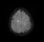
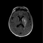
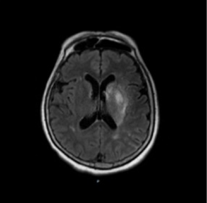
Lesion location data were obtained from MRI performed upon hospital admission. Two neurologists independently evaluated the images simultaneously and, by consensus, determined the lesion location, both of whom were blinded to the other's study procedures.
Lesion areas were subdivided considering laterality, tentorium, and cortical area, distributed as follows:
-
Laterality: Right hemisphere lesion / Left hemisphere lesion
-
Tentorium: Supratentorial and/or infratentorial lesion

-
Cortical area: Cortical, subcortical, and/or cortico-subcortical lesion
Stage 3 - Instrumental evaluation by videofluoroscopy
This stage was performed in the Contrast Radiology Department by a radiologist and a speech-language pathologist, both experts in the exam and blinded to dysphagia risk assessment results. The time interval between dysphagia risk assessment and videofluoroscopy was ≤ 48 hours.
Patients remained seated at a 90° angle and were evaluated in the lateral view. The anatomical limits observed in the exam extended from the oral cavity to the esophagus, demarcated anteriorly by the lips, posteriorly by the pharyngeal wall, superiorly by the nasopharynx, and inferiorly by the esophagus20. The Siemens Axiom R100 X-ray system and Siemens M44-2 monitor were used. Images were captured at 30 frames per second, recorded, and stored on a notebook computer for later analysis.
The exam protocol started with 100% barium sulfate diluted in food consistencies according to the International Dysphagia Diet Standardisation Initiative (IDDSI)21:
-
IDDSI-0 (thin liquid): still mineral water at room temperature + 100% barium sulfate gel;
-
IDDSI-2 (mildly thick): 100% barium sulfate gel thickened with 1.2 g (1 sachet) of ThickenUp Clear (51-350 cP);
-
IDDSI-4 (extremely thick): 100% barium sulfate gel thickened with 3.6 g (3 sachets) of ThickenUp Clear (>1750 cP).
Volumes of 5 ml and 10 ml of each food consistency were offered in a plastic tablespoon. Administration was self-fed, with patients instructed to bring the spoon to their mouth and swallow spontaneously, avoiding verbal command interference3. The sequence of food consistency presentation varied according to patient safety, and administration was interrupted if laryngotracheal aspiration with clinical risk occurred. Thus, not all volumes and consistencies were assessed in all participants.
Stage 4 - Analysis of videofluoroscopic exams
Images were independently analyzed by two speech-language pathologists, both of whom had over 10 years of experience in the field. The evaluators were blinded to participants’ clinical history and brain lesion location, ensuring unbiased analysis. For safety reasons, not all volumes of the three food consistencies were administered. A total of 163 swallows were analyzed, distributed as follows: IDDSI-0 (53 swallows), IDDSI-2 (59 swallows), and IDDSI-4 (51 swallows).
The site of pharyngeal response onset was evaluated based on the first vigorous anterosuperior movement of the hyoid bone, according to the Modified Barium Swallow Impairment Profile (MBSImPTM)22, adapted and translated into Brazilian Portuguese23. For analysis purposes, only component 6 of this protocol was considered, which classifies the site of pharyngeal response onset into five distinct anatomical points:
-
0 = Bolus head at the posterior ramus of the mandible (first hyoid excursion);
-
1 = Bolus head at the valleculae;
-
2 = Bolus head at the laryngeal surface of the epiglottis;
-
3 = Bolus head at the piriform sinus;
-
4 = No visible onset at any site.
Statistical analysis
Data were collected and tabulated in Excel spreadsheets (Microsoft, v.15.33, 2017). For descriptive analysis, categorical variables were presented as frequencies, prevalence, and percentages.
The frequency of occurrence between variables was compared using the chi-square test, with Standardized Residual Measure (SRM) applied to identify significant deviations, where SRM > 2 indicated higher incidence and SRM < -2 indicated lower incidence24. Correlations between variables were analyzed with Spearman’s test, considering correlations as null (≤ 0.10) to strong (≥ 0.7). Statistical significance was set at 5% (p < 0.05).
To assess inter-rater agreement, Cohen’s Kappa test was used, with interpretation values ranging from null (≤ 0) to excellent (0.81-1.00)25. Overall, inter-rater agreement in identifying the site of pharyngeal response onset varied by food consistency: excellent (0.81-1.00) for IDDSI-4, high (0.61-0.80) for IDDSI-2, and moderate (0.41-0.60) for the lowest viscosity consistency, IDDSI-0.
RESULTS
Table 1 details the general characterization of the study sample, which consisted of 30 patients. Among them, 11 (36.6%) were admitted on the same day as the ischemic stroke, while the remaining 19 (63.4%) were admitted the following day. The mean interval between ischemic stroke and the videofluoroscopic swallowing study (VFSS) was approximately four days. Regarding lesion characteristics, a relatively homogeneous distribution was observed regarding the lesion laterality, with most patients presenting supratentorial cortical lesions.

Comparison of the frequency of occurrence of the sites of onset of the pharyngeal swallowing response for 5 ml and 10 ml volumes of IDDSI-0, IDDSI-2, and IDDSI-4 consistencies
DISCUSSION
The neurophysiological control of the pharyngeal phase of swallowing is complex and coordinated by multiple centers represented in the central and peripheral nervous systems bilaterally. Although the importance of the cerebral cortex in triggering and modulating the pharyngeal phase of swallowing is recognized, issues related to laterality, cortical areas, and the role of supratentorial and infratentorial regions in the pharyngeal phase of swallowing remain under discussion in the literature15,26.
The results presented in Table 2 show that there was no correlation between the site of onset of the pharyngeal response and brain lesion location across the different food volumes and consistencies.
Recent studies have focused on the relationship between temporal parameters of swallowing and brain lesion location. Most findings indicate that infratentorial lesions may result in more significant impairments in the pharyngeal phase of swallowing compared to supratentorial lesions16,17. The pharyngeal response time tends to be prolonged, leading to delays and increasing the risk of tracheal aspiration15,26. Regarding laterality, right hemisphere lesions show more compromised pharyngeal components and are more frequently associated with laryngeal penetration and tracheal aspiration events than left hemisphere lesions11,12.
It is important to emphasize that the absence or prolongation of the pharyngeal response may be associated with a higher risk of aspiration in the post-stroke population. However, a delayed onset of the pharyngeal response alone does not necessarily imply the presence of dysphagia. Individual variability must be considered, such as age, underlying conditions, and other relevant aspects of swallowing. On the other hand, when such a delay is combined with other swallowing physiology impairments, it may represent a significant complication9,27.
The lack of correlation in this study between the site of onset of the pharyngeal response and lesion location may be related to sample size and the number of variables involved, including food consistencies, volumes, and distinct lesion sites. Furthermore, the heterogeneity of supratentorial and infratentorial lesion groups may have limited the ability to detect statistically significant correlations between the studied parameter and brain lesion location.
The results illustrated in Figure 4 show the comparison of occurrence frequency of pharyngeal response onset sites. Significant differences were observed in the proportion of responses across the food volumes and consistencies assessed (p < 0.03), except for 10 ml of IDDSI-2 (p = 0.19). Overall, pharyngeal response onset was more frequent in superior structures such as the Posterior Mandibular Angle (PMA) and Epiglottic Valleculae (EV) with higher viscosity consistency (IDDSI-4). Conversely, with the lowest viscosity consistency (IDDSI-0), the pharyngeal response onset occurred more inferiorly in the Piriform Sinuses (PS).
These findings are consistent with previous studies showing that pharyngeal motility changes according to the sensory characteristics of the swallowed bolus20,27. It is believed that swallowing higher viscosity consistencies requires greater neuromuscular control compared to lower viscosity consistencies, providing stronger proprioceptive stimuli that influence pharyngeal response onset in superior structures such as the tongue base, posterior mandibular angle, and epiglottic valleculae27.
Table 3 shows the comparison of response onset site frequencies across different food volumes and consistencies. Significant results were found (p < 0.01). For 5 ml and 10 ml of IDDSI-4, the pharyngeal response onset occurred more frequently at the PMA and EV. For 10 ml of IDDSI-2, onset was most frequent at the laryngeal surface of the epiglottis (LSE), and for 5 ml and 10 ml of IDDSI-0, at the PS. These findings suggest that, in addition to bolus viscosity, volume also influences pharyngeal response onset.
These findings are relevant to clinical practice in oropharyngeal dysphagia, providing valuable information to improve evaluation and rehabilitation of these individuals. However, the pharyngeal swallowing response depends on multiple influences28. Previous studies highlight that variables such as volume, food consistency, verbal commands, and dental losses may influence the site of pharyngeal response onset3,9,29. Therefore, this parameter cannot be used in isolation to distinguish between asymptomatic and dysphagic patients.
It is necessary to recognize this study’s limitations. First, sample selection took place during the COVID-19 pandemic, which impacted sample size. Additionally, the clinical instability of acute-phase patients further restricted the number of participants. For safety reasons, not all patients received all volumes and consistencies, limiting statistical power. The heterogeneity of brain lesion sites also restricted the generalization of identified correlations.
Despite these challenges, our results may pave the way for future research exploring the relationship between neuroimaging findings and pharyngeal aspects of swallowing, to improve dysphagia management after ischemic stroke.
CONCLUSION
Based on the results of this study, no correlation was identified between the site of onset of the pharyngeal swallowing response and the topography of the brain lesion.
ACKNOWLEDGMENTS
To the Hospital de Clínicas Complex of the Federal University of Paraná (UFPR) for the support, infrastructure, and knowledge provided throughout this study. The dedication of all professionals and researchers involved was fundamental to the development of this research, especially my advisor and co-advisor, whose support, guidance, and encouragement were essential at every stage of this work.
REFERENCES
-
1 Song W, Wu M, Wang H, Pang R, Zhu L. Prevalence, risk factors, and outcomes of dysphagia after stroke: A systematic review and meta-analysis. Front Neurol. 2024 Jul 17;15:1403610. https://doi.org/10.3389/fneur.2024.1403610 PMID: 39087010.
» https://doi.org/10.3389/fneur.2024.1403610 -
2 Suntrup-Krueger S, Kemmling A, Warnecke T, Hamacher C, Oelenberg S, Niederstadt T et al. The impact of lesion location on dysphagia incidence, pattern and complications in acute stroke. Part 2: Oropharyngeal residue, swallow and cough response, and pneumonia. Eur J Neurol. 2017;24(6):867-74. https://doi.org/10.1111/ene.13307 PMID: 25677582.
» https://doi.org/10.1111/ene.13307 -
3 Almeida RCA, Haguette RCB, Andrade ISN. Deglutição com e sem comando verbal: achados videofluoroscópicos. Rev Soc Bras Fonoaudiol. 2011;16(3):291-7. http://dx.doi.org/10.1590/S1516-80342011000300009
» http://dx.doi.org/10.1590/S1516-80342011000300009 -
4 Park T, Kim Y, Ko DH, McCullough G. Initiation and duration of laryngeal closure during the pharyngeal swallow in post-stroke patients. Dysphagia. 2010;25(3):177-82. https://doi.org/10.1007/s00455-009-9237-9 PMID: 19760459.
» https://doi.org/10.1007/s00455-009-9237-9 -
5 Zainaee S, Archer B, Scherer R, Bingman V, Ghasemi M. Revealing goal-directed neural control of the pharyngeal phase of swallowing. Dysphagia. 2025;40(3):528-40. https://doi.org/10.1007/s00455-024-10758-3 PMID: 39387924.
» https://doi.org/10.1007/s00455-024-10758-3 -
6 Zancan M, Luchesi KF, Mituuti CT, Furkim AM. Onset locations of the pharyngeal phase of swallowing: Meta-analysis. CoDAS. 2017;29(2):e20160067. https://doi.org/10.1590/2317-1782/20172016067 PMID: 28327783.
» https://doi.org/10.1590/2317-1782/20172016067 - 7 Logemann JA. Evaluation and treatment of swallowing disorders. 2nd ed. Austin: Pro-Ed; 1998.
-
8 Uchimura EMT, Barcelos IHK, Paiva DB, Mourão LF, Crespo JÁ. Evaluation of the location of capsules swallowed with food during the pharyngeal phase triggering in asymptomatic adults. CoDAS. 2014;26(6):476-80. http://dx.doi.org/10.1590/2317-1782/20140000045 PMID: 25590910.
» http://dx.doi.org/10.1590/2317-1782/20140000045 -
9 Gatto AR, Cola PC, da Silva RG, Ribeiro PW, Spadotto AA, Henry MAAC. Influence of thermal and gustatory stimulus in the initiation of the pharyngeal swallow and bolus location instroke. J Stroke Cerebrovasc Dis. 2021;30(4):105349. https://doi.org/10.1016/j.jstrokecerebrovasdis.2020.105349 PMID: 33549862.
» https://doi.org/10.1016/j.jstrokecerebrovasdis.2020.105349 -
10 Saconato M, Leite FC, Lederman HM, Chiari BM, Gonçalves MIR. Temporal and sequential analysis of the pharyngeal phase of swallowing in poststroke patients. Dysphagia. 2020;35(4):598-615. https://doi.org/10.1007/s00455-019-10069-y PMID: 31612287.
» https://doi.org/10.1007/s00455-019-10069-y -
11 Daniels SK, Pathak S, Mukhi SV, Stach CB, Morgan RO, Anderson JA. The relationship between lesion localization and dysphagia in acute stroke. Dysphagia. 2017;32(6):777-84. https://doi.org/10.1007/s00455-017-9824-0 PMID: 28748320.
» https://doi.org/10.1007/s00455-017-9824-0 -
12 Wilmskoetter J, Martin-Harris B, Pearson WG Jr, Bonilha L, Elm JJ, Horn Jn et al. Differences in swallow physiology in patients with left and right hemispheric strokes. Physiol Behav. 2018;194:144-52. https://doi.org/10.1016/j.physbeh.2018.05.010 PMID:29758228.
» https://doi.org/10.1016/j.physbeh.2018.05.010 -
13 Suntrup-Krueger S, Ringmaier C, Muhle P, Wollbrink A, Kemmling A, Hanning U et al. Randomized trial of transcranial direct current stimulation for poststroke dysphagia. Ann Neurol. 2018;83(2):328-40. https://doi.org/10.1002/ana.25151 PMID: 29350775.
» https://doi.org/10.1002/ana.25151 -
14 Hess F, Foerch C, Keil F, Seiler A, Lapa S. Association of lesion pattern and dysphagia in acute intracerebral hemorrhage. Stroke. 2021;52(9):2921-9. https://doi.org/10.1161/strokeaha.120.032615 PMID: 34000833.
» https://doi.org/10.1161/strokeaha.120.032615 -
15 Wilmskoetter J, Daniels SK, Miller AJ. Cortical and subcortical control of swallowing-can we use information from lesion locations to improve diagnosis and treatment for patients with stroke? Am J Speech Lang Pathol. 2020;29(2S):1030-43. https://doi.org/10.1044/2019_AJSLP-19-00068 PMID: 32650664.
» https://doi.org/10.1044/2019_AJSLP-19-00068 -
16 Wilmskoetter J, Martin-Harris B, Pearson WG Jr, Bonilha L, Elm JJ, Horn J et al. Differences in swallow physiology in patients with left and right hemispheric strokes. Physiol Behav. 2018;194:144-52. https://doi.org/10.1016/j.physbeh.2018.05.010 PMID:29758228.
» https://doi.org/10.1016/j.physbeh.2018.05.010 -
17 Suntrup-Krueger S, Ringmaier C, Muhle P, Wollbrink A, Kemmling A, Hanning U et al. Randomized trial of transcranial direct current stimulation for poststroke dysphagia. Ann Neurol. 2018;83(2):328-40. https://doi.org/10.1002/ana.25151 PMID: 29350775.
» https://doi.org/10.1002/ana.25151 -
18 Kim JH, Oh SH, Jeong HJ, Sim YJ, Kim DG, Kim GC. Association between duration of dysphagia recovery and lesion location on magnetic resonance imaging in patients with middle cerebral artery infarction. Ann Rehabil Med. 2019;43(2):142-8. https://doi.org/10.5535/arm.2019.43.2.142 PMID: 29350775.
» https://doi.org/10.5535/arm.2019.43.2.142 -
19 Warnecke T, Im S, Kaiser C, Hamacher C, Oelenberg S, Dziewas R. Aspiration and dysphagia screening in acute stroke - the Gugging Swallowing Screen revisited. Eur J Neurol. 2017;24(4):594-601. http://dx.doi.org/10.1111/ene.13251 PMID:28322006.
» http://dx.doi.org/10.1111/ene.13251 -
20 Martin-Harris B, Jones B. The videofluorographic swallowing study. Phys Med Rehabil Clin N Am. 2008;19(4):769-85, viii. https://doi.org/10.1016/j.pmr.2008.06.004 PMID: 18940640.
» https://doi.org/10.1016/j.pmr.2008.06.004 -
21 Steele CM, Namasivayam-McDonald AM, Guida BT, Cichero JA, Fuivestein J, Hanson B et al. Creation and initial validation of the International Dysphagia Diet Standardisation Initiative functional diet scale. Arch Phys Med Rehabil. 2018:99(5):934-44. https://doi.org/10.1016/j.apmr.2018.01.012 PMID: 29428348.
» https://doi.org/10.1016/j.apmr.2018.01.012 -
22 Martin-Harris B, Brodsky MB, Michel Y, Castell DO, Schleicher M, Sandidge J et al. MBS measurement tool for swallow impairment-MBSImp: Establishing a standard. Dysphagia. 2008;23(4):392-405. https://doi.org/10.1007/s00455-008-9185-9 PMID: 18855050.
» https://doi.org/10.1007/s00455-008-9185-9 -
23 Baraçal-Prado AC, de Lima DP, Mourão LF, Crespo AN, Martin-Harris B, Davidson K et al. Translation into Brazilian Portuguese and cultural adaptation of the component scores definition from the Modified Barium Swallow Impairment Profile (MBSImP(tm)). CoDAS. 2021;33(6):e20200263. https://doi.org/10.1590/2317-1782/20202020263 PMID: 34468632.
» https://doi.org/10.1590/2317-1782/20202020263 - 24 Haberman SJ. The analysis of residuals in cross-classified tables. Biometrics. 1973;29(1):205.
- 25 Altman DG. Practical statistics for medical research. New York, NY: Chapman & Hall/CRC Press; 1999.
-
26 Yang QL, Chen Y, Wang XJ, Qiu HY, Chen MT, Zhou XH et al. Correlation between lesion location and dysphagia characteristics in post-stroke patients. J Stroke Cerebrovasc Dis. 2024;33(6):107682. https://doi.org/10.1016/j.jstrokecerebrovasdis.2024.107682 PMID: 38522758.
» https://doi.org/10.1016/j.jstrokecerebrovasdis.2024.107682 -
27 Yamada EK, Siqueira KO, Xerez D, Koch HA, Costa MMB. The influence of oral and pharyngeal phases on the swallowing dynamic. Arq Gastroenterol. 2004;41(1):18-23. https://doi.org/10.1590/s0004-28032004000100004 PMID: 15499419.
» https://doi.org/10.1590/s0004-28032004000100004 -
28 Stephen JR, Taves DH, Smith RC, Martin RE. Bolus location at the initiation of the pharyngeal stage of swallowing in healthy older adults. Dysphagia. 2005;20(4):266-72. https://doi.org/10.1007/s00455-005-0023-z PMID: 16633870.
» https://doi.org/10.1007/s00455-005-0023-z -
29 Yamamoto H, Furuya J, Tamada Y, Kondo H. Impacts of wearing complete dentures on bolus transport during feeding in elderly edentulous. J Oral Rehabil. 2013;40(12):923-31. http://dx.doi.org/10.1111/joor.12107 PMID: 24237359.
» http://dx.doi.org/10.1111/joor.12107
We declare that the data used in this study will not be available for sharing.

 Source: the author
 Source: the author
 Source: the author
 Captions: PMA = Posterior mandibular angle; EV = Epiglottic valleculae; HS = Superior hypopharynx/Laryngeal surface of the epiglottis; PS = Piriform sinus; AR = Absence of response. (Chi-square test).