 Total en bloc sacrectomy through a posterior-only approach with the “Modified Cathedral” technique: technical note
ABSTRACT
Objective: To describe the technique of total en bloc sacrectomy through a posterior-only approach, discussing indications, technical aspects, complications, and oncological outcomes available in the literature. Methods: Technical report based on detailed surgical description and narrative literature review. Expected results: The posterior-only approach may reduce operative time, intraoperative blood loss, and morbidity compared to the combined approach. Conclusion: Total en bloc sacrectomy via a posterior-only approach with the modified Cathedral reconstruction is a feasible and safe alternative in selected cases of primary malignant sacral tumors. Level of Evidence V; Technical Note.
Keywords:
Sacrum; Surgical Oncology; Spinal Neoplasms; Hemangioendothelioma; Bone Neoplasms
RESUMO:
Objetivo: Descrever a técnica de sacrectomia total em bloco por abordagem exclusivamente posterior, discutindo indicações, aspectos técnicos, complicações e desfechos oncológicos disponíveis na literatura. Métodos: Relato técnico baseado em descrição cirúrgica detalhada e revisão narrativa da literatura. Resultados esperados: A abordagem exclusivamente posterior pode reduzir o tempo operatório, a perda sanguínea intraoperatória e a morbidade em comparação com a abordagem combinada. Conclusão: A sacrectomia total em bloco por via exclusivamente posterior, com reconstrução tipo catedral modificada, é uma alternativa viável e segura em casos selecionados de tumores sacrais malignos primários. Nível de Evidência V; Nota Técnica.
Descritores:
Sacro; Oncologia Cirúrgica; Neoplasias da Coluna Vertebral; Hemangioendotelioma; Neoplasias Ósseas
RESUMEN:
Objetivo: Describir la técnica de sacrectomía total en bloque mediante un abordaje exclusivamente posterior, discutiendo indicaciones, aspectos técnicos, complicaciones y resultados oncológicos disponibles en la literatura. Métodos: Informe técnico basado en una descripción quirúrgica detallada y revisión narrativa de la literatura. Resultados esperados: El abordaje exclusivamente posterior puede reducir el tiempo quirúrgico, la pérdida de sangre intraoperatoria y la morbilidad en comparación con el abordaje combinado. Conclusión: La sacrectomía total en bloque mediante un abordaje posterior único con reconstrucción tipo catedral modificada es una alternativa factible y segura en casos seleccionados de tumores sacros malignos primarios. Nivel de Evidencia V; Nota Técnica.
Descriptores:
Sacro; Oncología Quirúrgica; Neoplasias de la Columna Vertebral; Hemangioendotelioma; Neoplasias Óseas
INTRODUCTION
En bloc total sacrectomy is a surgical procedure that involves the complete removal of a portion or the entirety of the sacrum in a single specimen, with the goal of achieving wide oncologic margins for malignant or aggressive benign tumors. While traditionally performed using combined anterior and posterior approaches, selected cases, typically tumors confined to the sacrum without significant anterior visceral or vascular involvement, can be safely addressed through a posterior-only approach.1,2
Reconstruction after sacrectomy is essential to restore spinopelvic stability; however, the literature does not demonstrate the superiority of any specific reconstruction technique over others.3 The Cathedral technique is a posterior reconstruction method that uses fibular struts, either autologous or allogenic, docked in the vertebral body of the lumbar spine, forming an anterior load-sharing “arch,” which supports the spine, offloads posterior instrumentation, and facilitates long-term fusion. The main difference between the traditional technique and the Cathedral reconstruction is that, in the latter, the load is transmitted through the anterior column rather than solely through the posterior fixation.
This approach allows tumor resection and reconstruction to be completed entirely through the posterior corridor, reducing surgical morbidity while maintaining mechanical stability.4-6
This technical note describes the posterior en bloc total sacrectomy procedure and details the application of the Cathedral reconstruction method. In our case, a total sacrectomy was performed through a posterior-only approach using a wire saw (Gigli saw), and three fibular struts were used for reconstruction.
MATERIALS AND METHODS
Case Description
A 38-year-old female with a 5-year history of sacral pain, left sciatica, and sphincter dysfunction was treated at Instituto Nacional de Traumatologia e Ortopedia - INTO. The initial biopsy was epithelioid hemangioendothelioma. A preoperative attempt at embolization was made, but no primary feeding artery suitable for embolization was identified, and an en bloc total sacrectomy was performed to comply with oncological principles.7 Intraoperatively, the tumor was found to extend intradurally up to the L5 level. The final diagnosis confirmed epithelioid hemangioendothelioma, with clear margins achieved after total sacrectomy. The reconstruction was performed in a second stage to reduce the risk of complications and improve patient outcomes.4,8 It was performed using pedicle and iliac screws, allografts, and fibular grafts. A vascularized fibula was used on the left side and anastomosed to a branch of the left superior gluteal artery, while two allogenic fibular struts from the tissue bank were placed on the right. Interference screws were also applied in the iliac wings to enhance fixation. The patient developed a surgical site infection, which was managed with surgical debridement, antibiotic therapy, and VAC therapy.9
Surgical Technique
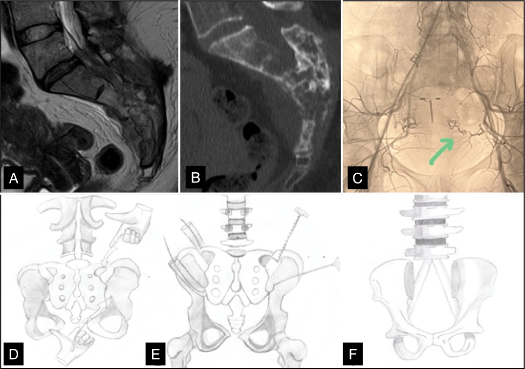
En bloc total sacrectomy is indicated for primary malignant sacral tumors, such as chordomas and sarcomas, and selected metastatic lesions that require wide oncologic margins. En bloc total sacrectomy performed through a posterior-only approach requires meticulous dissection of neurovascular and dural structures before tumor resection.10,11 After a wide midline exposure extending from the caudal lumbar spine to the posterior iliac crests, the lumbosacral fascia is released, and the posterior elements of the sacrum are skeletonized. The dural sac is identified proximally at L4-L5 and mobilized caudally to the level of the planned osteotomy. The laminectomy at this level is performed, and the dural sac is ligated immediately after the emergence of the L5 nerve root. The dural sac is ligated with nonabsorbable sutures and reinforced with dural sealants to prevent cerebrospinal fluid leakage. Intraoperative Valsalva maneuvers confirm watertight closure. Sacral nerve roots are carefully isolated within their foramina: S1–S5 roots are usually sacrificed bilaterally when the tumor involves the central sacrum. Attention is then directed to vascular control. From the posterior approach, the presacral venous plexus and lateral sacral veins are particularly at risk. Meticulous subperiosteal dissection along the anterior surface of the sacrum, performed from the posterior window through the foramina and lateral gutters, allows controlled identification of these structures. Bipolar cautery, hemoclips, and absorbable hemostatic agents are used to manage the plexus. The median sacral vessels, if accessible from the posterior corridor, are ligated early. Internal iliac branches encountered laterally during exposure of the sacroiliac joints are clipped and divided. Blunt digital dissection is performed proximally and distally using the index fingers to create an anterior plane separating the vascular structures and the lumbosacral trunk. The surgeon attempts to bring the fingertips together to confirm that the passage has been fully created (Figure 1). Through this space, a silicone tube is introduced, followed by the passage of a wire saw through the tube. The Gigli saw handles are then attached, and the osteotomy is performed in a posterolateral direction. Once neural and vascular elements are secured, osteotomies are performed. Transverse cuts are made with a wire saw at the predetermined level, commonly through the S1 body for total sacrectomy. High-speed burrs and osteotomes can be used under constant irrigation to minimize thermal injury, but we prefer to use the wire saw to diminish the risk of lesion of the iliac vessels and the lumbosacral trunk.1 In the posterior-only approach, bilateral iliac osteotomies through the iliac bone, with the resected segment including the medial portion of the iliac bone, allow gradual sacral mobilization and L5-S1 disc distraction, providing anterior exposure for safe dissection and ligation of the internal iliac vessels. The remaining disc is removed through a posteriorly created anterolateral corridor, enabling complete en bloc tumor resection while avoiding the morbidity of a traditional anterior laparotomy.1,12 Dissection of the piriformis includes 2.5 cm proximally due to the frequent recurrence in this area, along with the sacrotuberous and sacrospinous ligaments, to facilitate lateral release. En bloc delivery of the specimen is achieved by lifting the sacrum posteriorly, with assistants applying countertraction to protect residual dura and vessels.
38-years old female with low back pain and sphincter dysfunction was diagnosed epithelioid hemangioendothelioma. A - CT in sagittal plane revealing multiple expansible lytic lesions. B – MRI showing tumor expansion into the epidural space. C – Arteriography study of the sacral tumor. D - Blunt digital dissection is performed proximally and distally using the index fingers to create an anterior plane separating the vascular structures and the lumbosacral trunk. E - A silicone tube is introduced, followed by the passage of a wire saw through the tube. The Gigli saw handles are then attached, and the osteotomy is performed in a posterolateral direction. F - For the Cathedral reconstruction, docking sites are created centrally in the lowest remaining vertebral body and bilaterally in the iliac wings.
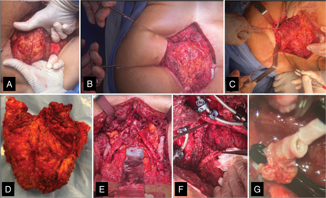
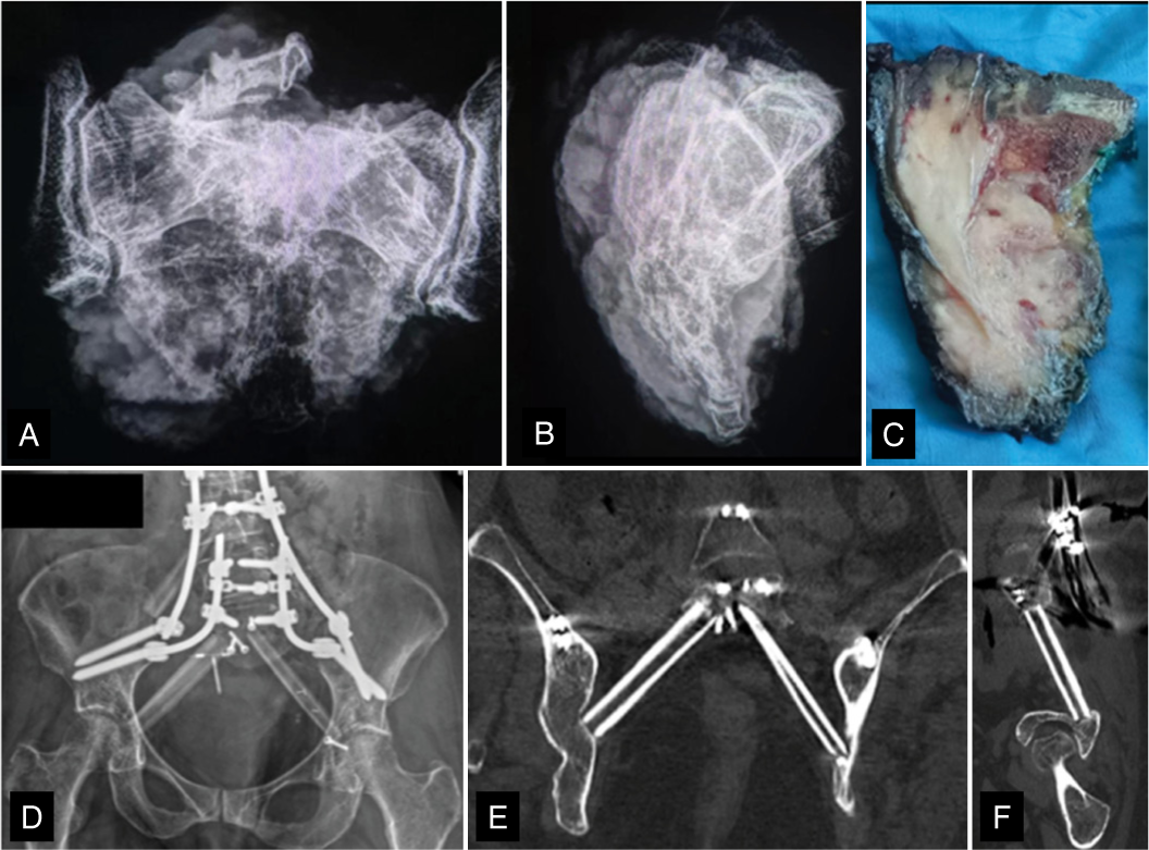
Reconstruction proceeds with posterior instrumentation. Pedicle screws are inserted into the lumbar vertebras, ensuring maximal cortical purchase, and iliac screws are placed bilaterally with long supra-acetabular trajectories. Fixation was extended from L2 to the ilium using a four-rod construct. For the Cathedral reconstruction, docking sites are created centrally in the lowest remaining vertebral body and bilaterally in the iliac wings. Structural fibular grafts, autologous or banked, are fashioned with tapered ends and impacted into the docking sockets, forming a bilateral arch spanning from each ilium to a vertebral body of the lumbar spine. In this case, three fibular grafts were used: one vascularized fibula on the left and two allogenic fibular struts from the tissue bank on the right, including an additional graft spanning from L4 to the ilium to enhance posterior stability. The vascularized fibula was incorporated on the left side to promote biological incorporation and enhance the fusion rate between the lumbar spine and the ilium13-15 (Figure 2). The additional third fibula was positioned to enhance posterior stability and improve load distribution across the spinopelvic junction, providing a more robust construct and facilitating long-term fusion and mechanical stability.6 This arch provides an anterior load-sharing strut, analogous to a buttress, reducing strain on posterior instrumentation. The four-rod construct was assembled, followed by placement of the fibular graft, and compression was applied to ensure intimate graft-host contact. Cross-links enhance torsional rigidity. (Figure 3)
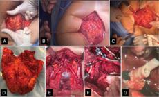
Intraoperative images of En Bloc Total Sacrectomy. A – Blunt digital dissection. B and C - Gigli saw handles are then attached, and the osteotomy is performed. D – Intraoperative view of the tumor specimen. E – Preparation of docking sites in vertebral body for Cathedral reconstruction. F and G - A vascularized fibula was used on the left side and anastomosed to a branch of the left superior gluteal artery.
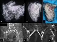
A, B and C - Digital photos of the surgical specimen and macroscopic specimen of pathological anatomy. D – Four-rods construction and Modified Cathedral technique with a third fibula graft spanning from L4 to the ilium to enhance posterior stability with interference screws. E and F – Coronal and sagittal views of the fibular structural grafts from L5 to iliac ring.
Throughout the procedure, the surgeon must be prepared for massive blood loss, cerebrospinal fluid fistulas, and intraoperative instability. The most common vascular hazard is uncontrolled bleeding from the presacral venous plexus, while neurological risks are linked to the sacrifice of sacral roots. Careful mobilization of the dura, precise ligation of roots, and hemostatic control are essential technical maneuvers to minimize these complications. We advocate for the use of two drains with delayed removal combined with a multidisciplinary approach involving microsurgery to minimize dead space; in our case, a gluteal advancement flap was performed.
Literature Results
The evolution of en bloc total sacrectomy has its roots in the late 20th century, when initial attempts were dominated by combined anterior–posterior approaches due to concerns about limited exposure and vascular or visceral injury. One of the first reports of posterior-only resections was published in 1987, describing sacrococcygeal chordomas removed via a posterior corridor up to S2, proving the feasibility of radical excision through this route, though with a notable rate of local recurrence when margins were close.16 In the following decades, most large tumors or those involving the upper sacrum were still managed with combined approaches where sacrectomy for chordoma carried high morbidity, frequent wound complications, and functional compromise, but laid the foundation for en bloc principles.17 The real shift came in the 2000s, when advances in imaging, neuromonitoring, embolization, and fixation allowed surgeons to reconsider a posterior-only strategy even for higher-level resections. It was demonstrated in 2008 and 2010 that total sacrectomy can be performed entirely via a posterior approach in selected cases, with oncologic outcomes comparable to those of combined approaches.11,12 In 2012, a series of 36 patients reported one of the largest early posterior-only sacrectomy cohorts, achieving negative margins in most cases, but also noting wound complications in nearly one-third, highlighting the morbidity associated with extensive posterior dissection.18 Posterior-only total sacrectomies have also been evaluated, confirming the approach is technically safe and provides adequate local tumor control, with perioperative morbidity remaining manageable.1 Across all these series, functional outcomes have correlated directly with the level of sacral root sacrifice: bilateral S2–S4 resection almost invariably leads to bowel and bladder dysfunction, while preservation of at least one S2 root substantially improves continence, and S1 preservation remains critical for lower limb motor power. The most frequent complications reported remain wound dehiscence and infection, with incidences between 20–40%, followed by intraoperative bleeding occasionally totaling up to 6 L1, with a median amount of 7.1 units of blood transfused, often mitigated today by preoperative embolization. Differences among studies reflect heterogeneity in patient cohorts and technical strategies: some included only low or mid-sacrectomies with lower morbidity, while others pushed the limits with total resections; reconstruction methods evolved from simple posterior fixation to more complex constructs incorporating strut grafts or the Cathedral technique to resist mechanical failure. Despite these variations, the collective evidence from the last 15 years supports posterior-only en bloc total sacrectomy as a reproducible, oncologically sound, and functionally acceptable option in specialized centers, provided margins can be achieved and neurovascular risks are meticulously managed.19,20
DISCUSSION
Posterior-only en bloc total sacrectomy is a feasible approach for selected sacral tumors, allowing complete tumor resection while avoiding an anterior surgical corridor. Preoperative embolization of feeding branches from the internal iliac system has been advocated to reduce intraoperative blood loss, though this remains center-dependent. The technique requires careful mobilization of the dural sac, identification and selective sacrifice of sacral nerve roots according to tumor involvement, control of presacral and iliac vessels, and precise osteotomies to achieve en bloc removal. Reconstruction using the Cathedral technique, with fibular struts docked in the vertebral body of the lumbar spine, provides structural support and offloads posterior instrumentation.21 Literature data show that posterior-only resections can achieve negative margins and local control comparable to combined approaches when patient selection is appropriate. Series that used the Cathedral reconstruction report improved mechanical stability and lower rates of hardware failure, although wound complications remain frequent. Functional outcomes are closely linked to the level of sacral root sacrifice, with S2–S4 resection commonly leading to bowel and bladder deficits, while preservation of S1 maintains lower limb motor function. In our case, the patient experienced a postoperative infection that needed treatment with debridement, antibiotics, and VAC therapy. It was ambulating with a walker on postoperative day one, with pain adequately controlled by medication. The patient received five units of blood, which is lower than the average of 7.1 units reported in some studies. Overall, posterior-only en bloc total sacrectomy with Cathedral reconstruction appears to be a reproducible technique that balances oncologic control with biomechanical stability, representing a viable option in centers experienced in complex sacral tumor surgery.
Literature and surgical experience support its reproducibility and effectiveness, although wound complications and functional deficits related to sacral root sacrifice remain common. Careful patient selection, thorough preoperative planning, and appropriate reconstruction strategy are essential to optimize both oncologic and functional outcomes.
CONCLUSION
Posterior-only en bloc total sacrectomy with Cathedral reconstruction is a technically demanding yet feasible approach for selected sacral tumors. It allows for adequate oncologic margins while restoring biomechanical stability.
DATA AVAILABILITY DECLARATION
The contents underlying the research are available in the manuscript.
ACKNOWLEDGMENTS
We gratefully acknowledge Mr. Ubaldo Junior for generously providing the hand-drawn illustrations for this paper.
REFERENCES
- 1 Zang J, Guo W, Yang R, Tang X, Li D. Is total en bloc sacrectomy using a posterior-only approach feasible and safe for patients with malignant sacral tumors?. J Neurosurg Spine. 2015;22(6):563-70.
- 2 Farrokhi MR, Nouraei H, Hosseini SV, Tarokh A, Mousavi SR, Taheri R, et al. Sacrectomy with posterior-only approach in sacral tumors: An analysis of 26 cases and review of literature. World Neurosurg. 2023:179:e288-e295.
- 3 Bederman SS, Shah KN, Hassan JM, Hoang BH, Kiester PD, Bhatia NN. Surgical techniques for spinopelvic reconstruction following total sacrectomy: a systematic review. Eur Spine J. 2014;23(2):305-19.
- 4 Rose P, Yaszemski M, Sim F. A biomechanical approach to advances in sacropelvic reconstruction. Hamdan Med J. 2018;11(4):193.
- 5 Kuklo TR, Bridwell KH, Lewis SJ, Baldus C, Blanke K, Iffrig TM, et al. Minimum 2-Year Analysis of Sacropelvic Fixation and L5–S1 Fusion Using S1 and Iliac Screws. Spine. 2001;26(18):1976-83.
- 6 Lewis SJ, Mohanty C, Gazendam AM, Kato S, Keshen SG, Lewis ND, et al. Posterior column reconstruction improves fusion rates at the level of osteotomy in three-column posterior-based osteotomies. Eur Spine J. 2018;27(3):636-43.
- 7 Boriani S. En bloc resection in the spine: a procedure of surgical oncology. J Spine Surg. 2018;4(3):668-76.
- 8 Brown MJ, Kor DJ, Curry TB, Warner MA, Rodrigues ES, Rose SH, et al. Sacral Tumor Resection: The Effect of Surgical Staging on Patient Outcomes, Resource Management, and Hospital Cost. Spine. 2011;36(19):1570-8.
- 9 Kim JE, Pang J, Christensen JM, Coon D, Zadnik PL, Wolinsky JP, et al. Soft-tissue reconstruction after total en bloc sacrectomy. J Neurosurg Spine. 2015;22(6):571-81.
- 10 Asavamongkolkul A, Waikakul S. Wide resection of sacral chordoma via a posterior approach. Int Orthop. 2012;36(3):607-12.
- 11 Varga PP, Lazary A. Chordoma of the sacrum: “en bloc” total sacrectomy and lumbopelvic reconstruction. Eur Spine J. 2010;19(6):1039-40.
- 12 McLoughlin GS, Sciubba DM, Suk I, Witham T, Bydon A, Gokaslan ZL, et al. En bloc total sacrectomy performed in a single stage through a posterior approach. Oper Neurosurg. 2008;63(1):ONS115–20.
- 13 Moran SL, Bakri K, Mardini S, Shin AY, Bishop AT. The use of vascularized fibular grafts for the reconstruction of spinal and sacral defects. Microsurgery. 2009;29(5):393-400.
- 14 Bongers MER, Ogink PT, Chu KF, Patel A, Rosenthal B, Shin JH, et al. The use of autologous free vascularized fibula grafts in reconstruction of the mobile spine following tumor resection: surgical technique and outcomes. J Neurosurg Spine. 2021;34(2):283-92.
- 15 Asaad M, Mericli AF, Hanasono MM, Roubaud MS, Bird JE, Rhines LD. Free Vascularized Fibula Flap Reconstruction of Total and Near-total Destabilizing Resections of the Sacrum. Ann Plast Surg. 2021;86(6):661-7.
- 16 Gennari L, Azzarelli A, Quagliuolo V. A posterior approach for the excision of sacral chordoma. J Bone Joint Surg Br. 1987;69-B(4):565-8.
- 17 Hulen CA, Temple HT, Fox WP, Sama AA, Green BA, Eismont FJ. Oncologic and Functional Outcome Following Sacrectomy for Sacral Chordoma. J Bone Joint Surg Am. 2006;88(7):1532-9.
- 18 Clarke MJ, Dasenbrock H, Bydon A, Sciubba DM, McGirt MJ, Hsieh PC, et al. Posterior-Only Approach for En Bloc Sacrectomy: Clinical Outcomes in 36 Consecutive Patients. Neurosurgery. 2012;71(2):357-64.
- 19 Wuisman P, Lieshout O, Sugihara S, Van Dijk M. Total Sacrectomy and Reconstruction: Oncologic and Functional Outcome. Clin Orthop. 2000;(381):192-203.
- 20 Fourney DR, Rhines LD, Hentschel SJ, Skibber JM, Wolinsky JP, Weber KL, et al. En bloc resection of primary sacral tumors: classification of surgical approaches and outcome. J Neurosurg Spine. 2005;3(2):111-22.
- 21 Araújo TPF, Narazaki DK, Teixeira WGJ, Busnardo F, Cristante AF, Barros Filho TEPD. SACRECTOMY ASSOCIATED WITH VERTEBRECTOMY: A NEW TECHNIQUE USING DOWEL GRAFTS FROM CADAVERS. Acta Ortopédica Bras. 2018;26(4):260-4.