
 Radiofrequency in Obstructive Hypertrophic Cardiomyopathy: The Role of Imaging in the Assessment of Septal Thickness and Gradient Reduction
Keywords
Radiofrequency Ablation; Cardiomyopathies; Left Ventricular Outflow Obstruction
Palavras-chave
Ablação por Radiofrequência; Cardiomiopatias; Obstrução da Via de Saída Ventricular Esquerda
Keywords
Radiofrequency Ablation; Cardiomyopathies; Left Ventricular Outflow Obstruction
Palavras-chave
Ablação por Radiofrequência; Cardiomiopatias; Obstrução da Via de Saída Ventricular Esquerda
Obstructive hypertrophic cardiomyopathy (HCM) is a genetic disorder characterized by pronounced myocardial hypertrophy and varying degrees of fibrosis. The obstruction of the left ventricular outflow tract (LVOT) is an anatomical condition with dynamic behavior that can lead to symptoms such as exercise intolerance and reduced functional class, and is also associated with cases of sudden cardiac death.1
The indication for invasive treatment aimed at reducing the intraventricular gradient is reserved for symptomatic patients —those who are refractory to medical therapy — with a gradient ≥ 50 mmHg. Common interventional therapies for this condition include surgical myectomy or, in a less invasive approach, alcohol septal ablation of coronary septal branches. However, these interventions have shown highly variable outcomes in the literature and remain infrequently used in clinical practice.2 , 3
In view of this scenario, catheter-based radiofrequency septal ablation (RFSA), using electroanatomical mapping for improved characterization of the interventricular septum, has been increasingly investigated as a less invasive therapeutic alternative with greater potential for lesion extent control.4
In this context, detailed echocardiographic evaluation is essential for procedural selection and follow-up, assessing morphological aspects (distribution of hypertrophy and degree of septal thickening) and functional/hemodynamic aspects (LVOT obstruction, systolic anterior motion of the mitral valve, and consequent mitral regurgitation).5 Moreover, it enables the diagnosis of potential complications and aids in monitoring the clinical course of patients undergoing interventional therapies, including RFSA.2 , 4
The study by Vilela et al.,6 published in this issue of ABC Imagem Cardiovascular , evaluated 22 patients diagnosed with obstructive HCM who underwent RFSA, aiming to correlate septal thickness with the hemodynamic response achieved through the procedure. To this end, the authors used transthoracic echocardiography for pre-procedural assessments and six-month follow-up, as well as transesophageal echocardiography for intra-procedural measurements.
The primary endpoint was the reduction of the maximal intraventricular gradient, as measured by continuous-wave Doppler at the LVOT. Procedural success was defined by composite criteria: an immediate gradient reduction of ≥ 50%, maintenance of the reduced gradient at six months, and improvement in functional class according to the NYHA classification. In the immediate post-procedural period, 72.7% of patients achieved the proposed gradient reduction, and 60% maintained this result during outpatient follow-up. A significant improvement in functional class was also observed throughout the follow-up period.
The authors found that patients with septal thickness ≥ 18 mm had significantly higher immediate success rates (p = 0.050) compared to those with an average thickness of 15 mm. However, no significant reduction in septal thickness was observed over time, suggesting that the gradient reduction was more likely related to functional modification of the treated segment rather than anatomical remodeling. Nonetheless, these findings should be interpreted with caution, given the small sample size (n = 22), which limits the generalization of results and restricts subgroup analysis of specific clinical or anatomical profiles.
The results of this study highlight the crucial contribution of cardiovascular imaging, particularly echocardiography, in supporting invasive decision-making in patients with HCM. Septal thickness measurement, accurate documentation of the intraventricular gradient, and functional assessment over time together provide a robust set of data, strengthening the clinical application of RFSA as an emerging therapeutic strategy. Although limited by sample size, the findings reinforce the technical feasibility of the method and its association with favorable clinical outcomes. In a context of ongoing advances in structural therapies, the integration of clinical reasoning with imaging modalities remains central to individualized case selection and management.
Despite the positive findings, the small sample size and retrospective design limit the generalizability of the results. The lack of direct comparison with traditional invasive techniques, such as surgical myectomy or alcohol septal ablation, also prevents definitive conclusions regarding the superiority or equivalence of the method. Prospective, randomized studies with long-term follow-up and objective criteria for clinical and functional success are essential to firmly establish RFSA within the therapeutic algorithm for HCM. The evolution of imaging techniques and their integration with electrophysiology represent a promising pathway toward the individualization of invasive approaches.
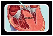
Schematic illustration of the catheter-based approach for RFSA, highlighting the thickened interventricular septum frequently involved in LVOT obstruction in obstructive HCM. The image shows the position of the ablation catheter relative to the mitral valve apparatus and the LV outflow tract, the target region for RFSA therapy.
References
-
1 Maron BJ, Ommen SR, Semsarian C, Spirito P, Olivotto I, Maron MS. Hypertrophic Cardiomyopathy: Present and Future, with Translation Into Contemporary Cardiovascular Medicine. J Am Coll Cardiol. 2014;64(1):83-99. doi: 10.1016/j.jacc.2014.05.003.
» https://doi.org/10.1016/j.jacc.2014.05.003 -
2 Ommen SR, Mital S, Burke MA, Day SM, Deswal A, Elliott P, et al. 2020 AHA/ACC Guideline for the Diagnosis and Treatment of Patients with Hypertrophic Cardiomyopathy: A Report of the American College of Cardiology/American Heart Association Joint Committee on Clinical Practice Guidelines. J Am Coll Cardiol. 2020;76(25):159-240. doi: 10.1016/j.jacc.2020.08.045.
» https://doi.org/10.1016/j.jacc.2020.08.045 -
3 Elliott PM, Anastasakis A, Borger MA, Borggrefe M, Cecchi F, Charron P, et al. 2014 ESC Guidelines on Diagnosis and Management of Hypertrophic Cardiomyopathy: the Task Force for the Diagnosis and Management of Hypertrophic Cardiomyopathy of the European Society of Cardiology (ESC). Eur Heart J. 2014;35(39):2733-79. doi: 10.1093/eurheartj/ehu284.
» https://doi.org/10.1093/eurheartj/ehu284 -
4 Valdigem BP, Correia EB, Moreira DAR, Bihan DL, Pinto IMF, Abizaid AAC, et al. Septal Ablation with Radiofrequency Catheters Guided by Echocardiography for Treatment of Patients with Obstructive Hypertrophic Cardiomyopathy: Initial Experience. Arq Bras Cardiol. 2022;118(5):861-72. doi: 10.36660/abc.20200732.
» https://doi.org/10.36660/abc.20200732 -
5 Gersh BJ, Maron BJ, Bonow RO, Dearani JA, Fifer MA, Link MS, et al. 2011 ACCF/AHA Guideline for the Diagnosis and Treatment of Hypertrophic Cardiomyopathy: A Report of the American College of Cardiology Foundation/American Heart Association Task Force on Practice Guidelines. Developed in Collaboration with the American Association for Thoracic Surgery, American Society of Echocardiography, American Society of Nuclear Cardiology, Heart Failure Society of America, Heart Rhythm Society, Society for Cardiovascular Angiography and Interventions, and Society of Thoracic Surgeons. J Am Coll Cardiol. 2011;58(25):212-60. doi: 10.1016/j.jacc.2011.06.011.
» https://doi.org/10.1016/j.jacc.2011.06.011 -
6 Vilela AA, Shinzato MH, Valdigem B, Paladino Filho AT, Correia EB, Assef JE. Cardiomiopatia Hipertrófica: Análise da Espessura Septal com a Redução do Gradiente em Pacientes Submetidos à Ablação Septal por Radiofrequência. Arq Bras Cardiol: Imagem cardiovasc. 2025;38(2):e20240097. doi: 10.36660/abcimg.20240097.
» https://doi.org/10.36660/abcimg.20240097

