My Approach To Echocardiographic Evaluation in Pediatric Patients with Sickle Cell Disease
Abstract
Sickle cell disease (SCD) is the most prevalent hereditary genetic disorder worldwide, and cardiovascular alterations are the main cause of death among patients with this disease.
Therefore, the early identification of markers of sickle cell cardiomyopathy using echocardiographic parameters (Central Illustration) is essential for the appropriate management of the cardiovascular condition in these patients.
This review provides a comprehensive overview of the use of echocardiography in pediatric patients with SCD, highlighting the main characteristics of the condition and the importance of regular follow-up.
Keywords
Echocardiography; Sickle Cell Anemia; Child

Resumo
A Doença Falciforme (DF) é a doença genética hereditária de maior prevalência no mundo, e as complicações cardiovasculares são uma das principais causas de óbito na população falcêmica.
Diante disso, o reconhecimento precoce dos marcadores da miocardiopatia falcêmica, através dos parâmetros ecocardiográficos (Figura Central), é fundamental para a conduta adequada da condição cardiovascular destes doentes.
Essa revisão abordará de forma objetiva e didática o uso da ecocardiografia em pacientes pediátricos com DF, mostrando as principais particularidades nesta doença e a importância do acompanhamento regular.
Palavras-chave
Ecocardiografia; Anemia Falciforme; Criança

Introduction
Sickle cell disease (SCD) is considered the most prevalent hereditary genetic disorder worldwide.
In Brazil, this condition affects between 60,000 and 100,000 individuals and has a higher incidence among the Afro-descendant population (8%). The distribution across the Brazilian territory is heterogeneous, with a higher incidence in the states of Bahia, Distrito Federal, and Piauí. The national incidence is approximately 1:1,200 live births, while it reaches 1:650 live births in Bahia.1
SCD is characterized by a recessive autosomal hereditary mutation, in which the glutamic acid is replaced by valine at the sixth position of the hemoglobin (Hb) beta chain. As a result, the organism produces Hb S , an abnormal type of Hb responsible for the physiopathology of SCD. Hb S has a shorter half-life (approximately one-sixth of the normal Hb A) due to its increased susceptibility to hemolysis and increased adhesiveness, contributing to vaso-occlusive events.
The disease may manifest as a homozygous form (Hb SS), known as sickle cell anemia, which is the most severe and prevalent form (approximately 60% to 75% of cases), with symptoms typically beginning within the first year of life. The heterozygous form occurs in approximately 25% to 40% of cases, tends to be milder, and results from the association of Hb S with other abnormal Hb, such as Hb SC and Hb SE. On the other hand, the sickle cell trait occurs when there is an association between Hb S and Hb A.2 Individuals with sickle cell trait are usually asymptomatic.
Under stress conditions (e.g., extreme temperatures, infection, or dehydration), the deoxygenated Hb S undergoes polymerization, causing red blood cells to assume a sickle shape. This condition makes Hb more prone to hemolysis, leading to chronic anemia. Moreover, the increased Hb rigidity impairs its passage through the microvasculature, triggering repeated painful vasoconstriction, endothelial dysfunction, inflammation, and ischemia-reperfusion injury.
The cardiac involvement in SCD arises from multiple mechanisms:
-
–
Chronic hemolytic anemia leads to reflex peripheral vasodilation to improve tissue oxygen delivery. This condition activates the renin-angiotensin-aldosterone system to retain sodium and water and, consequently, increase preload and chronic cardiac chamber dilation , particularly in the left chambers. Over time, this results in compensatory eccentric hypertrophy .
-
–
Chronic vaso-occlusive crises may cause microvascular myocardial dysfunction, renal failure, and systemic arterial hypertension. These conditions promote myocardial fibrosis and consequent systolic (less common) and diastolic dysfunction (more frequent).
-
–
Pulmonary hypertension in patients with SCD is multifactorial (Group 5): it may present with pre-capillary (arterial) characteristics secondary to intravascular hemolysis and Hb release into the plasma, which consumes nitric oxide and causes vasoconstriction, and post-capillary (venous) characteristics, in which diastolic dysfunction increases ventricular filling pressure, impairing blood flow through pulmonary veins and increasing pulmonary venous pressure. Additionally, chronic hypoxia and pulmonary thromboembolism may further contribute to elevated pulmonary pressure in patients with SCD.
Given these alterations, qualified and regular echocardiographic assessments are essential in patients with SCD.
Echocardiographic evaluation in patients with SCD
Dilation of cardiac chambers
The dilation of cardiac chambers, particularly the left chamber, occurs in approximately 30% to 65% of patients with SCD and is more prevalent in adults and those with the most severe form.
Atrium dilation is the earliest cardiac alteration resulting from volume overload secondary to compensatory vasodilation in response to anemia as a mechanism to improve oxygen delivery to tissues.
Sabatini et al. demonstrated an inverse association between serum Hb levels (degree of anemia) and left atrial dilation, and a direct association between reticulocyte levels (a marker for hemolytic anemia) and left atrial dilation3 ( Figure 1 ).

Eccentric hypertrophy
Eccentric hypertrophy (more common in the left chambers) occurs in 25% to 45% of patients with SCD and is more prevalent among older adults and individuals with more severe hemolytic anemia.
Koyunku et al. conducted a retrospective study on 146 patients with SCD aged over 18 years and found that 45 (30.8%) patients presented eccentric hypertrophy. After a 5-year follow-up, 31 (21.2%) patients had died. Survival analysis showed that patients presenting eccentric hypertrophy had a worse prognosis (the greater the eccentric hypertrophy, the higher the risk of death).4
Cardiac dysfunction
Chronic hemolytic anemia and recurrent vaso-occlusive episodes are physiopathological mechanisms for microvascular dysfunction and myocardial fibrosis. Moreover, disease-related systemic alterations, such as renal dysfunction, systemic arterial hypertension, and iron overload (which may occur in patients receiving periodical blood transfusions), contribute to the development or worsening of cardiac dysfunction, or both.
Diastolic dysfunction is common in adults with SCD (approximately 15% to 20% of cases) and is associated with an increased risk of premature death.5 Evaluating diastolic function in patients with SCD is needed but also challenging for echocardiographers.
In children, the assessment of diastolic function is limited because the Doppler parameters vary significantly according to age, body surface area, and heart rate. Unlike systolic function, which can be evaluated using one parameter (e.g., ejection fraction), there is no single gold-standard marker for diastolic function.
Even invasive measurements obtained by cardiac catheterization have limitations and provide only partial information on ventricular diastolic characteristics. Also, the parameters used to define diastolic dysfunction may be affected by the underlying disease physiopathology without truly reflecting diastolic function alterations, such as atrial dilation.6 , 7
In the general population, atrial dilation is associated with increased ventricular filling pressures. However, this dilation in patients with SCD occurs early in the development of the disease due to volume overload from the compensatory physiopathology of the condition and may not reflect elevated filling pressures. Hammoudi et al. evaluated 127 patients with SCD and found that most presented with left atrial dilation, but this finding was not associated with diastolic dysfunction. Instead, it correlated with disease severity and duration.5
Tricuspid regurgitation velocity peak is another parameter used to assess diastolic function. However, an increase in this parameter in patients with SCD may not necessarily indicate elevated pulmonary arterial pressure but rather increased cardiac preload or pre-capillary pulmonary hypertension. Hence, tricuspid regurgitation velocity data must be interpreted with caution in this population.5
The ratio between early diastolic transmitral flow velocity and tissue Doppler velocity (E/e′) is also used to estimate the left ventricular filling pressure in SCD. Sachdev et al. assessed 436 patients with SCD (homozygous for Hb S) and found that the E/e′ ratio independently correlated with reduced exercise capacity measured using the six-minute walk test.8
Another parameter used to evaluate diastolic function is the ratio between early and late diastolic flow velocity (E/A ratio). In a study of 141 patients with SCD, Sachdev et al. found that a low E/A ratio (i.e., indicative of diastolic dysfunction) was associated with increased mortality risk (hazard ratio of 3.5).9
Systolic dysfunction (assessed by conventional echocardiography) is rare in children with SCD.4 Poludasu et al. performed a meta-analysis of 19 case-control studies and found no significant difference in systolic function between 841 patients with SCD (homozygous Hb S) and 554 controls when evaluated using echocardiographic parameters10 ( Figure 2 ).

Apical 2- and 4-chamber views for the analysis of ventricular function during systole and diastole. Ejection fraction was calculated using Simpson's method. CVEd/VDFVE: Left Ventricular End-Diastolic Volume; MOD: Simpson's Method; FEVE/FE: Left Ventricular Ejection Fraction; CVE/VSFVE: Left Ventricular End-Systolic Volume; SV: Stroke Volume.
Strain analysis in SCD: what is new?
The development of 2D strain imaging enabled the early detection of cardiac damage in many chronic diseases and has become an increasingly important tool in prognostic stratification. The aim is to highlight the contribution of 2D strain in detecting subclinical ventricular myocardial damage in patients with SCD.
Cardiac dilation and eccentric ventricular hypertrophy in patients with SCD occur in response to increased preload and may progressively alter the cardiac function. These alterations contribute to morbidity and mortality in patients with SCD.
The global longitudinal strain of the left ventricle is a more specific predictor of myocardial remodeling than the left ventricle ejection fraction (LVEF), making it a sensitive tool for detecting early systolic dysfunction even when LVEF is preserved.
Studies have shown that patients with SCD presenting lower Hb levels (Hb < 9 g/dl) and increased preload are more likely to develop long-term subclinical systolic dysfunction in the left ventricle due to the limited adaptability of the heart to chronic preload increases. Resende et al. followed 219 patients with SCD for 30 months and observed that those with abnormal strain had worse clinical outcomes (pain crises, acute chest syndrome, and SCD-related death), independent of age, tricuspid regurgitation velocity peak, and ejection fraction assessed using conventional echocardiography. Thus, patients with SCD who present abnormal strain are at greater risk for adverse outcomes11 ( Figure 3 ).
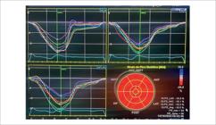
Left ventricle global longitudinal strain of −19.5% in a child with SCD. Sept: Septal wall; Ant: Anterior wall; Inf: Inferior wall; Lat: Lateral wall; Post: Posterior wall; GLPS: Global Longitudinal Peak Strain; FR: Frame Rate
Pulmonary hypertension
Pulmonary hypertension is characterized by reduced blood flow through the pulmonary arterial circulation due to increased pulmonary vascular resistance and elevated arterial pressures.
The prevalence of pulmonary hypertension is higher in patients with Hb SS disease than in those with SC, Sβ+, or Sβ° thalassemia.
Studies using echocardiography to measure tricuspid regurgitation velocity peak as an index of systolic pulmonary artery pressure demonstrated a high prevalence (30%) of pulmonary hypertension in patients with SCD.5
The physiopathology of pulmonary hypertension varies. First, since pulmonary pressure is a product of flow and pulmonary vascular resistance, the high cardiac output in SCD leads to increased pulmonary pressure regardless of altered pulmonary vascular resistance. Second, chronic volume overload may lead to left ventricular failure and subsequent pulmonary venous hypertension. Third, intravascular hemolysis may induce pulmonary arterial vasculopathy, mainly driven by nitric oxide depletion due to free plasma Hb.
Last, several other mechanisms may contribute, including hypoxemia, post-embolic pulmonary hypertension, lung injury related to SCD, and chronic liver disease. Importantly, several of these factors often occur simultaneously in the same patient with SCD and pulmonary hypertension. This represents a major challenge in the clinical management of the disease. Therefore, these patients are currently classified into Group 5 of the WHO classification system for pulmonary hypertension.5
Mehari et al. studied 531 patients and found associations between pulmonary pressure and reduced exercise tolerance and increased mortality rate in patients with SCD.12
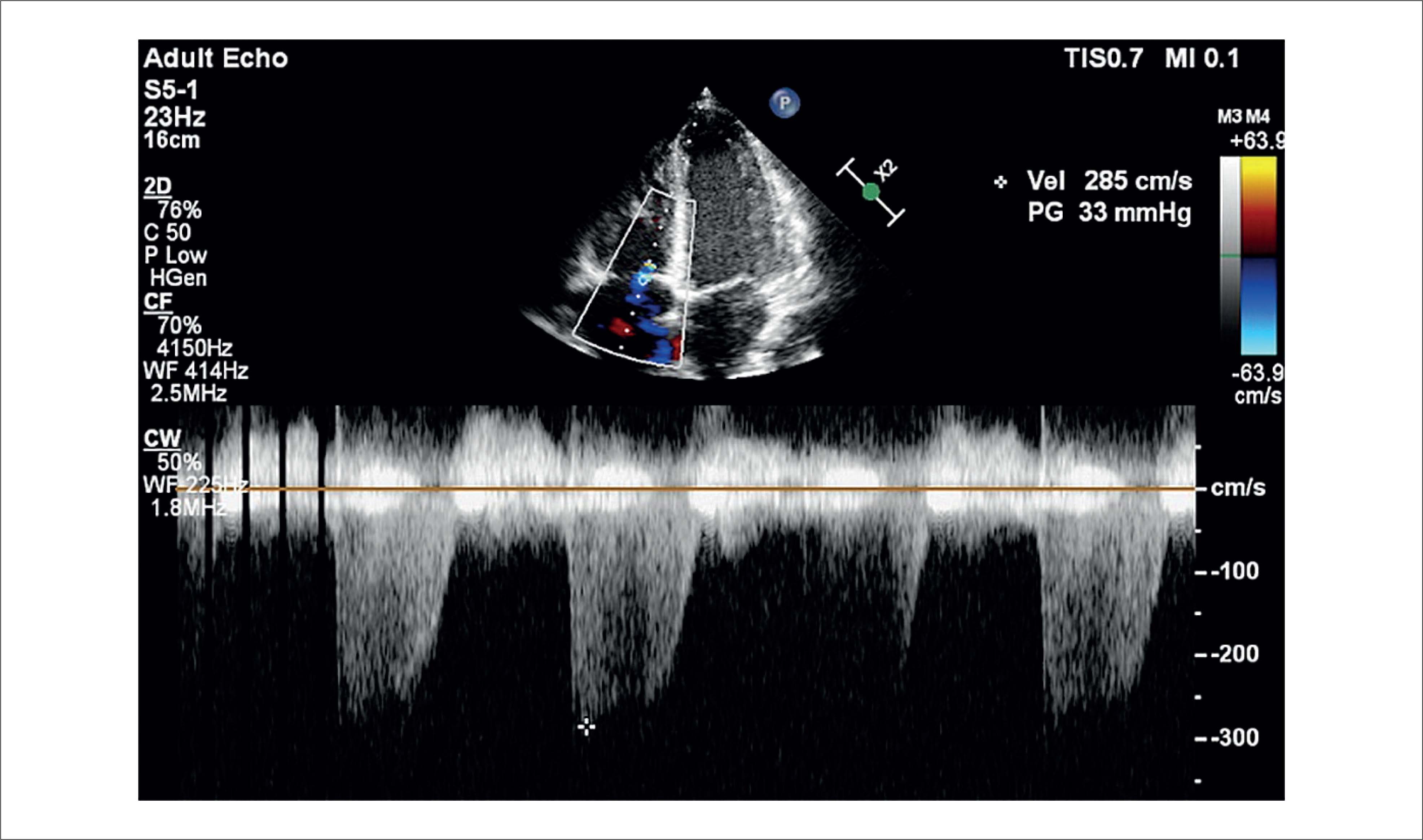
Given the impact of pulmonary hypertension on the morbidity and mortality of patients with SCD, the American Thoracic Association published a management guideline in 2014 recommending diagnostic cardiac catheterization when tricuspid regurgitation velocity was ≥ 3 m/s or between 2.5 and 2.9 m/s in symptomatic patients (e.g., reduced performance in the six-minute walk test or elevated serum NT-pro-BNP) 13 ( Figure 4 ).

Apical 4-chamber view for the analysis of tricuspid regurgitation using continuous Doppler in a patient with SCD. PG: Pressure Gradient; CW: Continuous Wave Doppler; WF: Waveform; CF: Color Flow Doppler
Echocardiographic assessment during SCD treatment
Symptomatic patients with SCD or those with pulmonary hypertension are candidates for disease-modifying treatments.
Hydroxyurea is one of the drugs used to treat SCD. One of its mechanisms of action is to increase fetal Hb levels; thus, reducing pathological Hb S levels and its physiopathological consequences.
Dhar et al. analyzed 100 patients with SCD, including 60 who received hydroxyurea, and observed that left ventricular dilation and hypertrophy significantly improved with hydroxyurea therapy. Additionally, left ventricular volume and mass were inversely correlated with treatment duration, demonstrating that pharmacological therapy may lead to cardiac remodeling.14
Another recommended therapy in symptomatic patients with SCD is chronic blood transfusion, aimed at reducing levels of pathological Hb S.
Turpin et al. evaluated 13 patients with SCD (homozygous Hb S) before and after chronic blood transfusion therapy and observed improved functional class and reduced tricuspid regurgitation jet velocity following transfusion.15
Therefore, patients with SCD undergoing disease-modifying treatment should also be regularly evaluated by echocardiography for a better definition of the cardiovascular status.
Echocardiographic assessment during SCD crises
Patients with SCD may present numerous acute events throughout life, such as pain crises, acute chest syndrome, splenic sequestration, and stroke.
Onalo et al. followed 176 patients for two years (92 without crisis and 84 with at least one crisis) and found a higher prevalence of pulmonary hypertension and cardiac dysfunction in those who experienced a crisis during this period.16
Therefore, patients with SCD who present with a sickle cell crisis must have their cardiovascular status re-evaluated to ensure appropriate cardiac management during these episodes.
Conclusion
Routine echocardiogram for asymptomatic pediatric patients with SCD is not yet a reality in clinical practice. However, the significant impact of cardiac alterations can be already observed from childhood.
Serial echocardiography exams must be considered to allow an early diagnosis of cardiac complications, improve clinical care for patients with SCD, and reduce cardiovascular morbidity and mortality in this population.
-
Sources of Funding
There were no external funding sources for this study.
-
Study Association
This study is not associated with any thesis or dissertation work.
-
Ethics Approval and Consent to Participate
This article does not contain any studies with human participants or animals performed by any of the authors.
-
Use of Artificial Intelligence
The authors did not use any artificial intelligence tools in the development of this work.
-
Availability of Research Data
The underlying content of the research text is contained within the manuscript.
References
- 1 Agência Nacional de Vigilância Sanitária. Manual de Diagnóstico e Tratamento de Doenças Falciformes. Brasília: ANVISA; 2001.
-
2 Brasil. Ministério da Saúde. Doença Falciforme [Internet]. Brasília: Ministério da Saúde; 2025 [cited 2025 May 19]. Available from: https://www.gov.br/saude/pt-br/assuntos/saude-de-a-a-z/d/doenca-falciforme
» https://www.gov.br/saude/pt-br/assuntos/saude-de-a-a-z/d/doenca-falciforme -
3 Sabatini L, Chinali M, Franceschini A, Di Mauro M, Marchesani S, Fini F, et al. Echocardiographic Evaluation in Paediatric Sickle Cell Disease Patients: A Pilot Study. J Clin Med. 2022;12(1):7. doi: 10.3390/jcm12010007.
» https://doi.org/10.3390/jcm12010007 -
4 Koyuncu MB, Tombak A, Orscelik O, Koseci T, Turker A, Basir H, et al. Cardiac Chamber Quantification by Echocardiography in Adults with Sickle Cell Disease: Need Attention to Eccentric Hypertrophy. Cureus. 2021;13(6):e15592. doi: 10.7759/cureus.15592.
» https://doi.org/10.7759/cureus.15592 -
5 Hammoudi N, Lionnet F, Redheuil A, Montalescot G. Cardiovascular Manifestations of Sickle Cell Disease. Eur Heart J. 2020;41(13):1365-73. doi: 10.1093/eurheartj/ehz217.
» https://doi.org/10.1093/eurheartj/ehz217 -
6 Rosa LM, Morhy SS. My Approach to Managing Left Ventricular Diastolic Function of Children and in Congenital Heart Disease. Arq Bras Cardiol: Imagem Cardiovasc. 2022;35(3):ecom29. doi: 10.47593/2675-312X/20223503ecom29.
» https://doi.org/10.47593/2675-312X/20223503ecom29 -
7 Hammoudi N, Charbonnier M, Levy P, Djebbar M, Stojanovic KS, Ederhy S, et al. Left Atrial Volume is Not an Index of Left Ventricular Diastolic Dysfunction in Patients with Sickle Cell Anaemia. Arch Cardiovasc Dis. 2015;108(3):156-62. doi: 10.1016/j.acvd.2014.09.010.
» https://doi.org/10.1016/j.acvd.2014.09.010 -
8 Sachdev V, Kato GJ, Gibbs JS, Barst RJ, Machado RF, Nouraie M, et al. Echocardiographic Markers of Elevated Pulmonary Pressure and Left Ventricular Diastolic Dysfunction are Associated with Exercise Intolerance in Adults and Adolescents with Homozygous Sickle Cell Anemia in the United States and United Kingdom. Circulation. 2011;124(13):1452-60. doi: 10.1161/CIRCULATIONAHA.111.032920.
» https://doi.org/10.1161/CIRCULATIONAHA.111.032920 -
9 Sachdev V, Machado RF, Shizukuda Y, Rao YN, Sidenko S, Ernst I, et al. Diastolic Dysfunction is an Independent Risk Factor for Death in Patients with Sickle Cell Disease. J Am Coll Cardiol. 2007;49(4):472-9. doi: 10.1016/j.jacc.2006.09.038.
» https://doi.org/10.1016/j.jacc.2006.09.038 -
10 Poludasu S, Ramkissoon K, Salciccioli L, Kamran H, Lazar JM. Left Ventricular Systolic Function in Sickle Cell Anemia: A Meta-Analysis. J Card Fail. 2013;19(5):333-41. doi: 10.1016/j.cardfail.2013.03.009.
» https://doi.org/10.1016/j.cardfail.2013.03.009 -
11 Resende MBS, Ferrari TCA, Araujo CG, Vasconcelos MCM, Tupinambás JT, Dias RCTM, et al. Prognostic Value of Left Ventricular Longitudinal Strain by Speckle-Tracking Echocardiography in Patients with Sickle Cell Disease. Int J Cardiovasc Imaging. 2020;36(11):2145-53. doi: 10.1007/s10554-020-01924-3.
» https://doi.org/10.1007/s10554-020-01924-3 -
12 Mehari A, Gladwin MT, Tian X, Machado RF, Kato GJ. Mortality in Adults with Sickle Cell Disease and Pulmonary Hypertension. JAMA. 2012;307(12):1254-6. doi: 10.1001/jama.2012.358.
» https://doi.org/10.1001/jama.2012.358 -
13 Klings ES, Machado RF, Barst RJ, Morris CR, Mubarak KK, Gordeuk VR, et al. An Official American Thoracic Society Clinical Practice Guideline: Diagnosis, Risk Stratification, and Management of Pulmonary Hypertension of Sickle Cell Disease. Am J Respir Crit Care Med. 2014;189(6):727-40. doi: 10.1164/rccm.201401-0065ST.
» https://doi.org/10.1164/rccm.201401-0065ST -
14 Dhar A, Leung TM, Appiah-Kubi A, Gruber D, Aygun B, Serigano O, et al. Longitudinal Analysis of Cardiac Abnormalities in Pediatric Patients with Sickle Cell Anemia and Effect of Hydroxyurea Therapy. Blood Adv. 2021;5(21):4406-12. doi: 10.1182/bloodadvances.2021005076.
» https://doi.org/10.1182/bloodadvances.2021005076 -
15 Turpin M, Chantalat-Auger C, Parent F, Driss F, Lionnet F, Habibi A, et al. Chronic Blood Exchange Transfusions in the Management of Pre-Capillary Pulmonary Hypertension Complicating Sickle Cell Disease. Eur Respir J. 2018;52(4):1800272. doi: 10.1183/13993003.00272-2018.
» https://doi.org/10.1183/13993003.00272-2018 -
16 Onalo R, Cooper P, Cilliers A, Nnebe-Agumadu U. Cardiovascular Changes in Children with Sickle Cell Crisis. Cardiol Young. 2020;30(2):162-70. doi: 10.1017/S1047951120000037.
» https://doi.org/10.1017/S1047951120000037
Edited by
-
Editor responsible for the review:
Marcelo Tavares
The underlying content of the research text is contained within the manuscript.

 Echocardiographic evaluation in patients with sickle cell disease.