Hypertrophic Cardiomyopathy: Analysis of Septal Thickness with Gradient Reduction in Patients Undergoing Radiofrequency Septal Ablation
Abstract
Background: Hypertrophic obstructive cardiomyopathy (HOCM) is an autosomal dominant genetic disorder. Invasive treatment for intraventricular gradient reduction is indicated in patients (p) who are refractory to clinical treatment with gradient ≥ 50 mmHg. Radiofrequency ablation (RFA) is one of the current options for invasive management.
Objectives: To evaluate the correlation between interventricular septal diameter and procedural success, defined as immediate post-procedural gradient reduction, improvement in functional class (FC), and sustained gradient reduction at 6-month follow-up.
Methods: Twenty-two patients with HOCM were included. Transthoracic echocardiography was performed before and six months after RFA, while transesophageal echocardiography (TEE) was carried out intraprocedure.
Results: The mean age was 56.64 years (± 12.23), with 68.18% of patients being female and 73% hypertensive. An immediate gradient reduction greater than 50% was observed in 72.7% of patients. At the 6-month follow-up, 60% of patients showed a ≥ 50% gradient reduction along with an improvement in FC. A median septal thickness of 18 mm was associated with greater procedural success compared to a median of 15 mm, and no significant change in septal thickness was observed over the 6-month follow-up period.
Conclusion: Radiofrequency septal ablation (RFSA) is an effective and safe technique for the invasive management of HOCM. The procedure enables a significant reduction in the intraventricular gradient, accompanied by improved FC and sustained short-term results. Thicker septal were associated with greater intraprocedural gradient reductions; however, there was no reduction in thickness during the 6-month follow-up.
Keywords:
Hypertrophic Cardiomyopathy; Radiofrequency Ablation
Resumo
Fundamento: A cardiomiopatia hipertrófica obstrutiva (CMHO) é uma doença genética autossômica dominante. A indicação de tratamento invasivo para a redução do gradiente intraventricular ocorre nos pacientes (p) refratários ao tratamento clínico com gradiente ≥ 50 mmHg. A ablação por radiofrequência (ARF) é, atualmente, uma das opções disponíveis para o manejo invasivo.
Objetivos: Correlacionar o diâmetro do septo interventricular com o sucesso do procedimento, definido como a redução do gradiente no pós-procedimento imediato, melhora da classe funcional e redução do gradiente seis meses após o procedimento.
Métodos: Foram incluídos 22 pacientes com CMHO. O ecocardiograma transtorácico foi realizado antes e seis meses após a ARF, enquanto o ecocardiograma transesofágico (ETE) foi realizado no intraprocedimento.
Resultados: A idade média foi de 56,64 anos (± 12,23), com 68,18% dos pacientes do sexo feminino e 73% hipertensos. A redução imediata do gradiente em mais de 50% foi observada em 72,7% dos pacientes. Aos seis meses, também houve redução do gradiente ≥ 50% em 60% dos casos, além de melhora da classe funcional. Os valores de espessura septal com mediana de 18 mm estiveram associados a maior sucesso no procedimento em comparação com os de mediana de 15 mm. Não houve alteração significativa na espessura septal durante o seguimento de seis meses.
Conclusão: A ablação septal por radiofrequência (ASRF) é uma técnica eficaz e segura no manejo invasivo da CMHO. Proporciona redução significativa do gradiente intraventricular com melhora da classe funcional e manutenção de resultados no curto prazo de seguimento. Septos mais espessos mostraram correlação com maiores reduções de gradientes no intraprocedimento, mas não houve redução da espessura septal no seguimento de seis meses.
Palavras-chave:
Cardiomiopatia Hipertrófica; Ablação por Radiofrequência

Introduction
Hypertrophic cardiomyopathy (HCM) is an autosomal dominant genetic disorder that affects one in 500 individuals1 and is considered the most common isolated form of hereditary heart disease.2 In addition to its genetic basis, the disease's pathophysiology involves a combination of diastolic dysfunction, left ventricular outflow tract (LVOT) obstruction, mitral regurgitation, and myocardial ischemia.3
This pathophysiological mechanism explains the development of clinical manifestations, which not only present with a highly heterogeneous clinical picture, but can begin at any stage of life.4 - 7 This diversity in clinical presentations is largely due to genetic heterogeneity itself, with progression ranging from asymptomatic cases to sudden death as the initial diagnosis.8
In the presence of symptoms, treatment for HCM is indicated.8 Pharmacological therapy is the first-line treatment used to relieve heart failure symptoms. However, in cases of significant obstruction and severe symptoms refractory to drug treatment, more invasive measures may be necessary to improve symptoms and reduce the intraventricular gradient.9
Two invasive methods, surgical myectomy and alcohol septal ablation, are used to relieve LVOT obstruction in patients refractory to clinical treatment. Recent studies have reported favorable results of a new interventional procedure, radiofrequency septal ablation (RFSA).
There are few studies in the literature correlating myocardial thickness diameters with the success of this procedure. Therefore, our objective was to assess whether the degree of myocardial wall thickening prior to the intervention was associated with greater success in RFSA in patients with hypertrophic obstructive cardiomyopathy (HOCM).
Methods
Study Sample
This is a retrospective observational study based on a review of medical records, approved by our institutional research ethics committee under protocol No. 5133. Patients of both sexes, aged 18 years or older, diagnosed with HOCM, and who underwent RFSA treatment were included. The procedure was performed between May 2017 and October 2019.
Invasive therapy was indicated for patients with HOCM with a maximum gradient at rest or after provocative maneuver ≥ 50 mmHg, were symptomatic, and refractory to clinical treatment. Patients who underwent another concomitant invasive procedure or declined participation were excluded from the study.
RFSA Protocol
The procedure protocol was described by Valdigem et al.10 RFSAs were performed in an electrophysiology room, with the patient under general anesthesia and mechanical ventilation. The right femoral artery was accessed using an 8F introducer for retroaortic access to the left ventricular septal region. Two additional punctures were made in the right femoral vein (6F introducer) to place one quadripolar catheter in the right septum (to identify the His bundle electrocardiographically) and another in the right ventricular apex.
With the aid of transesophageal echocardiography (TEE), the ventricle-aortic gradient was measured, and the area of greatest flow acceleration—corresponding to the point of greatest obstruction — was identified.
Thus, using TEE morphofunctional analysis and electroanatomical mapping, the therapeutic catheter was positioned at the site of greatest septal obstruction, and radiofrequency energy was applied for 120 seconds (80W, 60 °C). After each application, the gradient was measured by means of TEE. Immediately after the procedure, TEE was used to measure the maximum gradient in the LVOT and to rule out complications such as pericardial effusion, mitral valve injury, or significant mitral regurgitation.
After the sheaths were removed, patients remained in the intensive care unit (ICU) for 24 hours and were then transferred to the inpatient ward for 3 to 5 days.
Research Design Flowchart
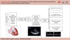
Medical records of eligible patients were reviewed according to the established inclusion and exclusion criteria. Clinical data and interventricular septal diameters (measured by pre-procedure transthoracic echocardiography) were analyzed. Procedural success was defined as immediate gradient reduction, absence of reintervention within six months, improvement in NYHA functional class (FC), and gradient reduction at 6-month follow-up (Central Illustration).
Statistical Analysis
Frequency distribution was used to describe categorical variables. Central trend measures (mean and median) and variability (minimum, maximum, and standard deviation) were used to describe numerical variables.
Given the variability in hemodynamic conditions, two assessment settings were defined: intraprocedure and outpatient. Intraprocedure: considering the gradients immediately before ablation and immediately after ablation, with the patient intubated under the effect of positive pressure. Outpatient: considering the gradients estimated by the echocardiogram before the procedure and six months after the procedure.
The nonparametric Mann-Whitney U test was used to verify the association between the septal measurement numerical variable and procedural success. This test was chosen because the normal distribution of the septal measurements was not observed.
A graph was constructed for the evolution of the septal thickness at the time points before the procedure, three months, and six months after the ablation. A mixed linear regression model was used to assess changes in septal thickness over time, treating repeated patient measurements as a random effect.
The paired Student's t -test was applied to compare the numerical variable gradient before the procedure and at 6 months, where the normal distribution of the data was identified.
The nonparametric Wilcoxon paired test was applied to compare the numerical variable immediate gradient before the procedure and immediately after, and no normality was observed in the variables data.
The Shapiro-Wilk test was conducted to verify data normality (normal distribution) for each numerical variable.
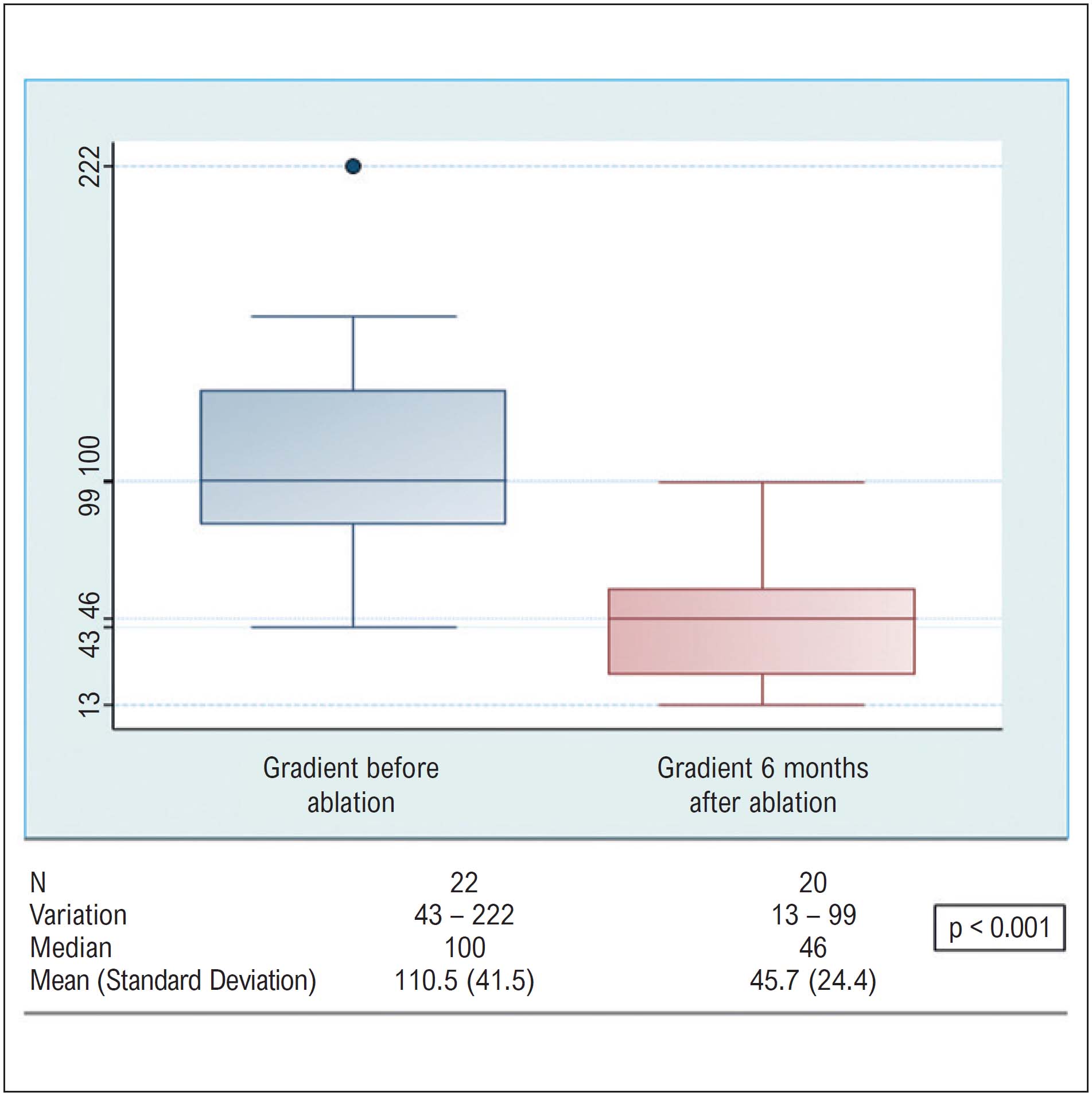
Box-Plot graphs were used to represent the distribution of the pre and post-immediate gradient at timepoint zero and six months later.
A significance level of 5% was adopted for all statistical tests. The statistical software STATA version 16.0 was used to perform the statistical analyses (ref: StataCorp. 2019. Stata Statistical Software: Release 16.0. College Station, Texas: Stata Corporation).
Results
The study included 22 patients with HOCM who underwent RFSA treatment at a tertiary cardiology hospital between May 2017 and October 2019.
Table 1 shows the clinical characteristics observed before the procedure. Regarding gender, the sample was predominantly female, with seven men (32%) and 15 women (68%). Patient age ranged from 23 to 79 years, with a mean of 56.73 ± 12.20 years. The mean body mass index (BMI) was 29.69 ± 5.46 kg/m².
Regarding medical history, 16 patients (73%) were hypertensive, 11 (50%) had dyslipidemia, six (27%) were diabetic, and three (14%) had a diagnosis of hypothyroidism. Three patients (14%) had associated coronary artery disease. A known family history of HCM was documented in approximately 23% of the cases.
Among the patients with previous coronary artery disease, only one had undergone prior myocardial revascularization.
Regarding symptoms ( Table 2 ), all 22 patients (100%) had dyspnea, 12 (55%) had angina, six (27%) experienced palpitation, seven (32%) had syncope, and five (23%) reported fainting. Nearly all patients (91%) were in FC III-IV despite clinical treatment. All 22 patients (100%) used beta-blockers, seven (32%) were on calcium channel blockers and two (9%) were using antiarrhythmics ( Table 3 ).
Mean heart rate was 62.45 bpm ± 6.99. The electrocardiogram performed before the procedure showed left ventricular overload in 100% of patients, and eight of them (36.4%) had left bundle branch block.
The mean septal thickness before the procedure was 18.59 mm (± 4.63), ranging from 13 mm to 32 mm ( Table 4 ). Diastolic dysfunction (grades I to III) and mitral insufficiency due to systolic anterior motion of the mitral valve were notable findings. All patients had preserved systolic function, with a mean ejection fraction estimated by Simpson of 68% ± 4.3. As expected, ventricular mass was increased, with a mean value of 341.86 g (± 113.85). The mean, median, and standard deviation values of the echocardiographic parameters are shown in Table 5 .
The mean initial maximum gradient estimated during the procedure was 78.18 mmHg (± 33.05). At the end of the procedure, the mean decreased to 34.09 mmHg (± 28.48).
Two patients developed complications at the puncture site, one pseudoaneurysm and two hematomas, which were resolved conservatively. Seven patients developed left bundle branch block immediately after the procedure and, and two patients had hypotension. There were no deaths or complications requiring surgical intervention during hospitalization. No cases of pericardial effusion or clinical cerebral embolic events were observed.
The mean length of hospital stay was 4.14 days, ranging from two to 10 days.
Before the radioablation procedure, 68% of the patients were in NYHA FC III, and 23% reported FC IV. At the end of one year after the procedure, 77% were in FC I, and 9% were in FC II ( Figure 1 ).

Percentage distribution of patients with FC I, II, III and IV before and after 6 months of the RFSA procedure. FC: functional class.
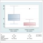
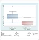
LVOT gradient measurements were performed immediately before and after radioablation, considering the same hemodynamic status (patient sedated, on mechanical ventilation with PEEP of 5, with the same pressure level and inferior vena cava diameter) for accurate analysis of gradient variation related to the procedure ( Figure 2 ). A statistically significant reduction in the LVOT gradient was observed, with the median pre-procedure value at 68 mmHg and the median immediate post-procedure value at 27 mmHg (p < 0.001). Obstructive gradients recorded in the outpatient setting prior to the procedure (mean = 110.5 mmHg) were higher than those recorded in the procedure room, as shown in Figure 3 , and higher than those measured in the outpatient setting six months post-procedure (45.7 mmHg) compared to the immediate post-procedure values. This difference is likely due to the hemodynamic response under sedation during the radioablation procedure. In both assessments, a significant reduction in the gradient was observed: a mean relative reduction of 58.9% immediately after the procedure and 52% at the 6-month follow-up ( Table 6 ).
There is no established definition of successful outcomes for RFSA in the literature. Ifor this study, success was defined as an immediate gradient reduction of ≥ 50%, with this reduction maintained six months after the procedure. Gradients lower than 50 mmHg and lower than 30 mmHg immediately after the procedure were also considered parameters of procedural success. Table 7 shows the proportion of patients who presented a reduced gradient (immediately after RFSA and at six months) according to success-defining parameters. The nonparametric Mann-Whitney U test was used to verify the association between ventricular septum diameter and RFSA success. Table 8 shows the distribution of septal measurements based on procedural outcomes. When defining success as a gradient reduction of ≥ 50%, septa with a median thickness of 18 mm were associated with better outcomes compared to those with 15 mm (p < 0.05). When success was defined as a gradient reduction of ≥ 50% and an absolute gradient below 50 mmHg, septa with a median thickness of 18 mm again showed better outcomes than those with 15 mm (p < 0.05). No significant associations were found in the other success categories (p > 0.05).
Proportion of patients who reduced gradients comparing pre and immediate post, and comparing pre-outpatient and 6-months post-outpatient
The measurement of septal thickness at the pre-procedure, three-months and six-months’ time points is shown in Figure 4 . The red line represents the mean values. A mixed linear model was used to evaluate the effect of time on septal thickness, considering the dependence between participants. No evidence was found to suggest that the measurements changed over time.

Discussion
To date, this study has shown that RFSA presents favorable results in reducing the ventricular gradient, without fatal complications.
To minimize hemodynamic variability, we compared gradients measured immediately before and after the procedure while the patient was sedated and on mechanical ventilation in the catheterization lab. Similarly, we compared outpatient gradients measured before the procedure with those measured six months post-procedure.
The procedure was largely successful, whether defined as a gradient reduction of more than 50%, an absolute gradient below 50 mmHg or 30 mmHg, or a combination of these criteria. Immediate gradient reduction above 50% occurred in 72.7% of patients.
The result at 6 months also demonstrated a gradient reduction ≥ 50% in 60% of the patients. At six months, the patients benefited from the procedure, as demonstrated by improved FC in 100% of the patients, without the need for reintervention.
The results analysis showed that septa with a median of 18 mm tended to have greater success than those with a median of 15 mm. This can be explained by the fact that greater septal thicknesses are related to higher gradients, and the reduction becomes more noticeable in patients who already have greater gradients.
Over the course of six months, the analysis of the septal diameter evolution was influenced by the loss of data, but there was no significant change during this period. These findings suggest that the gradient reduction probably occurs due to the lower motility of the septal segment treated by radioablation, rather than a reduction in its thickness.
Study Limitations
This was a retrospective study with a short follow-up period. As a retrospective study, we acknowledge that including a validated quality of life questionnaire pre- and post-procedure (six months) would have provided additional insight beyond NYHA FC. Likewise, the six-minute walk test and cardiopulmonary test would have offered quantitative parameters, which would certainly contribute more valuable data.
The sample size was small, which limits statistical correlation. The small sample size and retrospective design restricted our ability to perform comparative analyses with other established methods, such as alcohol septal ablation or surgical myectomy.
Since data were obtained from medical records, some cases were excluded due to incomplete information.
Conclusion
RFSA is an effective and safe technique in the invasive management of HOCM. The procedure allows for a significant reduction in intraventricular gradient, improvement in FC, and maintenance of results in short-term follow-up.
Multicenter studies comparing RFSA to traditional invasive techniques, with long-term follow-up, are required for this new modality to be considered an alternative for invasive management in HOCM, as outlined in the guidelines.
-
Sources of Funding
There were no external funding sources for this study.
-
Study Association
This article is part of the thesis of Doctoral submitted by Mariane Higa Shinzato, from Dante Pazzanese Institute/São Paulo University.
-
Ethics Approval and Consent to Participate
This study was approved by the Ethics Committee of the Dante Pazzanese Heart Institute under the protocol number 5133. All the procedures in this study were in accordance with the 1975 Helsinki Declaration, updated in 2013. Informed consent was obtained from all participants included in the study.
-
Use of Artificial Intelligence
The authors did not use any artificial intelligence tools in the development of this work.
-
Availability of Research
The underlying content of the research text is contained within the manuscript.
References
-
1 Maron BJ. Hypertrophic Cardiomyopathy. Lancet. 1997;350(9071):127-33. doi: 10.1016/S0140-6736(97)01282-8.
» https://doi.org/10.1016/S0140-6736(97)01282-8 -
2 Howell N, Bradlow W. Surgical Management of Left Ventricular Outflow Obstruction in Hypertrophic Cardiomyopathy. Echo Res Pract. 2015;2(1):37-44. doi: 10.1530/ERP-15-0005.
» https://doi.org/10.1530/ERP-15-0005 -
3 Bazan SGZ, Oliveira GO, Silveira CFSMP, Reis FM, Malagutte KNDS, Tinasi LSN, et al. Hypertrophic Cardiomyopathy: A Review. Arq Bras Cardiol. 2020;115(5):927-35. doi: 10.36660/abc.20190802.
» https://doi.org/10.36660/abc.20190802 -
4 Elliott PM, Anastasakis A, Borger MA, Borggrefe M, Cecchi F, Charron P, et al. 2014 ESC Guidelines on Diagnosis and Management of Hypertrophic Cardiomyopathy: The Task Force for the Diagnosis and Management of Hypertrophic Cardiomyopathy of the European Society of Cardiology (ESC). Eur Heart J. 2014;35(39):2733-79. doi: 10.1093/eurheartj/ehu284.
» https://doi.org/10.1093/eurheartj/ehu284 -
5 Maron BJ, Ommen SR, Semsarian C, Spirito P, Olivotto I, Maron MS. Hypertrophic Cardiomyopathy: Present and Future, with Translation Into Contemporary Cardiovascular Medicine. J Am Coll Cardiol. 2014;64(1):83-99. doi: 10.1016/j.jacc.2014.05.003.
» https://doi.org/10.1016/j.jacc.2014.05.003 -
6 Maron MS, Maron BJ. Hypertrophic Cardiomyopathy - Authors’ Reply. Lancet. 2013;381(9876):1457-8. doi: 10.1016/S0140-6736(13)60922-8.
» https://doi.org/10.1016/S0140-6736(13)60922-8 -
7 Maron BJ. Hypertrophic Cardiomyopathy: A Systematic Review. JAMA. 2002;287(10):1308-20. doi: 10.1001/jama.287.10.1308.
» https://doi.org/10.1001/jama.287.10.1308 - 8 Bittencourt MI, Rocha RM, Albanesi FM Filho. Hypertrophic Cardiomyopathy. Rev Bras Cardiol. 2010;23(1):17-24.
-
9 Cooper RM, Shahzad A, Hasleton J, Digiovanni J, Hall MC, Todd DM, et al. Radiofrequency Ablation of the Interventricular Septum to Treat Outflow Tract Gradients in Hypertrophic Obstructive Cardiomyopathy: A Novel Use of CARTOSound® Technology to Guide Ablation. Europace. 2016;18(1):113-20. doi: 10.1093/europace/euv302.
» https://doi.org/10.1093/europace/euv302 -
10 Valdigem BP, Correia EB, Moreira DAR, Bihan DL, Pinto IMF, Abizaid AAC, et al. Septal Ablation with Radiofrequency Catheters Guided by Echocardiography for Treatment of Patients with Obstructive Hypertrophic Cardiomyopathy: Initial Experience. Arq Bras Cardiol. 2022;118(5):861-72. doi: 10.36660/abc.20200732.
» https://doi.org/10.36660/abc.20200732
Edited by
-
Editor responsible for the review:
Marcelo Tavares
The underlying content of the research text is contained within the manuscript.

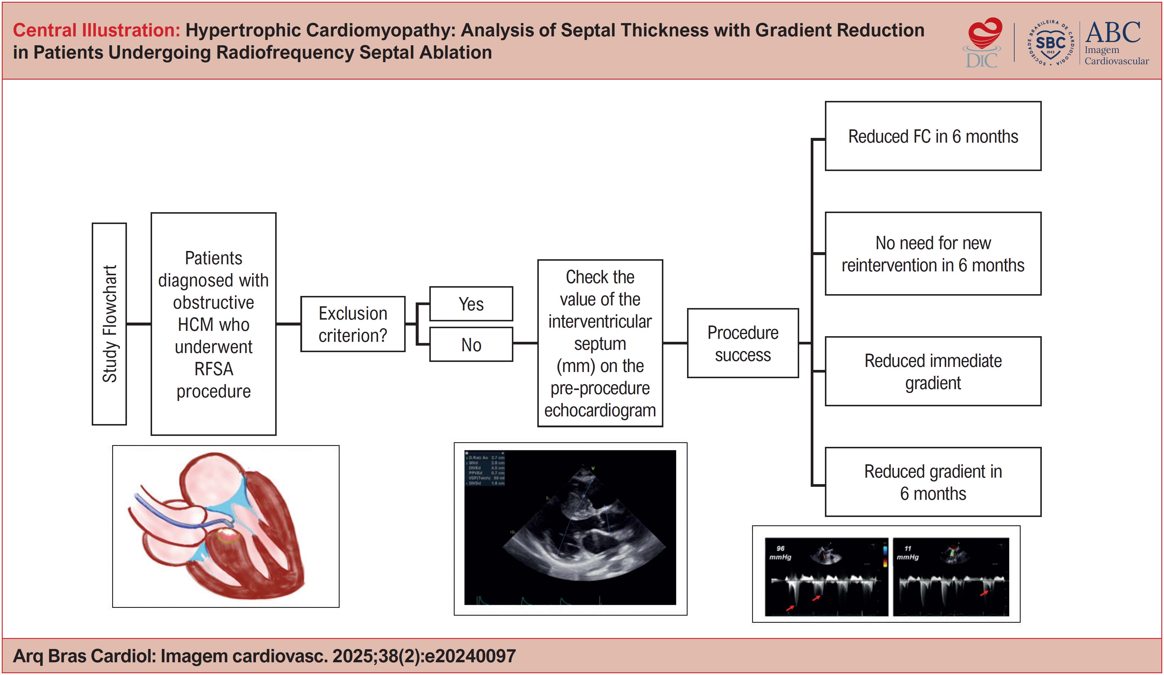 HCM: Hypertrophic cardiomyopathy; RFSA: Radiofrequency septal ablation; FC: functional class.