Safety of dobutamine stress echocardiography using a modified protocol in a large unselected population
Abstract
Background Dobutamine stress echocardiography (DSE) using the conventional protocol (CP-DSE) may lead to important adverse effects.
Objective This study was aimed at assessing the safety of DSE using a modified protocol (MP-DSE).
Methods Data were collected from our institutional database to compare MP-DSE with the CP-DSE. In the CP-DSE, atropine could be administered during the fourth stage or its extension. In the MP-DSE, atropine was initiated at the beginning of the third stage, and there was no stage extension. Upon completion of DSE or for arrhythmia control, metoprolol was administered in the CP-DSE and esmolol in the MP-DSE. In cases of typical angina, nitroglycerin was administered at the examiner’s discretion in the CP-DSE, whereas its use was predetermined in the MP-DSE. A p-value < 0.05 was considered statistically significant.
Results Of 17,811 tests performed, 9,121 were conducted using the MP-DSE. During DSE, myocardial oxygen consumption, represented by the rate-pressure product, was significantly higher in the MP-DSE group (22,530 ± 4,575 vs 23,037 ± 4,072 bpm.mmHg; p < 0.001). Hypertensive peak (1% vs 0.4%; p = 0.0001) and nonsustained ventricular tachycardia (0.6% vs 0.1%; p = 0.0001) were more frequent in the CP-DSE, while supraventricular tachyarrhythmia (2.1% vs 3.78%; p = 0.0001) and atrial fibrillation (0.8% vs 1.3%; p = 0.003) were more common in the MP-DSE. These arrhythmias resolved spontaneously or with medication. In the CP-DSE, two patients had ventricular fibrillation and one, acute coronary syndrome.
Conclusion In our study, the modified protocol for DSE proved to be a safe option, with no severe adverse effects observed, despite the presence of an elevated rate-pressure product.
Keywords:
Stress Echocardiography; Safety; Drug-Related Side Effects and Adverse Reactions

Resumo
Fundamento A Ecocardiografia de Estresse com Dobutamina (EED) utilizando o Protocolo Convencional (PC-EED) pode causar efeitos adversos graves.
Objetivo Este estudo teve como objetivo avaliar a segurança da EED utilizando um Protocolo Modificado (PM-EED).
Métodos Os dados foram coletados do banco de dados da nossa instituição para comparação entre o PM-EED e o PC-EED. No PC-EED, a atropina podia ser administrada durante o quarto estágio ou sua extensão. No PM-EED, a atropina foi iniciada no início do terceiro estágio, sem extensão de estágio. Após a conclusão da EED ou para controle de arritmias, o metoprolol foi administrado no PC-EED e o esmolol, no PM-EED. Em caso de angina típica, a nitroglicerina era administrada a critério do examinador no PC-EED, enquanto no PM-EED seu uso era pré-determinado. Um valor de p < 0,05 foi considerado estatisticamente significativo.
Resultados Dos 17 811 testes realizados, 9 121 foram conduzidos utilizando o protocolo PM-EED. Durante a EED, o consumo de oxigênio miocárdico – representado pelo produto pressão-frequência – foi significativamente maior no grupo PM-EED (22.530 ± 4.575 vs 23.037 ± 4.072 bpm·mmHg; p < 0,001). Pico hipertensivo (1% vs. 0,4%; p = 0,0001) e taquicardia ventricular não sustentada (0,6% vs. 0,1%; p = 0,0001) foram mais frequentes no grupo PC-EED, enquanto taquiarritmia supraventricular (2,1% vs 3,78%; p = 0,0001) e fibrilação atrial (0,8% vs. 1,3%; p = 0,003) foram mais comuns no grupo PM-EED. Essas arritmias se resolveram espontaneamente ou com intervenção medicamentosa. No grupo PC-EED, dois pacientes apresentaram fibrilação ventricular e um desenvolveu síndrome coronariana aguda.
Conclusão Em nosso estudo, o protocolo modificado para EED mostrou-se uma opção segura, sem ocorrência de efeitos adversos graves, mesmo na presença de um produto pressão-frequência elevado.
Palavras-chaves:
Ecocardiografia sob Estresse; Segurança; Efeitos Colaterais e Reações Adversas Relacionados a Medicamentos

Introduction
Dobutamine stress echocardiography (DSE) protocols have traditionally involved exposure to large doses of dobutamine and a significant incidence of adverse effects. However, the development of newer protocols for this diagnostic method has progressively reduced these risks.1-6
The addition of atropine to DSE protocols at an earlier stage has enhanced the test’s sensitivity without compromising its specificity and safety. As a result, current guidelines limit the maximum dose of dobutamine and atropine is initiated before or even during the last stage of dobutamine infusion.6,7
The intravenous administration of an ultra-short-acting beta-blocker at the end of the DSE is essential for controlling heart rate (HR), arrhythmias and myocardial ischemia, and may even enhance the accuracy of the test.2,3-8 However, the literature does not indicate a preference between esmolol and metoprolol. Ischemia occurring during DSE might require the use of coronary vasodilator, administered either orally or intravenously, although current guidelines do not specify the drug formulation or route of administration.3-6
Reports of adverse effects associated with DSE have been essential to establish contraindications to the test, emphasizing its higher risk compared to echocardiography with other stressors, such as exercise, dipyridamole, or adenosine.9-11 Thus, ongoing monitoring and the development of new protocols are necessary to minimize complications, particularly considering that dobutamine is the most widely available pharmacologic agent for stress echocardiography. Therefore, this study aimed at assessing the safety of DSE by applying a modified protocol to a large, unselected population.
Methods
This retrospective study compared the conventional protocol for DSE (CP-DSE) with a modified protocol (MP-DSE), using data collected from our institutional database about patients referred for DSE to assess myocardial ischemia with an adequate echocardiographic window. Patients submitted to the CP-DSE, constituting the CP-DSE group, were enrolled between 1997 and 2007, and those who underwent MP-DSE, constituting the MP-DSE group, were enrolled between 2008 and 2021.
Patient demographic data, coronary artery disease risk factors and history of myocardial revascularization were assessed through medical history, clinical evaluation and laboratory tests. All patients were informed about the objectives and potential risks of DSE and provided consent to undergo the procedure. Patients referred for myocardial viability assessment were excluded from the analysis.
This study’s protocol complies with the ethical principles outlined in the 1975 Declaration of Helsinki ethical principles and was submitted to and approved by our institutional Human Research Ethics Committee.
In both groups, dobutamine was continuously infused in up to four stages at incremental doses of 10, 20, 30 and 40 μcg.kg-1.min-1. When indicated, atropine was administered in boluses of 0.25- to 0.50-mg, up to the maximum cumulative dose of 2 mg.2-4,12 In cases of paradoxical sinus deceleration, defined as a 5- to 10- beat reduction in HR, atropine could be administered during the initial stages, at the examiner’s discretion. Upon test conclusion, an intravenous beta-blocker or coronary vasodilator was used according to each group’s protocol. However, when coronary spasm was suspected, priority was given to vasodilator administration.
Absolute contraindications to dobutamine use were unstable angina, recent myocardial infarction, severe and symptomatic aortic stenosis, acute aortic dissection, decompensated heart failure, ventricular pseudoaneurysm and obstructive hypertrophic cardiomyopathy. Atropine was contraindicated in cases of myasthenia gravis, pyloric stenosis, prostate disease determining urinary retention and narrow-angle glaucoma.9,11
To increase the number of tests completed, beta-blocker withdrawal was recommended two to three days before the test for CP-DSE patients and 5 days before for MP-DSE patients. A quad-screen display was used for comparative analysis, with CX 200 (Apogee) and Vingmed System Five (General Electric) equipment utilized in the CP-DSE group, and Vivid 7 and Vivid E9 (General Electric) equipment in the MP-DSE group.
Regarding important adverse effects, hypertensive peak was defined as blood pressure higher than 230/120 mm Hg, while hypotension was defined as systolic blood pressure (SBP) lower than 100 mm Hg. We retrieved the following arrhythmias from the database: supraventricular tachyarrhythmia (junctional or atrial), atrial fibrillation, sustained ventricular tachycardia (duration > 30 seconds), nonsustained ventricular tachycardia and ventricular fibrillation. Additional events recorded included paradoxical sinus deceleration, acute coronary syndrome, acute myocardial infarction, ventricular rupture, asystole and death. DSE should be discontinued in cases of potential life-threatening conditions or intolerance to the test medications.
Blood pressure was measured at the beginning of each stage and during the recovery phase. Electrocardiographic monitoring was continuous, and 12-lead electrocardiography was performed before and throughout the procedure. DSE was considered complete when at least 85% of the age-predicted maximal HR (220 minus age in years) was achieved and/or myocardial ischemia was detected. Ischemia was defined as either the appearance of wall motion abnormalities in at least two contiguous myocardial segments that were previously normal, or the worsening of a preexisting abnormality – excluding cases where an akinetic segment became dyskinetic.
In the early version of the CP-DSE, dobutamine was infused in three-minute stages and atropine was initiated at the end of the fourth stage (40-μcg.kg-1.min-1 of dobutamine), which was then extended to allow for atropine bolus administration. However, the CP-DSE protocol evolved over time, and in its later version, atropine began to be administered starting from the fourth stage.13
An intravenous bolus of metoprolol (5 to 10 mg) was administered upon DSE completion, regardless of whether the result was positive or negative for myocardial ischemia, or in the presence of tachyarrhythmias. Additional boluses could be given if arrhythmias or angina persisted. If angina continued, nitroglycerin could be administered as a sublingual spray or tablet, or as an intravenous infusion, at the examiner’s discretion.
In the MP-DSE protocol, all stages had a maximum duration of three minutes, and atropine infusion began during the third stage alongside dobutamine administration – except in cases where the examiner identified an exaggerated chronotropic response to dobutamine.13
Upon DSE completion, esmolol was intravenously administered in 30-mg bolus, independently of a positive or negative result for myocardial ischemia. If arrhythmia or typical angina occurred, additional bolus could be repeated at 3-minute intervals. When typical angina did not subside within six minutes, nitroglycerin solution (100 µcg/mL) would be intravenously administered in 3-mL bolus, followed by intravenous infusion (1 mL/minute) that would be maintained while angina persisted, as long as SBP > 110 mm Hg.14,15
For the statistical analysis, categorical variables were presented in tables as point estimates of prevalence, and quantitative variables were expressed as mean ± standard deviation, and minimum and maximum values. The variables “adverse effects”, “risk factors” and “type of therapy” were organized in contingency tables according to protocol groups, test results (negative or positive for myocardial ischemia) and age ranges. Associations between protocol groups and these variables were analyzed using Pearson’s chi-square test or Fisher’s exact test, as appropriate. For the quantitative variables, normal distribution was analyzed using Shapiro-Wilk test, and then, bivariate analysis was performed using Mann-Whitney test. A 5% significance level was adopted for all statistical tests, and analyses were performed using SPSS version 20.
Results
This study assessed 17,811 completed DSE out of 18,531 tests performed. The sample was distributed as follows: 8,690 tests in the CP-DSE group and 9,121 in the MP-DSE group. Female sex and young patients predominated in both groups. In the MP-DSE group, older men (≥65 years), use of anti-hypertensive, lipid-lowering and hypoglycemic medications, as well as known coronary artery disease, were more common than in the CP-DSE group. Discontinuation of oral beta-blocker prior to testing was more frequent in the MP-DSE group, while CP-DSE patients more often underwent DSE while still on beta-blocker therapy (Table 1). In the CP-DSE group, 14.4% of the tests (n = 1,251) were concluded before the fourth stage, compared to 66% (n = 5,994) in the MP-DSE group. There were 434 (4.99%) inconclusive tests in the CP-DSE group and 286 (3.1%) in the MP-DSE group. None of the inconclusive tests reached the target HR or met the criteria for myocardial ischemia (Figure 1).

– Flowchart of the patients; none of the inconclusive tests reached the target heart rate nor met the criteria for myocardial ischemia.
In the entire sample and considering both positive and negative DSE results for myocardial ischemia, HR was higher in the CP-DSE group at baseline, but, during stress, HR did not differ between the groups. At baseline, SBP and rate-pressure product (HR x SBP) of negative and positive DSE results for myocardial ischemia were higher in the CP-DSE group. However, during stress, both SBP and rate-pressure product were higher in the MP-DSE group in both negative and positive tests for myocardial ischemia (Table 2).
In the CP-DSE group, as compared to the MP-DSE group, peak hypertensive effect, typical angina and nonsustained ventricular tachycardia were more frequently found in both positive and negative tests for myocardial ischemia. Isolated ventricular ectopics, supraventricular tachyarrhythmias and atrial fibrillation were more frequently found in the positive tests of the CP-DSE group and in the negative tests of the MP-DSE group (Table 3).
The most severe adverse effects occurred in the CP-DSE patients with positive test results for ischemia and included ventricular fibrillation (2 patients) and acute coronary syndrome (1 patient). These patients were admitted for hemodynamic evaluation and subsequently underwent surgical myocardial revascularization, with no complications reported (Table 3). In the MP-DSE group, nitroglycerin solution was administered to 76 patients presenting with typical angina.
Atropine was administered to 83% of CP-DSE patients and 92.6% of MP-DSE patients. The mean dose of atropine was higher in the MP-DSE group, including among patients with negative ischemia results, regardless of age, and among non-elderly patients with positive ischemia results. However, the mean dose of atropine did not differ between groups among elderly patients with positive ischemia results. Lower doses of atropine were administered to elderly patients, regardless of ischemia status (Table 4). Early administration of atropine to reverse paradoxical sinus deceleration occurred in 16 CP-DSE patients and 64 MP-DSE patients.
Discussion
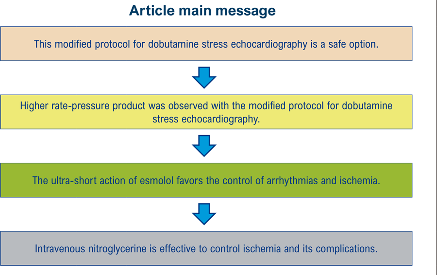
In this study with two large unselected populations, we assessed the safety of two protocols for DSE. In the modified protocol, a lower amount of dobutamine was administered and esmolol was established as the ultra-short-acting beta-blocker to control arrhythmia or ischemia. In patients with persistent angina, the control was achieved with predetermined doses of nitroglycerin, as shown in the Central Illustration.
Over the past four decades, dobutamine infusion duration and dosage have varied. The frequent use of high doses has led to high dobutamine exposure and important adverse effects. Subsequent protocols have maintained 3-minute intervals per stage, and atropine administration could be initiated along with the 20-, 30-, or 40-μcg.kg-1.min-1dobutamine infusion.1,3,4,6,7,12,16 In the CP-DSE group, there was greater exposure to dobutamine, and the test was concluded from the fourth stage on in 85.6% of the patients, while, in the MP-DSE group, only 44% needed to reach the fourth stage for conclusion.
According to Anthenoffer et al.,13 paradoxical sinus deceleration during DSE may represent the Bezold-Jarisch reflex, triggered by parasympathetic stimulation in the posterior wall of the left ventricle or by baroreceptor activation. This deceleration can be either gradual or sudden and abrupt. Based on data retrieved from the database, this reflex was observed in the CP-DSE group and was followed by atropine administration at the examiner’s discretion to reverse the deceleration. However, only 16 such cases were recorded, suggesting possible underreporting or a less stringent diagnostic criterion. In the MP-DSE group, early and immediate administration of atropine was strictly implemented in 64 patients to prevent atrioventricular block or asystole.
Intravenous administration of an ultra-short-acting beta-blocker at the end of DSE is essential for controlling or preventing arrhythmias and ischemic manifestations, as well as for facilitating a rapid return to baseline heart rate. Either esmolol or metoprolol may be used, although current guidelines do not specify a preferred agent.2,6
In the CP-DSE group, metoprolol was used, and its effectiveness in controlling arrhythmias, ischemic manifestations and reducing HR was confirmed.1,7,17 However, we observed that in the MP-DSE group, esmolol not only contributed to the control of ischemic manifestations and arrhythmias but also appeared to reduce HR more rapidly. Notably, in the management of supraventricular arrhythmias, esmolol has been shown to act faster and more effectively than amiodarone in cases of acute atrial fibrillation.3,18,19
Metoprolol administered at the end of the test enhances the sensitivity of DSE for diagnosing myocardial ischemia without compromising specificity.8,20 In our study, esmolol revealed wall motion abnormalities consistent with ischemia during the recovery phase (Video 1), which may suggest a class effect of ultra-short-acting beta-blockers.
Video 1
– In the two-chamber apical view, during the peak of dobutamine stress echocardiography (168 bpm), a subtle contractility abnormality can be observed. Following the intravenous infusion of an esmolol bolus, during the recovery phase (116 bpm), the area of wall motion abnormality extends to the anterior and inferior walls, indicating intense and unequivocal myocardial ischemia.
Watch in: http://abcimaging.org/supplementary-material/2025/3803/ABCImag-2025-0054_AO_video_1.mp4
– In the two-chamber apical view, during the peak of dobutamine stress echocardiography (168 bpm), a subtle contractility abnormality can be observed. Following the intravenous infusion of an esmolol bolus, during the recovery phase (116 bpm), the area of wall motion abnormality extends to the anterior and inferior walls, indicating intense and unequivocal myocardial ischemia.
When myocardial ischemia presents with ST-segment elevation or is not promptly controlled with a beta-blocker, the use of nitrates becomes essential.21 In the CP-DSE group, sublingual nitrate was administered; however, its effect was not always sufficiently rapid due to impaired absorption caused by mouth dryness – a common side effect of atropine, particularly in elderly patients. Therefore, we opted to use lingual spray nitrate or intravenous isosorbide mononitrate. When these options were unavailable, intravenous nitroglycerin was administered at the examiner’s discretion. In the MP-DSE group, nitroglycerin infusion was routinely implemented with predetermined timing and dosage, which may have contributed to the prevention of potential ischemic complications.14,15,22
In our study, during stress, the rate-pressure product was higher in the MP-DSE group for both positive and negative ischemia results. This may have been influenced by a greater number of hypertensive patients, a lower proportion of patients on oral beta-blockers and earlier administration of atropine in this group. The rate-pressure product is known to correlate with myocardial oxygen consumption; therefore, when this parameter is elevated, the likelihood of ischemia increases. In the MP-DSE group, the higher rate-pressure product indicated increased oxygen demand. Evidence suggests that the rate-pressure product is a more reliable indicator of the adequacy of dobutamine stress than the achievement of target HR.23
It is worth noting that, despite higher systolic blood pressure (SBP) during stress in the MP-DSE group, hypertensive peaks were more frequent in the CP-DSE group. This may be related to alpha-adrenergic stimulation secondary to greater exposure to dobutamine. Similar findings were reported by Lee et al.24in their study involving 3,129 patients, in which atropine was administered at a later stage.
Typical angina is a late and less frequent manifestation than wall motion abnormalities in the ischemic cascade.3 Although the MP-DSE group had a higher percentage of patients with risk factors for probable or known coronary artery disease, typical angina was more frequent in the CP-DSE group, which may be consistent with the higher incidence of positive DSE results for ischemia observed in that group.
Isolated ectopic beats are common during DSE and become clinically relevant when they occur in bursts, as tachyarrhythmias, or as fibrillation. However, the vast majority can be suppressed following test completion and administration of a beta-blocker bolus. Supraventricular ectopic beats in recurrent bursts, nonsustained tachyarrhythmias and atrial fibrillation were more frequently observed in the MP-DSE group, particularly in tests with negative results for ischemia.
The incidence of atrial fibrillation during DSE has varied, on average, from 1% to 4%, with rates reported as 0.86% among 4,818 patients in the meta-analysis by Mansencal et al.25 and 2% among 3,800 patients in the study by Carasso et al.26 Some publications have not associated atropine use with a higher incidence of arrhythmia.1 Tsutsui et al.16 found no significant difference in atrial fibrillation incidence during DSE with or without early atropine administration (0.8% vs. 1.2%; p = NS). These percentages are very similar to those observed in our study (0.82% vs. 1.27%; p < 0.001), although we found a statistically significant difference in the MP-DSE group, which involved lower doses of dobutamine and more frequent use of atropine. This finding does not rule out a potential association between atropine use and atrial fibrillation, which warrants further investigation.
Considering the higher mean dose of atropine and the greater number of patients who had discontinued oral beta-blockers prior to testing, as previously recommended, the higher percentage of completed tests in the MP-DSE group was expected. Consistent with a previous study involving octogenarians, our findings showed that elderly patients required lower doses of atropine.10 Furthermore, supporting published data, reduced atropine use in elderly patients was observed in both positive and negative ischemia results.
Nonsustained ventricular tachycardia was more frequently observed in the CP-DSE group, regardless of whether the test result was positive or negative for ischemia, suggesting an effect related to the higher doses of dobutamine. At the examiner’s discretion, the test was interrupted and a beta-blocker bolus was administered, effectively preventing the progression to sustained ventricular tachycardia.
The most severe complications occurred exclusively in the CP-DSE group. One patient developed acute coronary syndrome during DSE, requiring prolonged intravenous nitroglycerin infusion. This patient subsequently underwent coronary angiography, which revealed severe coronary artery disease. Ventricular fibrillation was observed in two other patients – one during the fourth stage and the other during the recovery phase – both of whom tested positive for myocardial ischemia and had severe coronary artery disease. Defibrillation was successfully performed in both cases without sequelae, and surgical myocardial revascularization was carried out thereafter. No cases of cardioversion or electrical defibrillation were reported in the MP-DSE group.
In this study, none of the following complications was observed: Takotsubo syndrome, acute myocardial infarction, atrioventricular dissociation, asystole, cardiac rupture, death or sustained ventricular tachycardia. Geleijnse et al.11 reported that, in the absence of sustained ventricular tachycardia, the risk of severe complications during stress testing – whether with a vasodilator, dobutamine or exercise – is comparable.
The study by Rozanski et al.,27 using nuclear medicine imaging, demonstrated that over two decades, the percentage of tests yielding positive results for ischemia declined to 5%. Other studies have shown that, over four decades, the rate of inducible myocardial ischemia during stress echocardiography dropped from 60% to less than 10%, likely due to changes in patient profiles and improvements in anti-ischemic therapy.28,29 This may explain the lower percentage of positive ischemia tests in the MP-DSE group in our study. Additionally, it is worth noting the higher rate-pressure product observed in that group during stress. The decline in positive test rates has prompted the incorporation of new tools to enhance the diagnostic accuracy of stress echocardiography.
The potential occurrence of significant adverse effects should never be underestimated. Based on the findings of our study, we believe that the implementation of a well-defined protocol and prompt action to manage adverse events are essential for ensuring the safety of DSE. Key safety measures include strict attention to DSE contraindications, minimizing dobutamine exposure, immediate reversal of paradoxical sinus deceleration, use of esmolol as an ultra-short-acting beta-blocker, and intravenous nitroglycerin administration when necessary.
Our study has several limitations. It is a retrospective analysis, and neither the pretest probability of coronary artery disease nor its influence on test outcomes was calculated.27-30 Regarding the use of beta-blockers and coronary vasodilators, ischemia severity was not quantified when comparing the groups. There was no standardization of coronary vasodilator use, although this limitation was mitigated in the MP-DSE group. Hemodynamic assessment to evaluate DSE accuracy was not performed, and patients were not followed longitudinally to compare the prognostic value of CP-DSE versus MP-DSE; however, these were beyond the scope of this study. Although, in practice, heart rate reduction occurred more rapidly with esmolol than with metoprolol, a comparative study is needed to confirm this observation. Myocardial contrast echocardiography could potentially increase the number of diagnostic tests, but due to its cost, it remains uncommon in our setting.
In conclusion, the modified DSE protocol, featuring early administration of atropine and intravenous use of esmolol and nitroglycerin, proved to be a safe approach, with no severe adverse effects reported. It resulted in reduced exposure to dobutamine and a higher rate-pressure product, contributing to both safety and diagnostic efficacy.
References
-
1 Poldermans D, Fioretti PM, Boersma E, Forster T, van Urk H, Cornel JH, et al. Safety of Dobutamine-Atropine Stress Echocardiography in Patients with Suspected or Proven Coronary Artery Disease. Am J Cardiol. 1994;73(7):456-9. doi: 10.1016/0002-9149(94)90675-0.
» https://doi.org/10.1016/0002-9149(94)90675-0 -
2 Pellikka PA, Roger VL, Oh JK, Miller FA, Seward JB, Tajik AJ. Stress Echocardiography. Part II. Dobutamine Stress Echocardiography: Techniques, Implementation, Clinical Applications, and Correlations. Mayo Clin Proc. 1995;70(1):16-27. doi: 10.1016/S0025-6196(11)64660-0.
» https://doi.org/10.1016/S0025-6196(11)64660-0 -
3 Krahwinkel W, Ketteler T, Gödke J, Wolfertz J, Ulbricht LJ, Krakau I, et al. Dobutamine Stress Echocardiography. Eur Heart J. 1997;18(Suppl D):D9-15. doi: 10.1093/eurheartj/18.suppl_d.9.
» https://doi.org/10.1093/eurheartj/18.suppl_d.9 -
4 Pellikka PA, Nagueh SF, Elhendy AA, Kuehl CA, Sawada SG; American Society of Echocardiography. American Society of Echocardiography Recommendations for Performance, Interpretation, and Application of Stress Echocardiography. J Am Soc Echocardiogr. 2007;20(9):1021-41. doi: 10.1016/j.echo.2007.07.003.
» https://doi.org/10.1016/j.echo.2007.07.003 -
5 Sicari R, Nihoyannopoulos P, Evangelista A, Kasprzak J, Lancellotti P, Poldermans D, et al. Stress Echocardiography Expert Consensus Statement--Executive Summary: European Association of Echocardiography (EAE) (a Registered Branch of the ESC). Eur Heart J. 2009;30(3):278-89. doi: 10.1093/eurheartj/ehn492.
» https://doi.org/10.1093/eurheartj/ehn492 -
6 Pellikka PA, Arruda-Olson A, Chaudhry FA, Chen MH, Marshall JE, Porter TR, et al. Guidelines for Performance, Interpretation, and Application of Stress Echocardiography in Ischemic Heart Disease: From the American Society of Echocardiography. J Am Soc Echocardiogr. 2020;33(1):1-41.e8. doi: 10.1016/j.echo.2019.07.001.
» https://doi.org/10.1016/j.echo.2019.07.001 -
7 Steeds RP, Wheeler R, Bhattacharyya S, Reiken J, Nihoyannopoulos P, Senior R, et al. Stress Echocardiography in Coronary Artery Disease: A Practical Guideline from the British Society of Echocardiography. Echo Res Pract. 2019;6(2):G17-G33. doi: 10.1530/ERP-18-0068.
» https://doi.org/10.1530/ERP-18-0068 -
8 Mathias W Jr, Tsutsui JM, Andrade JL, Kowatsch I, Lemos PA, Leal SM, et al. Value of Rapid Beta-Blocker Injection at Peak Dobutamine-Atropine Stress Echocardiography for Detection of Coronary Artery Disease. J Am Coll Cardiol. 2003;41(9):1583-9. doi: 10.1016/s0735-1097(03)00242-0.
» https://doi.org/10.1016/s0735-1097(03)00242-0 -
9 Varga A, Garcia MA, Picano E; International Stress Echo Complication Registry. Safety of Stress Echocardiography (from the International Stress Echo Complication Registry). Am J Cardiol. 2006;98(4):541-3. doi: 10.1016/j.amjcard.2006.02.064.
» https://doi.org/10.1016/j.amjcard.2006.02.064 -
10 Abreu JS, Diógenes TC, Farias AG, Morais JM, Paes JN Jr. Safety and Feasibility of Dobutamine-Atropine Stress Echocardiography in Octogenarian Patients. Arq Bras Cardiol. 2005;85(3):198-204. doi: 10.1590/s0066-782x2005001600009.
» https://doi.org/10.1590/s0066-782x2005001600009 -
11 Geleijnse ML, Krenning BJ, Nemes A, van Dalen BM, Soliman OI, Ten Cate FJ, et al. Incidence, Pathophysiology, and Treatment of Complications during Dobutamine-Atropine Stress Echocardiography. Circulation. 2010;121(15):1756-67. doi: 10.1161/CIRCULATIONAHA.109.859264.
» https://doi.org/10.1161/CIRCULATIONAHA.109.859264 -
12 Geleijnse ML, Fioretti PM, Roelandt JR. Methodology, Feasibility, Safety and Diagnostic Accuracy of Dobutamine Stress Echocardiography. J Am Coll Cardiol. 1997;30(3):595-606. doi: 10.1016/s0735-1097(97)00206-4.
» https://doi.org/10.1016/s0735-1097(97)00206-4 -
13 Attenhofer CH, Pellikka PA, McCully RB, Roger VL, Seward JB. Paradoxical Sinus Deceleration during Dobutamine Stress Echocardiography: Description and Angiographic Correlation. J Am Coll Cardiol. 1997;29(5):994-9. doi: 10.1016/s0735-1097(97)00030-2.
» https://doi.org/10.1016/s0735-1097(97)00030-2 - 14 Abrams J. A Reappraisal of Nitrate Therapy. JAMA. 1988;259(3):396-401.
-
15 Curry SH, Lopez LM, Lambert CR, Kwon HR, Stack RK. Plasma Concentrations and Hemodynamic effects of Intravenous, Sublingual, and Aerosolized Nitroglycerin in Patients Undergoing Cardiac Catheterization. Biopharm Drug Dispos. 1993;14(2):107-18. doi: 10.1002/bdd.2510140203.
» https://doi.org/10.1002/bdd.2510140203 -
16 Tsutsui JM, Osório AF, Lario FA, Fernandes DR, Sodre G, Andrade JL, et al. Comparison of Safety and Efficacy of the Early Injection of Atropine during Dobutamine Stress Echocardiography with the Conventional Protocol. Am J Cardiol. 2004;94(11):1367-72. doi: 10.1016/j.amjcard.2004.07.141.
» https://doi.org/10.1016/j.amjcard.2004.07.141 -
17 Mathias W Jr, Arruda A, Santos FC, Arruda AL, Mattos E, Osório A, et al. Safety of Dobutamine-Atropine Stress Echocardiography: A Prospective Experience of 4,033 Consecutive Studies. J Am Soc Echocardiogr. 1999;12(10):785-91. doi: 10.1016/s0894-7317(99)70182-3.
» https://doi.org/10.1016/s0894-7317(99)70182-3 -
18 Maurovich-Horvat P, Károlyi M, Horváth T, Szilveszter B, Bartykowszki A, Jermendy ÁL, et al. Esmolol is Noninferior to Metoprolol in Achieving a Target Heart Rate of 65 Beats/Min in Patients Referred to Coronary CT Angiography: A Randomized Controlled Clinical Trial. J Cardiovasc Comput Tomogr. 2015;9(2):139-45. doi: 10.1016/j.jcct.2015.02.001.
» https://doi.org/10.1016/j.jcct.2015.02.001 -
19 Milojevic K, Beltramini A, Nagash M, Muret A, Richard O, Lambert Y. Esmolol Compared with Amiodarone in the Treatment of Recent-Onset Atrial Fibrillation (RAF): An Emergency Medicine External Validity Study. J Emerg Med. 2019;56(3):308-18. doi: 10.1016/j.jemermed.2018.12.010.
» https://doi.org/10.1016/j.jemermed.2018.12.010 -
20 Karagiannis SE, Bax JJ, Elhendy A, Feringa HH, Cokkinos DV, van Domburg R, et al. Enhanced Sensitivity of Dobutamine Stress Echocardiography by Observing Wall Motion Abnormalities during the Recovery Phase after Acute Beta-Blocker Administration. Am J Cardiol. 2006;97(4):462-5. doi: 10.1016/j.amjcard.2005.09.075.
» https://doi.org/10.1016/j.amjcard.2005.09.075 -
21 Haouzi AJ, Schwartz S, Liszka E. Coronary Artery Spasm Following Dobutamine Stress Echocardiogram. BMJ Case Rep. 2020;13(8):e235206. doi: 10.1136/bcr-2020-235206.
» https://doi.org/10.1136/bcr-2020-235206 -
22 Kim C, Ha M, Kim W, Park SJ, Hwang SH, Yong HS, et al. Nitrates Administered by Spray versus Tablet: Comparison of Coronary Vasodilation on CT Angiography. Eur Radiol. 2021;31(1):515-24. doi: 10.1007/s00330-020-07104-0.
» https://doi.org/10.1007/s00330-020-07104-0 -
23 Sawada SG. A Reappraisal of Dobutamine Echocardiography for Risk Stratification before Noncardiac Surgery. J Am Soc Echocardiogr. 2020;33(4):433-7. doi: 10.1016/j.echo.2020.01.017.
» https://doi.org/10.1016/j.echo.2020.01.017 -
24 Lee CY, Pellikka PA, Shub C, Sinak LJ, Seward JB. Hypertensive Response during Dobutamine Stress Echocardiography. Am J Cardiol. 1997;80(7):970-1. doi: 10.1016/s0002-9149(97)00561-4.
» https://doi.org/10.1016/s0002-9149(97)00561-4 -
25 Mansencal N, Mustafic H, Hauguel-Moreau M, Lannou S, Szymanski C, Dubourg O. Occurrence of Atrial Fibrillation during Dobutamine Stress Echocardiography. Am J Cardiol. 2019;123(8):1277-82. doi: 10.1016/j.amjcard.2019.01.022.
» https://doi.org/10.1016/j.amjcard.2019.01.022 -
26 Carasso S, Sandach A, Kuperstein R, Schwammenthal E, Glikson M, Luria D, et al. Atrial Fibrillation in Dobutamine Stress Echocardiography. Int J Cardiol. 2006;111(1):53-8. doi: 10.1016/j.ijcard.2005.07.001.
» https://doi.org/10.1016/j.ijcard.2005.07.001 -
27 Rozanski A, Gransar H, Hayes SW, Min J, Friedman JD, Thomson LE, et al. Temporal Trends in the Frequency of Inducible Myocardial Ischemia during Cardiac Stress Testing: 1991 to 2009. J Am Coll Cardiol. 2013;61(10):1054-65. doi: 10.1016/j.jacc.2012.11.056.
» https://doi.org/10.1016/j.jacc.2012.11.056 -
28 Carpeggiani C, Landi P, Michelassi C, Sicari R, Picano E. The Declining Frequency of Inducible Myocardial Ischemia during Stress Echocardiography Over 27 Consecutive Years (1983-2009). Int J Cardiol. 2016;224:57-61. doi: 10.1016/j.ijcard.2016.08.313.
» https://doi.org/10.1016/j.ijcard.2016.08.313 -
29 Picano E, Pierard L, Peteiro J, Djordjevic-Dikic A, Sade LE, Cortigiani L, et al. The Clinical Use of Stress Echocardiography in Chronic Coronary Syndromes and Beyond Coronary Artery Disease: A Clinical Consensus Statement from the European Association of Cardiovascular Imaging of the ESC. Eur Heart J Cardiovasc Imaging. 2024;25(2):e65-e90. doi: 10.1093/ehjci/jead250.
» https://doi.org/10.1093/ehjci/jead250 -
30 Cortigiani L, Bigi R, Bovenzi F, Molinaro S, Picano E, Sicari R. Prognostic Implication of Appropriateness Criteria for Pharmacologic Stress Echocardiography Performed in an Outpatient Clinic. Circ Cardiovasc Imaging. 2012;5(3):298-305. doi: 10.1161/CIRCIMAGING.111.971242.
» https://doi.org/10.1161/CIRCIMAGING.111.971242
-
Study Association:
This study is not associated with any thesis or dissertation work.
-
Ethics Approval and Consent to Participate:
This study was approved by the Ethics Committee of the Nome da Instituição under the protocol number CAAE: 70990923.6.0000.5534, opinion number 6.312.526. All the procedures in this study were in accordance with the 1975 Helsinki Declaration, updated in 2013. Informed consent was obtained from all participants included in the study.
-
Use of Artificial Intelligence:
The authors did not use any artificial intelligence tools in the development of this work.
-
Availability of Research Data:
The underlying content of the research text is contained within the manuscript.
-
Sources of Funding:
There were no external funding sources for this study.
Edited by
-
Editor responsible for the review:
Marcelo Tavares
The underlying content of the research text is contained within the manuscript.