
 My Approach to Point-of-Care Ultrasound for Dyspnea Assessment
Abstract
Dyspnea is a common and potentially serious symptom whose traditional diagnostic approach, based on physical examination and complementary tests, may present significant limitations. Point-of-care ultrasound (POCUS) has emerged as an effective tool for the rapid and accurate assessment of this symptom, both in outpatient settings and in emergency and critical care environments.
This article provides a practical, illustrated guide to the integrated application of POCUS in the evaluation of dyspnea, with an emphasis on distinguishing cardiac from pulmonary causes. It describes techniques for acquiring cardiac, pulmonary, and venous ultrasound images, highlights key sonographic findings, and outlines a step-by-step approach to clinical application. Lung POCUS has demonstrated high sensitivity in detecting differential diagnoses of dyspnea. Its integration with echocardiography and lower limb ultrasound further refines diagnosis by identifying specific clinical patterns such as acute pulmonary edema (APE), pulmonary embolism (PE), and acute respiratory distress syndrome (ARDS).
The structured use of POCUS represents a significant advancement in the modern physical exam, enabling faster and more accurate bedside clinical decision-making. Its adoption should be encouraged among cardiologists, intensivists, and healthcare professionals working in critical care settings.
Keywords
Ultrasonography; Echocardiography; Dyspnea

Resumo
A dispneia é um sintoma comum e potencialmente grave, cuja abordagem diagnóstica tradicional, baseada em exame físico e exames complementares, pode apresentar limitações importantes. O ultrassom point-of-care (POCUS) consolidou-se como uma ferramenta eficaz para a avaliação rápida e precisa desse sintoma, tanto em ambiente ambulatorial quanto em contextos de emergência e terapia intensiva.
Este artigo apresenta um guia prático e ilustrado sobre a aplicação integrada do POCUS na avaliação da dispneia, com ênfase na diferenciação entre causas cardíacas e pulmonares. São descritas técnicas de aquisição de imagens cardíacas, pulmonares e venosas, os principais achados ultrassonográficos e um passo a passo de sua aplicação na prática clínica. O POCUS pulmonar tem demonstrado alta sensibilidade na detecção do diagnóstico diferencial de dispneia. Sua integração com a ecocardiografia e o ultrassom de membros inferiores permite, ainda, refinar o diagnóstico, identificando padrões clínicos específicos, como edema agudo de pulmão (EAP), tromboembolismo pulmonar (TEP) e síndrome do desconforto respiratório agudo (SDRA).
O uso estruturado do POCUS representa um avanço relevante no exame físico moderno, permitindo decisões clínicas mais rápidas e precisas à beira-leito. Sua adoção deve ser incentivada entre cardiologistas, intensivistas e profissionais que atuam em ambientes de cuidados críticos.
Palavras-chave
Ultrassonografia; Ecocardiografia; Dispneia

Introduction
Dyspnea is a common and potentially serious complaint encountered in emergency departments, intensive care units, and outpatient settings. Traditionally, clinical reasoning around this symptom relies on correlating patient history, physical examination, and complementary tests such as chest X-ray (CXR), computed tomography, or biomarkers like D-dimer and BNP. However, these methods have significant limitations, including the low sensitivity of physical examination and delays in obtaining complementary test results.
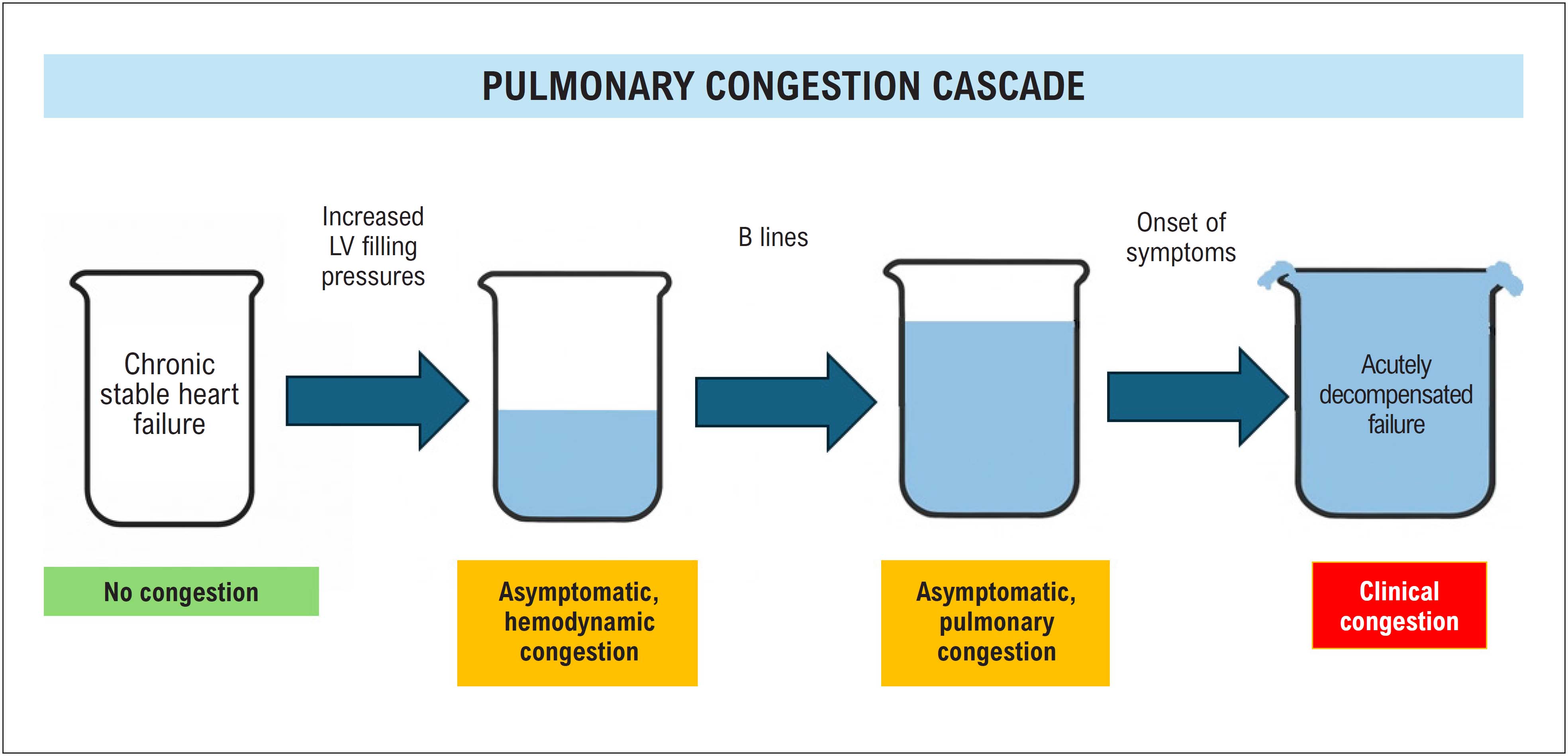
In this context, point-of-care ultrasound (POCUS) has become an essential tool for enhancing diagnostic sensitivity in the evaluation of dyspnea (Central Illustration).1 , 2 Several studies have shown that lung POCUS is more sensitive than auscultation and CXR for detecting pulmonary congestion,1 , 3 and can even identify abnormalities before the onset of clinical symptoms ( Figure 1 ).4 Moreover, ultrasound findings such as the presence of multiple B-lines correlate with invasive hemodynamic measurements, including elevated left ventricular (LV) filling pressures and pulmonary capillary wedge pressure, especially in patients with heart failure. Nonetheless, interpretation must always consider the clinical context, particularly differential diagnoses such as interstitial lung diseases or acute inflammatory injuries.1 , 3 , 4
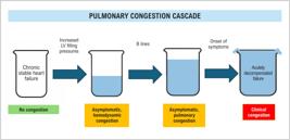
Hemodynamic congestion is the first step in the sequence leading to pulmonary congestion.Initially, exertion-induced B-lines emerge, marking a subclinical stage. Resting B-lines appear subsequently, preceding overt clinical signs that may take hours or days to develop. Therapeutic interventions are likely more effective when initiated during the early, asymptomatic stages of the preclinical and pre-radiological cascade. Adapted from Picano E, Scali MC, Ciampi Q, et al. LV: left ventricular
This article presents a step-by-step approach to evaluating patients with dyspnea of unclear etiology using an integrated POCUS strategy, including assessment of the lungs, heart, and deep venous system of the lower limbs. The aim is to demonstrate how this approach can efficiently support clinical reasoning.
For cardiologists seeking to go beyond traditional physical examination, mastering POCUS means embracing what Braunwald, Narula, and colleagues have termed the "fifth pillar of the modern physical exam": insonation, alongside inspection, palpation, percussion, and auscultation.5 This tool is transforming clinical practice by enabling real-time, portable, bedside visualization of conditions that previously depended on slower or unavailable complementary tests. In emergency settings, especially in the evaluation of dyspnea, POCUS has become a frontline modality for differentiating causes such as pulmonary edema, pneumonia, and pneumothorax.1
Just as the stethoscope revolutionized auscultation two centuries ago, the ultrasound probe is now beginning to play a comparable role in the physical exam of the modern cardiologist.
Technique and Probe Positioning
The POCUS assessment of a dyspneic patient involves different probes and scanning windows, tailored to the anatomical target.6
-
Heart: phased array transducer, with parasternal long- and short-axis, apical and subcostal windows being the most commonly used ( Figure 2 ).7
-
Lungs: For pulmonary ultrasound, using a high-frequency linear transducer (7–13 MHz) is preferable to obtain detailed views of the pleural line and its dynamic features, such as lung sliding.8 The convex transducer, with its greater penetration and wider field of view, is better suited for evaluating pulmonary artifacts such as A-lines and B-lines ( Figure 3 ).

Basic echocardiographic windows.Ao: aorta. LA: left atrium. RA: right atrium. LV: left ventricle. RV: right ventricle.

Lung ultrasound assessment in the lateral decubitus position: A-pattern. Left: B-mode image showing the A-pattern, with a hyperechoic pleural line and multiple horizontal A-lines (reverberation artifacts), indicating well-aerated lung. Also visible is the acoustic shadow of the rib (marked with an asterisk). Right: schematic representation of the ultrasound scan of the right hemithorax, with the probe positioned perpendicular to the rib cage. Blue arrows indicate the sequential scan direction along the intercostal spaces.
The scan begins in the longitudinal orientation with the probe marker facing cephalad, allowing identification of the rib spaces and pleural line.8 Next, a transverse sweep is performed by sliding the probe from anterior to lateral regions, either covering the entire thoracic surface or focusing on the zone of interest—a reasoning process analogous to auscultation during the traditional physical exam.
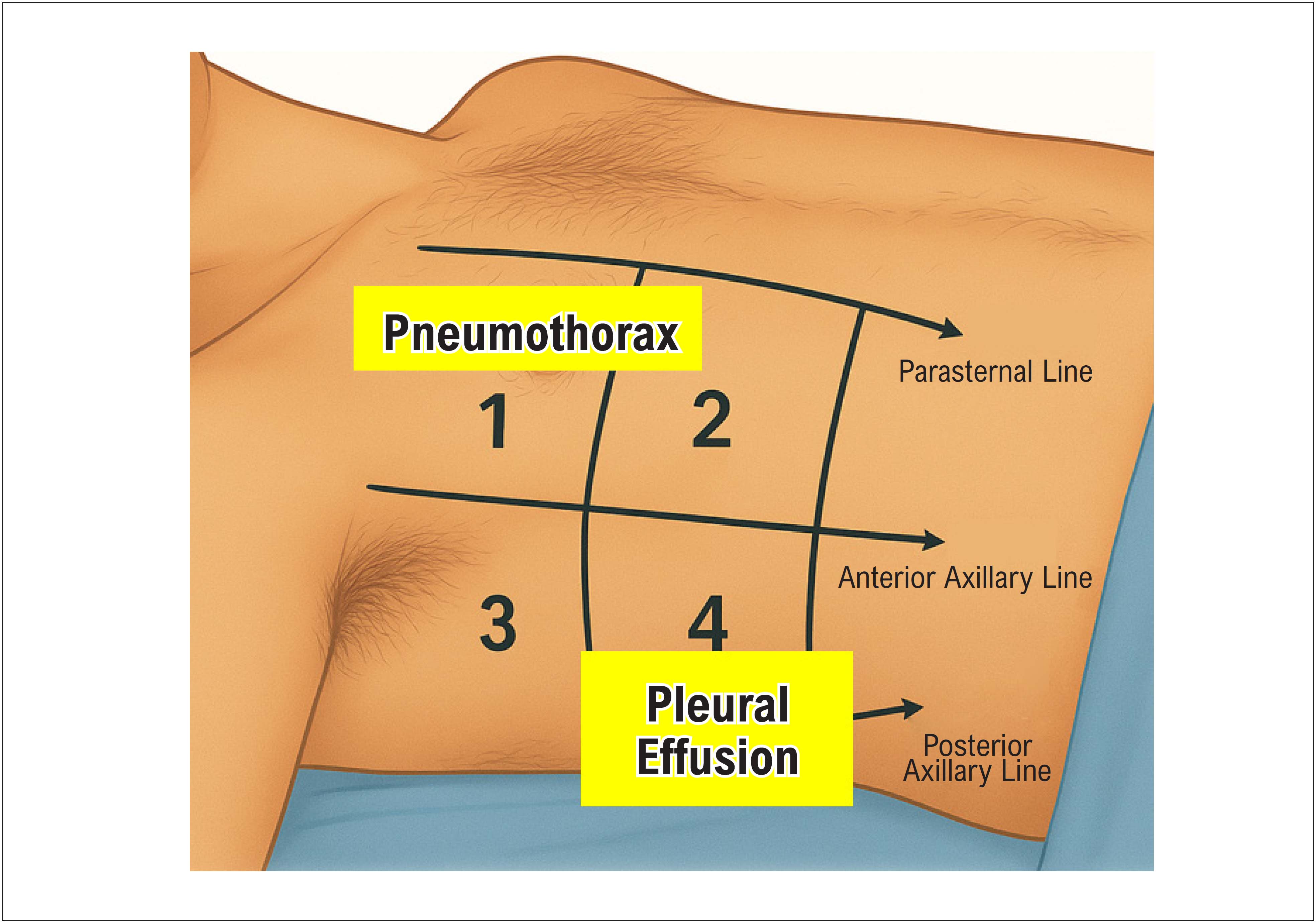
The anterosuperior thoracic quadrant is best for pneumothorax assessment, while the inferolateral quadrant along the posterior axillary line is preferred for evaluating pleural effusion and consolidations. The exam can be performed with the patient in the supine or semi-upright position, depending on clinical condition and diagnostic goal ( Figure 4 ).

Division of the hemithorax into quadrants for lung assessment, with the patient in supine position. 1: anterosuperior region; 2: anteroinferior region; 3: upper lateral region; 4: lower lateral region.
-
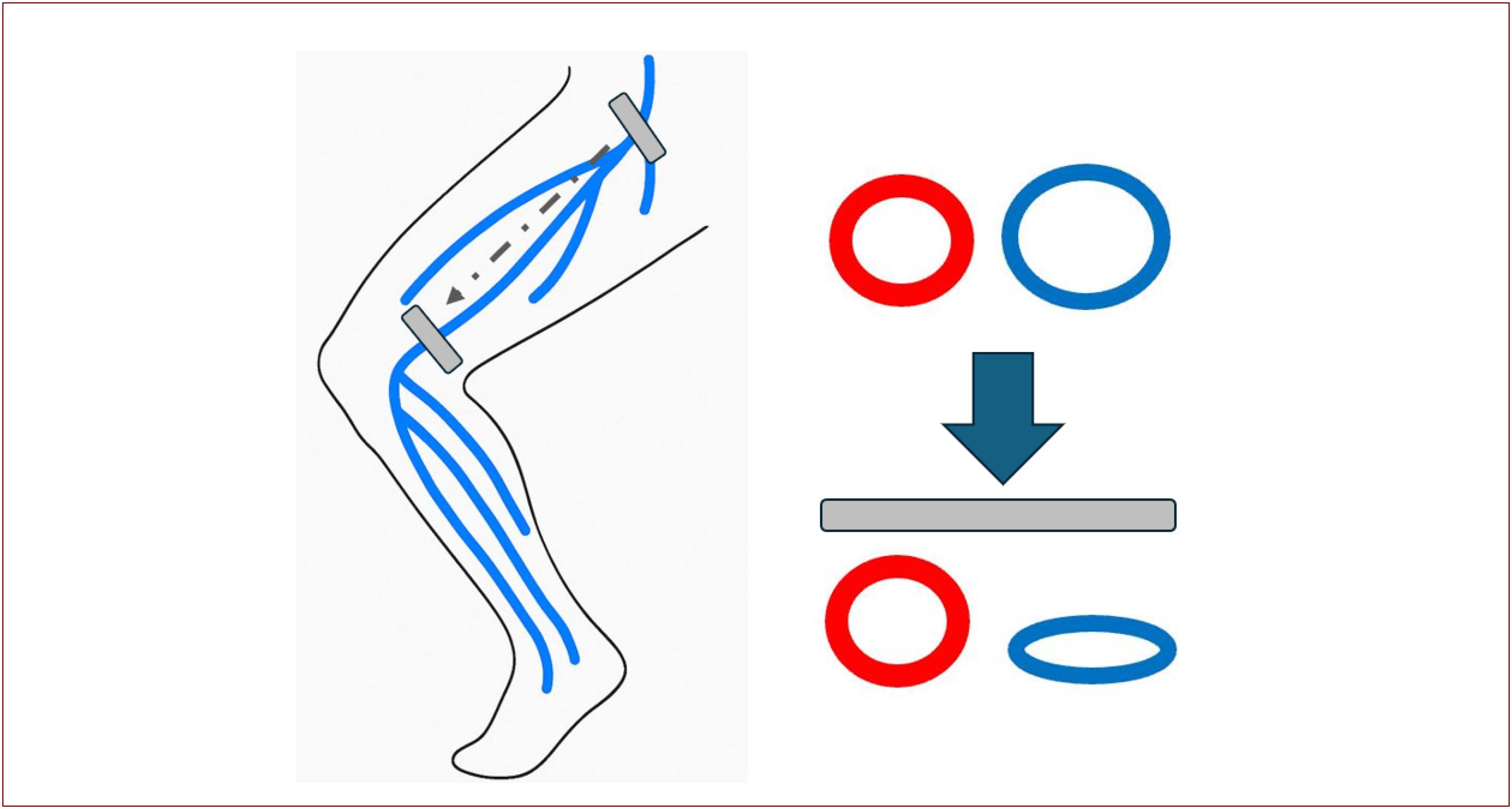
Lower limb deep veins: high-frequency linear transducer for compression ultrasound from the common femoral vein9 down to the popliteal vein ( Figure 5 ).

Venous compression protocol for deep vein thrombosis (DVT) evaluation—normal exam with vein collapse upon probe compression. This choice allows clear visualization of anatomical structures while respecting the regional anatomy. The gray arrow indicates the scan direction.
Why use POCUS? Advantages over physical exam and CXR
Standalone physical examination has low sensitivity for detecting pulmonary congestion, ranging from 17% to 33%, whereas POCUS can exceed 90% sensitivity for the same purpose.10 , 11 Studies have also shown that the presence of 15 or more B-lines strongly correlates with biomarkers such as NT-proBNP, the E/e’ ratio, and functional class in patients with decompensated heart failure, reinforcing its prognostic value4 , 12 , 13 ( Figure 6 ). Furthermore, a reduction in B-lines during treatment is associated with clinical improvement and hemodynamic compensation, validating POCUS as both a diagnostic and therapeutic monitoring tool.3 In contrast, CXR may show delayed or inconclusive findings in early stages. Pulmonary ultrasound can detect congestion before symptom onset and demonstrate early resolution after therapy, sometimes within a few hours.3 , 14 - 17

Schematic representation of interstitial pulmonary edema progression as evaluated by lung ultrasound, reflecting the gradual loss of aeration. Based on Gargani.18
It is essential to remember that POCUS should not replace the traditional physical exam or formal tests such as echocardiography, computered tomography, lower limb Doppler, or biomarkers, but rather serve as a complementary tool to support more informed clinical decision-making.
Bedside protocols: which one to choose?
Several POCUS protocols have been developed for evaluating dyspnea, including the Bedside Lung Ultrasound in Emergency (BLUE) protocol,1 used in emergency contexts, the FALLS protocol,19 for hemodynamic assessment in shock, and the Rapid Ultrasound for Shock and Hypotension (RUSH) protocol,20 for undifferentiated shock. Each offers a specific and structured approach tailored to different clinical scenarios.
The BLUE protocol is the most widely validated and utilized for ultrasonographic assessment of acute respiratory failure. A study involving over 300 patients demonstrated a sensitivity of 90.5% and specificity of 98.2% for identifying the main causes of respiratory failure, with an average scan time of under three minutes. Developed by Daniel Lichtenstein in 2008, its primary goal is to enable a rapid, structured, and accurate approach to diagnosing the most common causes of dyspnea in adults.
The BLUE protocol is based on syndromic reasoning, as it begins with assessing the presence or absence of lung sliding, followed by the analysis of pulmonary artifacts (A-lines and B-lines), subpleural and pulmonary consolidations (C-lines), and pleural effusions. Based on these findings, specific ultrasonographic profiles are generated that correlate with clinical syndromes such as Chronic obstructive pulmonary disease (COPD), asthma, heart failure, pneumonia, pulmonary embolism (PE), and pneumothorax.
It is important to remember that the goal of any protocol is to guide clinical reasoning, not to restrict it. Understanding the available tools, knowing when to apply them, and how to interpret the results is more valuable than rigidly following any given model.
Step-by-step guide to the BLUE protocol
The exam begins by placing the transducer on the anterosuperior chest wall, followed by the anteroinferior and finally the inferolateral regions of both hemithoraces, with the patient lying supine or semi-upright. The transducer is oriented longitudinally to clearly identify the pleural line5 between the intercostal spaces (also known as the "bat sign"). The following are assessed:
-
Lung sliding;
-
Horizontal artifacts (A lines);
-
Vertical artifacts (B lines);
-
Subpleural and pulmonary consolidations;
-
Evaluation of pleural effusion.
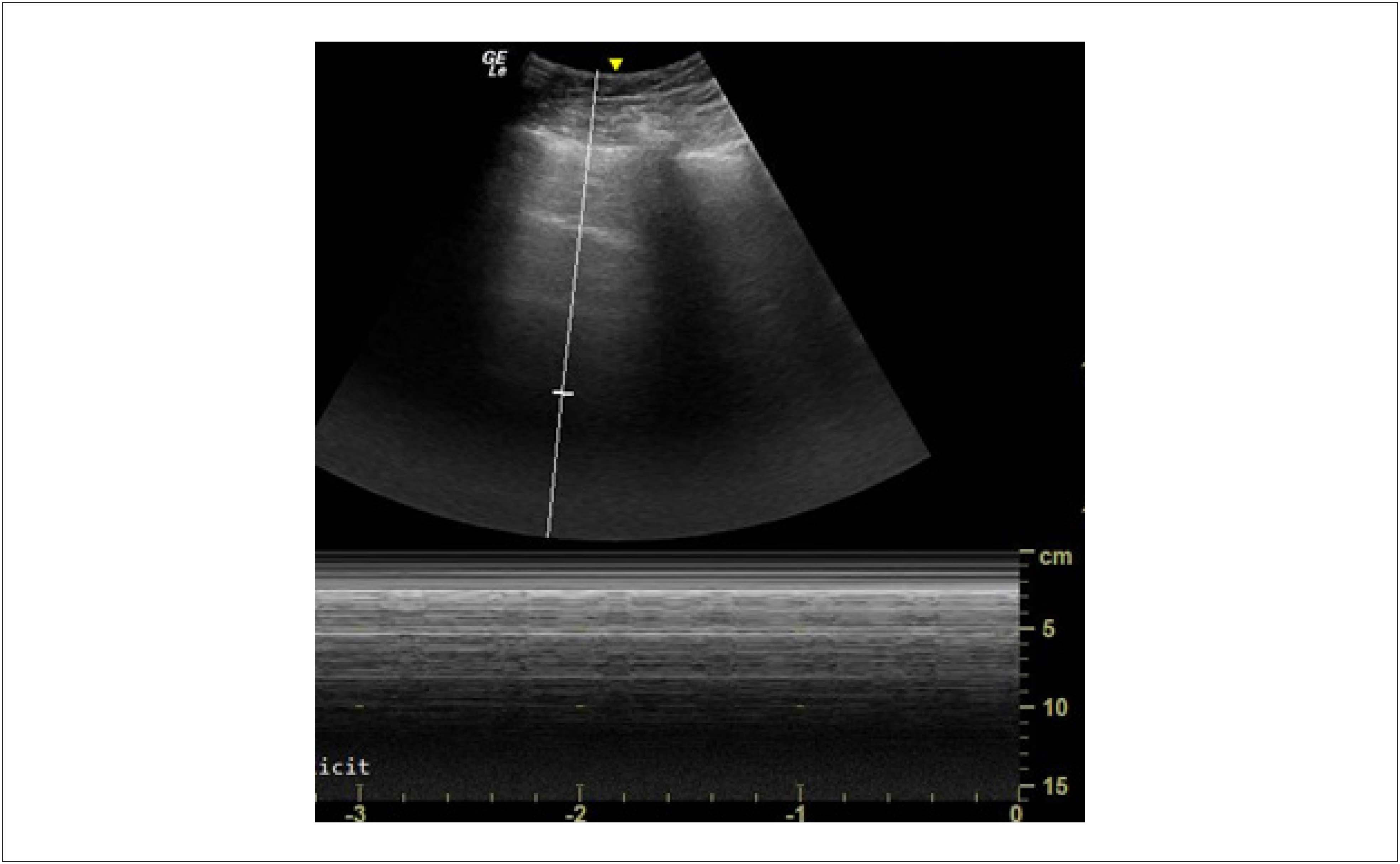
Lung sliding refers to the dynamic, horizontal movement of the pleural line seen on lung ultrasound, occurring in sync with respiration. It represents the visceral pleura gliding against the parietal pleura during breathing, indicating normal contact and well-aerated lungs. In B-mode, this movement appears as a horizontal shimmering of the pleural line; in M-mode, it creates the classic " sea-shore sign ", with a static upper section (chest wall) and a granular lower section (moving lung) ( Figure 7 ).
The presence or absence of lung sliding, combined with the analysis of pulmonary artifacts (A-lines and B-lines) or direct visualization of lung parenchyma in cases of consolidation, will define the BLUE protocol diagnostic profiles listed below.
These profiles should be complemented by evaluating DVT in the lower limbs, especially in patients with an A-profile and suspected PE. Compression of the common femoral and popliteal veins using a linear transducer allows for quick confirmation or exclusion of DVT.
Diagnostic profiles in the BLUE protocol
By assessing the structures described above, the following classic ultrasonographic profiles are identified, as defined by Lichtenstein et al.,1 , 19 with the patient in the supine or semi-upright position ( Figure 8 ):

-
A-profile: presence of repetitive horizontal A-lines originating from the pleural line, with bilateral lung sliding preserved. Suggests normally aerated lungs or patterns typical of COPD or asthma. When combined with evidence of DVT, it increases the likelihood of PE.
-
A’-profile: same as the A-profile, but with absent lung sliding and presence of the pathognomonic lung point—the transition zone between areas with and without lung sliding—indicative of pneumothorax with 100% specificity.
-
B-profile: bilateral, predominant B-lines (long, vertical, hyperechoic, well-defined, dynamic comet-tail artifacts that erase A-lines), with lung sliding preserved. It indicates pulmonary congestion.
-
B’-profile: same as B-profile, but with absent lung sliding. It may indicate interstitial pneumonia or ARDS.
-
A/B-profile: asymmetric pattern with B-lines on one hemithorax and A-lines on the other. It suggests mixed pathologies or early stages of congestion.
-
C-profile: presence of anterior alveolar consolidations, with or without air bronchograms, typically representing pneumonia.
-
PLAPS -profile: presence of posterior and/or lateral alveolar consolidations or pleural effusions.
-
Pleural Effusion Profile: an anechoic space between the lung and diaphragm and/or pleura, associated with conditions such as heart failure, pneumonia, or malignancy.
Image-Guided Clinical Reasoning
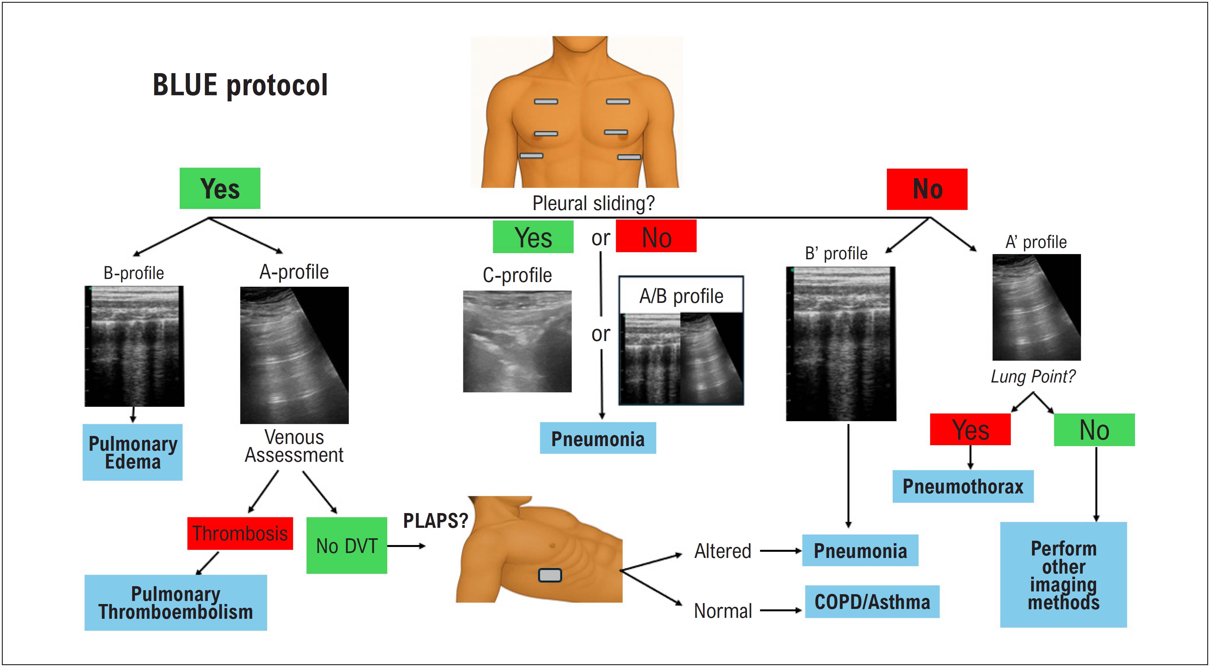
Once the main ultrasound findings have been identified and the pulmonary profiles classified according to the BLUE protocol,1 clinical reasoning can be organized into a systematic flowchart ( Flowchart 1 ). This approach streamlines the bedside differential diagnosis of dyspnea and speeds up decision-making by integrating pulmonary, cardiac, and venous data, as described below:

BLUE Protocol flowchart for the assessment of acute dyspnea by bedside lung ultrasound (adapted from Lichtenstein et al.).
Multisystem Integration: Lungs, Heart, and Vessels
Although the BLUE protocol prioritizes lung assessment, incorporating echocardiography and venous ultrasound enhances interpretation and expands the diagnostic capabilities of POCUS in patients with dyspnea. Two key questions can guide cardiac evaluation:
-
Is there LV dysfunction? Findings such as reduced anterior mitral valve leaflet systolic excursion toward the interventricular septum (EPSS, E-point septal separation), decreased mitral annular plane systolic excursion (MAPSE), ventricular dilation, and diffuse hypokinesia suggest LV systolic dysfunction.17
Additionally, the mitral E/e’ ratio12 plays a central role in indirectly estimating LV filling pressures. To obtain this ratio:
-
Use the apical four-chamber view with pulsed Doppler to measure the E wave of mitral inflow;
-
Then, using tissue Doppler imaging (TDI), place the cursor at the septal and lateral mitral annulus to measure the e’ wave;
-
An E/e’ ratio ≥ 15 strongly suggests elevated left-sided filling pressures, while values < 8 indicate normal pressures. Intermediate values require clinical correlation.
This parameter is especially useful in patients with diffuse B-lines on lung ultrasound, as it helps distinguish between cardiogenic pulmonary congestion and non-cardiogenic causes such as ARDS. When combined with pulmonary findings and clinical data, diagnostic accuracy improves, enabling more targeted and timely management.
-
-
Is there right ventricular (RV) overload or dysfunction? Key findings include:11
-
RV dilation (RV/LV ratio > 1);
-
Paradoxical septal motion ("D-shape");
-
McConnell's sign (hypokinesia of the mid and basal free RV wall with preserved apical contractility);
-
Tricuspid regurgitation with elevated estimated pulmonary artery systolic pressure.
-
These findings are strongly suggestive of acute cor pulmonale , particularly in the context of PE or decompensated pulmonary hypertension.
How to integrate lung ultrasound with echocardiography to identify the congestion pattern?
Echocardiography can be integrated with lung ultrasound, allowing different types of congestion to be distinguished: absent, purely hemodynamic, combined hemodynamic and pulmonary, or purely pulmonary with possible non-cardiac etiology.16
These four congestion profiles, resulting from the integration of echocardiography (presence or absence of increased filling pressures based on the E/e’ ratio) and lung ultrasound (presence or absence of pulmonary congestion based on the identification of diffuse B lines), are explained as follows:
-
No Congestion: When echocardiography shows no evidence of elevated LV filling pressure (normal E/e’) and lung ultrasound reveals no B-lines, the patient is classified as not congested ( Figure 9 ).
-
Hemodynamic Congestion: If echocardiography indicates elevated LV filling pressure (high E/e’) but lung ultrasound does not detect B-lines, the patient presents with hemodynamic congestion. This reflects increased pressure on the left side of the heart without a detectable fluid overload in the lungs, possibly an early stage of cardiac congestion ( Figure 10 ).
-
Hemodynamic and Pulmonary Congestion: When echocardiography reveals elevated LV filling pressure and lung ultrasound shows multiple, bilateral diffuse B-lines, the patient has both hemodynamic and pulmonary congestion. This reflects increased pressure on the left side of the heart that has resulted in fluid leakage into the pulmonary interstitium, as manifested by B-lines (clinical congestion) ( Figure 11 ).
-
Pulmonary Congestion without Hemodynamic Congestion: If echocardiography shows no signs of elevated LV filling pressure, but lung ultrasound reveals multiple, bilateral diffuse B-lines, the patient has pulmonary congestion without hemodynamic overload. This suggests a non-cardiac cause for the B-lines, such as acute lung injury (ALI) or acute respiratory distress syndrome (ARDS) ( Figure 12 and Table 2 ).

Integration of echocardiography with lung ultrasound: Normal E/e’ and A-profile on lung ultrasound indicate absence of congestion.

Integration of echocardiography with lung ultrasound: Elevated E/e’ and A-profile on lung ultrasound indicate hemodynamic congestion.

Integration of echocardiography with lung ultrasound: Elevated E/e’ and B-profile on lung ultrasound indicate both hemodynamic and pulmonary congestion.

Integration of echocardiography with lung ultrasound: Normal E/e’ ratio and B-profile on lung ultrasound indicate pulmonary involvement without hemodynamic congestion. ARDS: acute respiratory distress syndrome.
Conclusion
The integration of POCUS in the evaluation of dyspnea marks a major advance in contemporary clinical practice. Its high diagnostic accuracy, combined with the ability to provide dynamic monitoring, makes it an indispensable tool in emergency medicine, critical care, and even outpatient settings.
Despite these advantages, POCUS remains underused by cardiologists,6 even though it has proven reproducibility and a positive impact on diagnostic accuracy. Evidence suggests that the main barriers to adoption are related to medical training and access to proper education.
Mastery of ultrasound techniques and familiarity with sonographic profiles, such as those outlined in the BLUE protocol, will enable an objective and effective syndromic approach.
-
Sources of Funding
There were no external funding sources for this study.
-
Study Association
This study is not associated with any thesis or dissertation work.
-
Ethics Approval and Consent to Participate
This article does not contain any studies with human participants or animals performed by any of the authors.
-
Use of Artificial Intelligence
During the preparation of this work, the authors used ChatGPT for suggestions on restructuring and improving textual flow, as well as for generating and transforming illustrative medical images. Following the use of this tool, the authors reviewed and edited the content as necessary and take full responsibility for the content of the published article.
-
Availability of Research Data
The underlying content of the research text is contained within the manuscript.
Acknowledgments
To Dr. Marcelo Dutra and Dr. Natalia Tavares, for kindly providing the ultrasound images in Figure 8 .
References
-
1 Lichtenstein DA, Mezière GA. Relevance of Lung Ultrasound in the Diagnosis of Acute Respiratory Failure: The BLUE Protocol. Chest. 2008;134(1):117-25. doi: 10.1378/chest.07-2800.
» https://doi.org/10.1378/chest.07-2800 -
2 Francisco MJ Neto, Lira EB Filho, Queiroz MRG. Point-of-Care in Clinical Practice: Consolidated Reality. Arq Bras Cardiol. 2024;121(1):e20230688. doi: 10.36660/abc.20230688.
» https://doi.org/10.36660/abc.20230688 -
3 Coiro S, Rastogi T, Girerd N. How and When to Use Lung Ultrasound in Patients with Heart Failure? Rev Cardiovasc Med. 2022;23(6):198. doi: 10.31083/j.rcm2306198.
» https://doi.org/10.31083/j.rcm2306198 -
4 Picano E, Scali MC, Ciampi Q, Lichtenstein D. Lung Ultrasound for the Cardiologist. JACC Cardiovasc Imaging. 2018;11(11):1692-705. doi: 10.1016/j.jcmg.2018.06.023.
» https://doi.org/10.1016/j.jcmg.2018.06.023 -
5 Narula J, Chandrashekhar Y, Braunwald E. Time to Add a Fifth Pillar to Bedside Physical Examination: Inspection, Palpation, Percussion, Auscultation, and Insonation. JAMA Cardiol. 2018;3(4):346-50. doi: 10.1001/jamacardio.2018.0001.
» https://doi.org/10.1001/jamacardio.2018.0001 -
6 Mayo PH, Copetti R, Feller-Kopman D, Mathis G, Maury E, Mongodi S, et al. Thoracic Ultrasonography: A Narrative Review. Intensive Care Med. 2019;45(9):1200-11. doi: 10.1007/s00134-019-05725-8.
» https://doi.org/10.1007/s00134-019-05725-8 -
7 Accorsi TAD, Amorim EF. Princípios Básicos de Ultrassonografia para o Clínico. Rev Soc Cardiol Estado São Paulo. 2024;34(2):101-7. doi:10.29381/0103-8559/20243402101-7.
» https://doi.org/10.29381/0103-8559/20243402101-7 -
8 Volpicelli G, Elbarbary M, Blaivas M, Lichtenstein DA, Mathis G, Kirkpatrick AW, et al. International Evidence-Based Recommendations for Point-of-Care Lung Ultrasound. Intensive Care Med. 2012;38(4):577-91. doi: 10.1007/s00134-012-2513-4.
» https://doi.org/10.1007/s00134-012-2513-4 -
9 Martinez EC, Diarte E, Martinez DO, Reyes LR, Cano DAA, Navarro CC, et al. Point-of-Care Ultrasound for the Diagnosis of Frequent Cardiovascular Diseases: A Review. Cureus. 2023;15(12):e51032. doi: 10.7759/cureus.51032.
» https://doi.org/10.7759/cureus.51032 -
10 Pivetta E, Goffi A, Lupia E, Tizzani M, Porrino G, Ferreri E, et al. Lung Ultrasound-Implemented Diagnosis of Acute Decompensated Heart Failure in the ED: A SIMEU Multicenter Study. Chest. 2015;148(1):202-10. doi: 10.1378/chest.14-2608.
» https://doi.org/10.1378/chest.14-2608 -
11 Price S, Platz E, Cullen L, Tavazzi G, Christ M, Cowie MR, et al. Expert Consensus Document: Echocardiography and Lung Ultrasonography for the Assessment and Management of Acute Heart Failure. Nat Rev Cardiol. 2017;14(7):427-40. doi: 10.1038/nrcardio.2017.56.
» https://doi.org/10.1038/nrcardio.2017.56 -
12 Nagueh SF, Appleton CP, Gillebert TC, Marino PN, Oh JK, Smiseth OA, et al. Recommendations for the Evaluation of Left Ventricular Diastolic Function by Echocardiography. Eur J Echocardiogr. 2009;10(2):165-93. doi: 10.1093/ejechocard/jep007.
» https://doi.org/10.1093/ejechocard/jep007 -
13 Miglioranza MH, Gargani L, Sant’Anna RT, Rover MM, Martins VM, Mantovani A, et al. Lung Ultrasound for the Evaluation of Pulmonary Congestion in Outpatients: A Comparison with Clinical Assessment, Natriuretic Peptides, and Echocardiography. JACC Cardiovasc Imaging. 2013;6(11):1141-51. doi: 10.1016/j.jcmg.2013.08.004.
» https://doi.org/10.1016/j.jcmg.2013.08.004 -
14 Platz E, Merz AA, Jhund PS, Vazir A, Campbell R, McMurray JJ. Dynamic Changes and Prognostic Value of Pulmonary Congestion by Lung Ultrasound in Acute and Chronic Heart Failure: A Systematic Review. Eur J Heart Fail. 2017;19(9):1154-63. doi: 10.1002/ejhf.839.
» https://doi.org/10.1002/ejhf.839 -
15 Gargani L, Volpicelli G. How I do it: Lung Ultrasound. Cardiovasc Ultrasound. 2014;12:25. doi: 10.1186/1476-7120-12-25.
» https://doi.org/10.1186/1476-7120-12-25 -
16 Gargani L, Girerd N, Platz E, Pellicori P, Stankovic I, Palazzuoli A, et al. Lung Ultrasound in Acute and Chronic Heart Failure: A Clinical Consensus Statement of the European Association of Cardiovascular Imaging (EACVI). Eur Heart J Cardiovasc Imaging. 2023;24(12):1569-82. doi: 10.1093/ehjci/jead169.
» https://doi.org/10.1093/ehjci/jead169 -
17 Noritomi DT, Vieira ML, Mohovic T, Bastos JF, Cordioli RL, Akamine N, et al. Echocardiography for Hemodynamic Evaluation in the Intensive Care Unit. Shock. 2010;34 (Suppl 1):59-62. doi: 10.1097/SHK.0b013e3181e7e8ed.
» https://doi.org/10.1097/SHK.0b013e3181e7e8ed -
18 Gargani L. Lung Ultrasound: A New Tool for the Cardiologist. Cardiovasc Ultrasound. 2011;9:6. doi: 10.1186/1476-7120-9-6.
» https://doi.org/10.1186/1476-7120-9-6 - 19 Lichtenstein D. FALLS-Protocol: Lung Ultrasound in Hemodynamic Assessment of Shock. Heart Lung Vessel. 2013;5(3):142-7.
-
20 Perera P, Mailhot T, Riley D, Mandavia D. The RUSH Exam: Rapid Ultrasound in SHock in the Evaluation of the Critically lll. Emerg Med Clin North Am. 2010;28(1):29-56. doi: 10.1016/j.emc.2009.09.010.
» https://doi.org/10.1016/j.emc.2009.09.010 -
21 Szabó GV, Szigetváry C, Szabó L, Dembrovszky F, Rottler M, Ocskay K, et al. Point-of-Care Ultrasound Improves Clinical Outcomes in Patients with Acute Onset Dyspnea: A Systematic Review and Meta-Analysis. Intern Emerg Med. 2023;18(2):639-53. doi: 10.1007/s11739-022-03126-2.
» https://doi.org/10.1007/s11739-022-03126-2
Edited by
-
Editor responsible for the review:
Marcelo Tavares
The underlying content of the research text is contained within the manuscript.

 POCUS: Point-of-care ultrasound; ARDS: acute respiratory distress syndrome; PE: pulmonary embolism; APE: Acute Pulmonary Edema.




 1 BLUE: Bedside Lung Ultrasound in Emergency; DVT: deep vein thrombosis; PLAPS: Posterolateral Alveolar and/or Pleural Syndrome; COPD: Chronic obstructive pulmonary disease


