My Approach to HFpEF Considering Ventricular-Arterial Coupling and Ventricular Interdependence
Abstract
Heart failure (HF) is a serious public health issue that impacts the mortality and morbidity of the global population. The prevalence of heart failure with preserved ejection fraction (HFpEF) is clearly increasing, associated with population aging, obesity, sedentary lifestyle, and cardiometabolic diseases. It is one of the most urgent diagnostic and therapeutic challenges nowadays. Additionally, the obesity phenotype of HFpEF may be associated with HF and occurs as a result of several mechanisms that are deleterious to cardiac function, including the direct action of epicardial adipose tissue (EAT), causing direct restraint of the pericardium and ventricular interdependence, with significant hemodynamic repercussions, decline in functional capacity, and a worse prognosis. The estimation of ventricular-arterial coupling (VAC) in a non-invasive manner by echocardiography allows a better understanding of the interaction between the heart and the arterial system by monitoring hemodynamic changes, which can guide medical therapy and have an impact on prognosis. Although these tools and parameters are promising, new technologies, such as artificial intelligence and machine learning, must be used to enable the applicability of VAC in clinical practice. Further studies are key to standardizing new methods and values in this context.
Obesity Phenotipe; Ventricular-Arterial Coupling; Ventricular Interdependence

Abstract
A insuficiência cardíaca (IC) é um grave problema de saúde pública, com impacto na mortalidade e na morbidade populacional global. A prevalência da insuficiência cardíaca com fração de ejeção preservada (ICFEp) revela-se em evidente crescimento, associada a fatores como envelhecimento da população, obesidade, sedentarismo e doenças cardiometabólicas, caracterizando-se como um dos desafios diagnósticos e terapêuticos mais urgentes da atualidade. Além disso, a ICFEp fenótipo obesidade pode estar relacionada à IC, ocorrendo como resultado de diversos mecanismos deletérios à função cardíaca, incluindo a ação direta do tecido adiposo epicárdico (TAE), provocando restrição direta do pericárdio e interdependência ventricular, com intensa repercussão hemodinâmica, declínio da capacidade funcional e pior prognóstico. A estimativa do acoplamento ventrículo-arterial (AVA) de modo não invasivo por meio da ecocardiografia permite compreender melhor a interação entre o coração e o sistema arterial por meio do monitoramento das alterações hemodinâmicas, podendo guiar a terapia médica, com impacto no prognóstico. Embora essas ferramentas e parâmetros sejam promissores, faz-se necessária a incorporação cada vez maior de novas tecnologias, como a inteligência artificial e o machine learning , de forma a viabilizar a aplicabilidade do AVA na prática clínica. Estudos futuros são fundamentais para a normatização de novos métodos e valores nesse contexto.
Fenótipo Obesidade; Acoplamento Ventrículo-Arterial; Interdependência

Introduction
Heart failure (HF) is a serious public health issue that affects millions of individuals worldwide and has a considerable impact on mortality and quality of life. Although epidemiological data indicate that the global incidence of HF tends to decrease, the prevalence of heart failure with preserved ejection fraction (HFpEF) continues to grow, being associated with population aging, obesity, sedentary lifestyle, and cardiometabolic diseases.1 - 3
The pathophysiology of HFpEF is associated with the primary morbidities that cause cardiac and vascular aggression from a chronic pro-inflammatory state. More specifically, this cardiac structural and functional impairment results from a complex interaction between metabolic abnormalities, damage to microcirculation, changes in the functioning of cellular organelles, autonomic dysfunction, exacerbation of the activity of the renin-angiotensin-aldosterone system (RAAS), and maladaptive immune responses.4 , 5
The diagnosis of HFpEF is challenging in most cases and is usually obtained in the advanced stages of the disease, which limits the therapeutic results. In summary, HFpEF can be diagnosed in the presence of clinical evidence of HF, left ventricular ejection fraction (LVEF) ≥ 50%, and evidence of elevated filling pressures and/or cardiac structural alteration. Therefore, a thorough clinical evaluation, echocardiographic examination, and natriuretic peptide levels remain fundamental diagnostic resources.6
Thus, HFpEF is one of the most pressing diagnostic and therapeutic challenges nowadays, considering its increasing prevalence, underdiagnosis, poor prognosis, limited therapeutic options, and considerable impact on healthcare systems.
HFpEF with obesity phenotype
HFpEF is characterized by the coexistence of multiple cardiac and non-cardiac diseases. In this way, distinct clinical phenotypes can be identified and determined by different risk factors, comorbidities, left ventricular (LV) remodeling, hemodynamic pattern, and organ dysfunction ( Table 1 ).6 , 7 Obesity, in addition to being unequivocally associated with systemic arterial hypertension (SAH), diabetes mellitus (DM), and insulin resistance, has been identified as the primary cause of HF, mediated by several elements that are deleterious to cardiac function due to altered cardiometabolism and direct restraint of the pericardium (epicardial fat), as well as systemic effects on the lung, skeletal muscle, kidney, and liver because of systemic inflammation, neurohormonal activation, autonomic dysregulation, and altered hemodynamic load ( Table 1 ).7 - 13
Recent studies on obesity have given greater attention to the location of fat than to the amount of fat itself since subcutaneous adipose tissue (SAT), visceral adipose tissue (VAT), and epicardial adipose tissue (EAT) contribute differently to the overall cardiovascular risk ( Figure 1 ).12 , 14 , 15 EAT may favor the onset of HFpEF due to pro-inflammatory paracrine effects, lipid infiltration, and pericardial restraint ( Figure 1 ).12 , 15
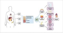
– Different locations where fat can be deposited and the pathological potential of EAT. Source: Adapted from Dronkers et al., 2024 .12 EAT: epicardial adipose tissue; VAT: visceral adipose tissue; SAT: subcutaneous adipose tissue; HFpEF: heart failure with preserved ejection fraction.
It is worth emphasizing that, in HFpEF, arterial hypertension and other comorbidities coexist and lead to a scenario of chronic afterload elevation as a consequence of increased arterial stiffness and impaired vasodilation (dysregulation of the NO-sGC-cGMP-PKG pathway - nitric oxide, soluble guanylate cyclase, cyclic guanosine monophosphate, and protein kinase G), associated with an increase in blood volume and the appearance of early reflected pulse waves in the aorta (AO) ( Figure 2 ). The stiffer (less compliant) artery causes a pulse wave that propagates at a higher speed, causing the corresponding reflex wave to return to the ascending AO earlier, still at the end of systole, with the aortic valve open. This results in an increase in systolic blood pressure (SBP), a reduction in diastolic blood pressure, and a reduction in coronary perfusion, i.e., a vicious cycle of progressive afterload increase and myocardial dysfunction ( Figure 2 ). Ventricular-arterial coupling (VAC) has been used to assess the efficiency of the ventricular-arterial interaction process, and it also appears to represent an accurate marker of early LV dysfunction and potential therapeutic guidance in HFpEF.13 , 16 - 22

– A: Pathophysiological mechanism that leads to LV dysfunction. B: Increased aortic stiffness and the emergence of early reflected waves that reach the heart at the end of systole, promoting increased pulse pressure, increased SBP, increased afterload, reduced diastolic blood pressure, and reduced coronary perfusion. Source: Adapted from Ikonomidis et al., 2019 .16 LV: left ventricle; SNS: sympathetic nervous system; SBP: systolic blood pressure; RAP: right atrial pressure.
VAC
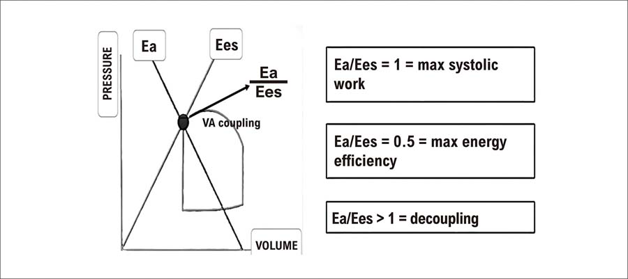
VAC assesses the relationship between two anatomically and functionally connected structures: the heart and the arteries. To investigate this interaction, a unit common to both systems is used: elastance, which measures changes in pressure for each unitary change in volume - unit: mmHg/ml. Originally, the elastance and VAC data came from the interpretation of the pressure-volume diagram of the cardiac cycle, derived from an invasive hemodynamic study ( Figure 3 ).16

– A classic Pressure-Volume diagram in the cardiac cycle demonstrating the Pressure-Volume relationship curves at the end of diastole and the Pressure-Volume relationship at the end of systole, determining Ea, Ees, and LV-AO VAC. Source: Adapted from Gamarra A et al., 2024.17 Ea: arterial elastance; Ees: end-systolic elastance; LV: left ventricle; AO: aorta; VAC: ventricular-arterial coupling.
Arterial elastance (Ea) is defined as the ratio between ventricular end-systolic pressure and Stroke volume (ESP/Stroke volume), being influenced by vascular resistance, pulsatile load, and heart rate. It is characterized as an index of arterial load for the left ventricle. End-systolic elastance (Ees), usually known as ventricular elastance, is an index of LV contractility, independent of the load, and reflects the slope of the pressure-volume relationship curve at the end of systole ( Figure 3 ), being the ratio between ventricular end-systolic pressure and ventricular end-systolic volume (ESP/ESV). The Ea/Ees ratio (ESP/Stroke volume)/(ESP/ESV) can be simplified as ESV/Stroke volume (removing ESP from the equation).22
VAC is defined as the ratio of Ea/Ees and investigates the contractile capacity of the heart and its ability to adapt to load impositions.21 Previous studies have shown that the ideal VAC value, derived from the Ea/Ees ratio, should vary between 0.5 and 1, reflecting the state in which the LV systolic work is ideal ( Figure 4 ). An Ea/Ees ratio > 1 suggests ventricular-arterial decoupling ( Figure 4 ). Such ventricular-arterial incompatibility is frequently seen in HFpEF, being attributed to diastolic dysfunction induced by increased afterload and subendocardial ischemia, and may be associated with a worse prognosis.16

– Assessment of energy efficiency in blood transfer from the ventricular to the arterial system by calculating VAC. Source: Adapted from Chirinos et al., 2013.18 Ea: arterial elastance; Ees: end-systolic elastance; VE: left ventricle; VA: ventricular-arterial
Left VAC
The gold standard echocardiographic method for assessing LV-AO VAC is the so-called single beat method , developed by Chen et al.23 ( Figure 5 ). Simple non-invasive parameters that can be easily collected should be obtained: systolic and diastolic blood pressure, ejection volume, ejection fraction, pre-ejection period, and total ejection period. As the formulas are relatively complex, it is recommended to use an easy-to-use application (iElastance© - Apple iOS application)24 ( Figure 6 ), which yields fast and reliable results.
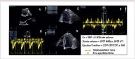
– (A) Two-dimensional echocardiographic image in parasternal view, long axis, zooming in on the LV outflow tract diameter measurement. (B) VTI image of the Doppler spectrum of the LV outflow tract flow, which is obtained by tracing the fullest line of the spectrum to calculate the stroke volume. (C)/(D) Two-dimensional echocardiographic image in apical 4-chamber (D) and 2-chamber (C) views to obtain the LVEF value (Simpson method). (E) Apical 5-chamber view of the Doppler spectrum of the LV outflow tract flow and mitral inflow to obtain pre-ejection time (green) and total ejection time (orange). Ea: arterial elastance; SBP: systolic blood pressure; LVOT: left ventricular outflow tract; EDV: end-diastolic volume; ESV: end-systolic volume; VTI: velocity-time integral; LVEF: left ventricular ejection fraction.

– (1) Image reproduced from the mobile application showing how easy it is to use. It is only necessary to fill in the fields with the data obtained, as shown in Figure 5 . (2) Practical example of filling in the fields. (3) Immediate result for Ea, Ees: end-systolic elastance, and VAC. Source: Bertini P. iElastance- Apple iOS App. Available at: https://apps.apple.com/br/app/ielastance/id556528864.24
The proposed normal value for Ea is 2.2 (±0.8) mmHg/ml, and for Ees is 2.3 (±1.0) mmHg/ml.
where DBP and SBP are the diastolic and systolic cuff blood pressures, measured through the upper arm; E(nor)(est) is the normalized ventricular elastance, estimated at the beginning of ejection; and Stroke volume is derived from the product of the VTI (velocity-time integral) of the Doppler spectrum of the LV outflow tract flow by the measured LV outflow tract diameter.22 Using the dedicated application avoids complex and time-consuming calculations. The user simply fills in the fields with the requested data: SBP (mmHg), diastolic blood pressure (mmHg), LV ejection fraction (%), stroke volume (ml), pre-ejection time (ms), and total ejection time (ms) ( Figure 5 ). The result is immediate with the values of: Ea, Ees, and VAC ( Figure 6 ).
Several methodologies have emerged to assess LV-AO VAC using echocardiography. The measurement of pulse wave velocity (PWV) appears to comprehensively represent arterial load (arterial stiffness, aortic impedance, and early reflected waves) ( Figure 2 ), associated with the measurement of the global longitudinal strain (GLS) of the LV as a marker of myocardial performance. Therefore, the use of the PWV/GLS ratio may be more appropriate in several scenarios to characterize VAC since it incorporates “gold standard” methods to assess arterial load (PWV) and LV contractility (GLS), with prognostic value.16 , 22 , 25
The use of 3D echocardiography to measure ventricular volumes, the use of myocardial work to analyze myocardial performance and the increasing incorporation of artificial intelligence and machine learning to calculate ventricular elastance (Ees) represent technical evolution and contribute to greater applicability of VAC in clinical practice in the future.16 , 17 Studies will be necessary to standardize methods and values in this context.
Right VAC
The TAPSE/PASP ratio (tricuspid annular plane systolic excursion/pulmonary artery systolic pressure) expresses the relationship between RV contractility and its afterload. It has been shown to have a good correlation with invasive methods deemed the “gold standard” for measuring RV (right ventricle)-PA (pulmonary artery) coupling. Consequently, the TAPSE/PASP ratio has been incorporated into the main guidelines for the assessment of RV and pulmonary hypertension.26 - 28
TAPSE is measured by M-mode echocardiography ( Figure 7A ) and assesses RV contractile function. To estimate RV afterload (PASP), the maximum velocity of the tricuspid regurgitation jet is determined by analyzing the continuous Doppler spectral curve and adding the right atrial pressure (RAP), which is estimated by observing the inferior vena cava. As a result, the RVSP (right ventricular systolic pressure) is obtained, which is calculated using the modified Bernoulli equation: 4 x peak velocity ( Figure 7B ). In the absence of right ventricular outflow tract obstruction or pulmonary stenosis, RVSP is equal to the PASP.

– (A) Tricuspid annular displacement measured by M mode echocardiography - TAPSE. (B) Continuous Doppler spectral curve, demonstrating tricuspid regurgitation flow, allowing the systolic pressure in the pulmonary artery to be estimated. The TAPSE/PASP ratio estimates the value of RV-PA VAC. TAPSE: tricuspid annular plane systolic excursion; PASP: pulmonary artery systolic pressure; RV: right ventricle; PA: pulmonary artery.
In HFpEF with obesity phenotype, right ventricular dysfunction results from several mechanisms acting on preload, afterload, and contractility of the right ventricle. Increased pulmonary pressure (combined pre- and post-capillary) is frequently present, as is right ventricular dilation, primary ventricular sarcomere dysfunction, pericardial constriction, and ventricular interdependence, which further aggravate biventricular filling and pulmonary hypertension.27
RV dysfunction is an independent predictor of mortality and hospitalization risk in HFpEF. Therefore, it should always be investigated. A TAPSE/PASP ratio < 0.48 mm/mmHg was associated with higher all-cause mortality and higher hospitalization for HF in a population with HF with preserved and reduced LVEF.29 Another study has demonstrated that, in the subgroup of patients with HFpEF and pre-capillary pulmonary hypertension, a TAPSE/PASP ratio < 0.36 mm/mmHg was associated with poor clinical outcomes, but suggested that these individuals could respond more favorably to interventions targeting RV afterload.30
It is important to highlight that the RV-GSL/PASP ratio has also been used as an appropriate methodology to assess RV-PA coupling and has proven to be an accurate instrument for assessing patients in different scenarios.26 - 28
VAC X HFpEF
In the context of HFpEF, the calculation of the LVEF does not characterize broader aspects of the ventricular-arterial connection, particularly those related to afterload (Ea) or contractility (Ees), which are highly relevant for therapeutic management in patients with clinical symptoms of HF and preserved LVEF. In this scenario, Ees may be normal, while Ea may be increased, resulting in Ea/Ees > 1.0 (VA decoupling). However, Ea/Ees can often occur with a normal result (between 0.5 and 1.0), despite Ea and Ees being elevated. Therefore, it is crucial to identify the value of the elastances individually22 ( Figures 5 and 6 ). Some interventions have shown potential impact on elastances, in addition to improving VAC: antihypertensive drugs,21 angiotensin receptor-neprilysin inhibitors (ARNIs),31 sodium-glucose cotransporter-2 (SGLT2) inhibitors,32 GLP-1 analogs,33 and interleukin IL-134 and IL-12 inhibitors.35
The use of VAC to assess the population with HFpEF has proven to be an accurate tool for stratifying those individuals at greater risk. In a recent study involving patients with LVEF > 40%, it was shown that the subgroup of patients, despite having higher LVEF, had a greater degree of LV hypertrophy, higher LV filling pressures, and a higher rate of LV-AO decoupling with a worse prognosis. On top of that, the subgroup that showed RV-PA decoupling, verified by the TAPSE/PASP ratio, also demonstrated a worse prognosis, regardless of LVEF values, E/e’ ratio, and presence of atrial fibrillation.34
Pericardial restraint (HFpEF) X ventricular interdependence
Obese patients with HFpEF have peculiar pathophysiological characteristics, including hypervolemia, RV-PA decoupling, biventricular hypertrophy, right ventricular dysfunction, pre- and post-pulmonary capillary hypertension, elevated filling pressures in left chambers, and increased EAT36 ( Figure 8 ).

– A proposed hemodynamic mechanism for HFpEF with obesity phenotype. LV: left ventricle; RV: right ventricle; O2: oxygen; LVEDP: left ventricular end-diastolic pressure; RVEDV: right ventricular end-diastolic volume.
Due to its proximity to the myocardium, excess EAT can promote local inflammatory and mechanical constrictive effects on the cardiac muscle. Pericardial restraint associated with hypervolemia can increase pericardial pressure, promote competition for filling between the RV and LV, cause flattening of the ventricular septum, highlight ventricular interdependence, and lead to RV distension, reduced LV preload, increased LV end-diastolic pressure, and reduced cardiac output ( Figure 8 ). On echocardiography, it is possible to quantify, using the parasternal short-axis plane, the LV eccentricity index, which becomes higher (LV eccentricity index > 1) ( Figure 9 ) as greater pericardial constriction and higher intracardiac pressures are observed, including right atrial, pulmonary arterial, and RV end-diastolic pressures, which are concomitant with a significant reduction in functional physical capacity.37 - 40

– Illustration showing an increase in the LV eccentricity index due to ventricular interdependence, as proposed for HFpEF with obesity phenotype. Source: modified Borlaug et al., 2019.39
Ventricular interdependence has been a therapeutic target of several current studies. Studies performing transcatheter interventions to reduce tricuspid regurgitation in patients with HFpEF have been designed to improve ventricular interdependence and increase LV filling.40 , 41 Similarly, a pilot study has demonstrated the potential for surgical pericardiotomy in patients with HFpEF and restraint EAT.42
Final considerations
The magnitude of the global public health issue of HFpEF, in which obesity has a prevalence of approximately 70% ( Table 1 ), has motivated, in recent years, a more specific investigation of the phenotype of subpopulations for better diagnostic, therapeutic, and prognostic results. Particular attention has been given to excess EAT in this population, which, associated with volume overload, can lead to pericardial restraint with ventricular interdependence, contributing to a more significant increase in left chamber filling pressures, worsening of pulmonary pressure, reduction in cardiac output, low physical capacity, and a worse prognosis. This hemodynamic condition can be identified by echocardiogram (LV eccentricity index > 1).
Non-invasive analysis of VAC by echocardiography appears to be a promising tool to assess hemodynamics in a more individualized manner and customize the treatment of patients with HFpEF. The single-beat echocardiographic method, coupled with a dedicated application, is becoming increasingly feasible. Besides, physicians are gaining more experience with this technology, having a better understanding of the complex interaction between the heart and the vessels. Yet, efforts to develop new methods must continue, both through advanced imaging techniques and new software solutions, artificial intelligence, and machine learning. It is clear that the earlier an accurate diagnosis, etiological definition, and severity estimation are made, and preventive and therapeutic efforts are implemented, the greater the benefits in terms of disease progression and the associated outcomes.
References
-
1 Maryniak A, Maisuradze N, Ahmed R, Biskupski P, Jayaraj J, Budzikowski AS. Heart Failure with Preserved Ejection Fraction Update: A Review of Clinical Trials and New Therapeutic Considerations. Cardiol J. 2022;29(4):670-9. doi: 10.5603/CJ.a2022.0051.
» https://doi.org/10.5603/CJ.a2022.0051 -
2 Kittleson M, Panjrath G, Amancherla K, Davis LL, Deswal A, Dixon DL, et al. 2023 ACC Expert Consensus Decision Pathway on Management of Heart Failure with Preserved Ejection Fraction: A Report of the American College of Cardiology Solution Set Oversight Committee. J Am Coll Cardiol. 2023;81(18):1835-78. doi: 10.1016/j.jacc.2023.03.393
» https://doi.org/10.1016/j.jacc.2023.03.393 -
3 Logeart D. Heart Failure with Preserved Ejection Fraction: New Challenges and New Hopes. Presse Med. 2024;53(1):104185. doi: 10.1016/j.lpm.2023.104185.
» https://doi.org/10.1016/j.lpm.2023.104185 -
4 Jasinska-Piadlo A, Campbell P. Management of Patients with Heart Failure and Preserved Ejection Fraction. Heart. 2023;109(11):874-83. doi: 10.1136/heartjnl-2022-321097.
» https://doi.org/10.1136/heartjnl-2022-321097 -
5 Bozkurt B, Coats AJ, Tsutsui H, Abdelhamid M, Adamopoulos S, Albert N, et al. Universal Definition and Classification of Heart Failure: A Report of the Heart Failure Society of America, Heart Failure Association of the European Society of Cardiology, Japanese Heart Failure Society and Writing Committee of the Universal Definition of Heart Failure. J Card Fail. 2021;27(4):387-413. doi: 10.1016/j.cardfail.2021.01.022.
» https://doi.org/10.1016/j.cardfail.2021.01.022 -
6 Hamo CE, De Jong C, Hartshorne-Evans N, Lund LH, Shah SJ, Solomon S, et al. Heart Failure with Preserved Ejection Fraction. Nat Rev Dis Primers. 2024;10(1):55. doi: 10.1038/s41572-024-00540-y.
» https://doi.org/10.1038/s41572-024-00540-y -
7 Redfield MM, Borlaug BA. Heart Failure with Preserved Ejection Fraction: A Review. JAMA. 2023;329(10):827-38. doi: 10.1001/jama.2023.2020.
» https://doi.org/10.1001/jama.2023.2020 -
8 Donal E, L'Official G, Kosmala W. Heart Failure with Preserved Ejection Fraction: Defining Phenotypes. J Card Fail. 2020;26(11):929-31. doi: 10.1016/j.cardfail.2020.09.013.
» https://doi.org/10.1016/j.cardfail.2020.09.013 -
9 Khan SS, Ning H, Wilkins JT, Allen N, Carnethon M, Berry JD, et al. Association of Body Mass Index with Lifetime Risk of Cardiovascular Disease and Compression of Morbidity. JAMA Cardiol. 2018;3(4):280-7. doi: 10.1001/jamacardio.2018.0022.
» https://doi.org/10.1001/jamacardio.2018.0022 -
10 Cercato C, Fonseca FA. Cardiovascular Risk and Obesity. Diabetol Metab Syndr. 2019;11:74. doi: 10.1186/s13098-019-0468-0.
» https://doi.org/10.1186/s13098-019-0468-0 -
11 Shams E, Kamalumpundi V, Peterson J, Gismondi RA, Oigman W, Correia MLG. Highlights of Mechanisms and Treatment of Obesity-Related Hypertension. J Hum Hypertens. 2022;36(9):785-93. doi: 10.1038/s41371-021-00644-y.
» https://doi.org/10.1038/s41371-021-00644-y -
12 Dronkers J, van Veldhuisen DJ, van der Meer P, Meems LMG. Heart Failure and Obesity: Unraveling Molecular Mechanisms of Excess Adipose Tissue. J Am Coll Cardiol. 2024;84(17):1666-77. doi: 10.1016/j.jacc.2024.07.016.
» https://doi.org/10.1016/j.jacc.2024.07.016 -
13 Borlaug BA, Sharma K, Shah SJ, Ho JE. Heart Failure with Preserved Ejection Fraction: JACC Scientific Statement. J Am Coll Cardiol. 2023;81(18):1810-34. doi: 10.1016/j.jacc.2023.01.049.
» https://doi.org/10.1016/j.jacc.2023.01.049 -
14 Ferrara D, Montecucco F, Dallegri F, Carbone F. Impact of Different Ectopic Fat Depots on Cardiovascular and Metabolic Diseases. J Cell Physiol. 2019;234(12):21630-41. doi: 10.1002/jcp.28821.
» https://doi.org/10.1002/jcp.28821 -
15 Gosh S. Ectopic Fat: The Potential Target for Obesity Management. J Obes Metab Res 2014;1(1):30-8. doi: 10.4103/2347-9906.123883.
» https://doi.org/10.4103/2347-9906.123883 -
16 Ikonomidis I, Aboyans V, Blacher J, Brodmann M, Brutsaert DL, Chirinos JA, et al. The Role of Ventricular-Arterial Coupling in Cardiac Disease and Heart Failure: Assessment, Clinical Implications and Therapeutic Interventions. A Consensus Document of the European Society of Cardiology Working Group on Aorta & Peripheral Vascular Diseases, European Association of Cardiovascular Imaging, and Heart Failure Association. Eur J Heart Fail. 2019;21(4):402-24. doi: 10.1002/ejhf.1436.
» https://doi.org/10.1002/ejhf.1436 -
17 Gamarra A, Díez-Villanueva P, Salamanca J, Aguilar R, Mahía P, Alfonso F. Development and Clinical Application of Left Ventricular-Arterial Coupling Non-Invasive Assessment Methods. J Cardiovasc Dev Dis. 2024;11(5):141. doi: 10.3390/jcdd11050141.
» https://doi.org/10.3390/jcdd11050141 -
18 Chirinos JA. Ventricular-Arterial Coupling: Invasive and Non-Invasive Assessment. Artery Res. 2013;7(1):1-24.doi: 10.1016/j.artres.2012.12.002.
» https://doi.org/10.1016/j.artres.2012.12.002 -
19 Laurent S, Cockcroft J, van Bortel L, Boutouyrie P, Giannattasio C, Hayoz D, et al. Expert Consensus Document on Arterial Stiffness: Methodological Issues and Clinical Applications. Eur Heart J. 2006;27(21):2588-605. doi: 10.1093/eurheartj/ehl254.
» https://doi.org/10.1093/eurheartj/ehl254 -
20 Shah AM, Claggett B, Sweitzer NK, Shah SJ, Anand IS, Liu L, et al. Prognostic Importance of Impaired Systolic Function in Heart Failure with Preserved Ejection Fraction and the Impact of Spironolactone. Circulation. 2015;132(5):402-14. doi: 10.1161/CIRCULATIONAHA.115.015884.
» https://doi.org/10.1161/CIRCULATIONAHA.115.015884 -
21 Iakovou I, Karpanou EA, Vyssoulis GP, Toutouzas PK, Cokkinos DV. Assessment of Arterial Ventricular Coupling Changes in Patients Under Therapy with Various Antihypertensive Agents by a Non-Invasive Echocardiographic Method. Int J Cardiol. 2004;96(3):355-60. doi: 10.1016/j.ijcard.2003.07.022.
» https://doi.org/10.1016/j.ijcard.2003.07.022 -
22 Holm H, Magnusson M, Jujic A, Bozec E, Girerd N. How to Calculate Ventricular-Arterial Coupling? Eur J Heart Fail. 2022;24(4):600-2. doi: 10.1002/ejhf.2456.
» https://doi.org/10.1002/ejhf.2456 -
23 Chen CH, Fetics B, Nevo E, Rochitte CE, Chiou KR, Ding PA, et al. Noninvasive Single-Beat Determination of Left Ventricular End-Systolic Elastance in Humans. J Am Coll Cardiol. 2001;38(7):2028-34. doi: 10.1016/s0735-1097(01)01651-5.
» https://doi.org/10.1016/s0735-1097(01)01651-5 -
24 Bertini P. iElastance - Apple iOS App [Internet]. Cupertino: Apple; 2024 [cited 2025 Jan 6]. Available from: https://apps.apple.com/br/app/ielastance/id556528864 .
» https://apps.apple.com/br/app/ielastance/id556528864 -
25 Ikonomidis I, Katsanos S, Triantafyllidi H, Parissis J, Tzortzis S, Pavlidis G, et al. Pulse Wave Velocity to Global Longitudinal Strain Ratio in Hypertension. Eur J Clin Invest. 2019;49(2):e13049. doi: 10.1111/eci.13049.
» https://doi.org/10.1111/eci.13049 -
26 Humbert M, Kovacs G, Hoeper MM, Badagliacca R, Berger RMF, Brida M, et al. 2022 ESC/ERS Guidelines for the Diagnosis and Treatment of Pulmonary Hypertension. Eur Heart J. 2022;43(38):3618-731. doi: 10.1093/eurheartj/ehac237.
» https://doi.org/10.1093/eurheartj/ehac237 -
27 Li H, Ye T, Su L, Wang J, Jia Z, Wu Q, et al. Assessment of Right Ventricular-Arterial Coupling by Echocardiography in Patients with Right Ventricular Pressure and Volume Overload. Rev Cardiovasc Med. 2023;24(12):366. doi: 10.31083/j.rcm2412366.
» https://doi.org/10.31083/j.rcm2412366 -
28 Houston BA, Brittain EL, Tedford RJ. Right Ventricular Failure. N Engl J Med. 2023;388(12):1111-25. doi: 10.1056/NEJMra2207410.
» https://doi.org/10.1056/NEJMra2207410 -
29 Bosch L, Lam CSP, Gong L, Chan SP, Sim D, Teo D, et al. Right Ventricular Dysfunction in Left-Sided Heart Failure with Preserved versus Reduced Ejection Fraction. Eur J Heart Fail. 2017;19(12):1664-71. doi: 10.1002/ejhf.873.
» https://doi.org/10.1002/ejhf.873 -
30 Gorter TM, van Veldhuisen DJ, Voors AA, Hummel YM, Lam CSP, Berger RMF, et al. Right Ventricular-Vascular Coupling in Heart Failure with Preserved Ejection Fraction and Pre- Vs. Post-Capillary Pulmonary Hypertension. Eur Heart J Cardiovasc Imaging. 2018;19(4):425-32. doi: 10.1093/ehjci/jex133.
» https://doi.org/10.1093/ehjci/jex133 -
31 Williams B, Cockcroft JR, Kario K, Zappe DH, Brunel PC, Wang Q, et al. Effects of Sacubitril/Valsartan versus Olmesartan on Central Hemodynamics in the Elderly with Systolic Hypertension: The PARAMETER Study. Hypertension. 2017;69(3):411-20. doi: 10.1161/HYPERTENSIONAHA.116.08556.
» https://doi.org/10.1161/HYPERTENSIONAHA.116.08556 -
32 Ikonomidis I, Papadavid E, Makavos G, Andreadou I, Varoudi M, Gravanis K, et al. Lowering Interleukin-12 Activity Improves Myocardial and Vascular Function Compared with Tumor Necrosis Factor-a Antagonism or Cyclosporine in Psoriasis. Circ Cardiovasc Imaging. 2017;10(9):e006283. doi: 10.1161/CIRCIMAGING.117.006283.
» https://doi.org/10.1161/CIRCIMAGING.117.006283 -
33 Kosiborod MN, Verma S, Borlaug BA, Butler J, Davies MJ, Jensen TJ, et al. Effects of Semaglutide on Symptoms, Function, and Quality of Life in Patients with Heart Failure with Preserved Ejection Fraction and Obesity: A Prespecified Analysis of the STEP-HFpEF Trial. Circulation. 2024;149(3):204-16. doi: 10.1161/CIRCULATIONAHA.123.067505.
» https://doi.org/10.1161/CIRCULATIONAHA.123.067505 -
34 Ikonomidis I, Tzortzis S, Lekakis J, Paraskevaidis I, Andreadou I, Nikolaou M, et al. Lowering Interleukin-1 Activity with Anakinra Improves Myocardial Deformation in Rheumatoid Arthritis. Heart. 2009;95(18):1502-7. doi: 10.1136/hrt.2009.168971.
» https://doi.org/10.1136/hrt.2009.168971 -
35 Reddy YNV, Carter RE, Sorimachi H, Omar M, Popovic D, Alogna A, et al. Effect of Dapagliflozin on Right Heart-Pulmonary Vascular Interaction in Heart Failure with Preserved Ejection Fraction: A Secondary Analysis of a Randomized Clinical Trial. JAMA Cardiol. 2024;9(9)843-51. doi: 10.1001/jamacardio.2024.1914.
» https://doi.org/10.1001/jamacardio.2024.1914 -
36 Ohte N, Kikuchi S, Iwahashi N, Kinugasa Y, Dohi K, Takase H, et al. Distinctive Left Ventricular-Arterial and Right Ventricular-Pulmonary Arterial Coupling Observed in Patients with Heart Failure and a Higher Left Ventricular Ejection Fraction Range. Eur Heart J Cardiovasc Imaging. 2024;25(6):774-81. doi: 10.1093/ehjci/jeae033.
» https://doi.org/10.1093/ehjci/jeae033 -
37 Obokata M, Reddy YNV, Pislaru SV, Melenovsky V, Borlaug BA. Evidence Supporting the Existence of a Distinct Obese Phenotype of Heart Failure with Preserved Ejection Fraction. Circulation. 2017;136(1):6-19. doi: 10.1161/CIRCULATIONAHA.116.026807.
» https://doi.org/10.1161/CIRCULATIONAHA.116.026807 -
38 Oduah MT, Sundaram V, Reddy YN. Epicardial Fat in Heart Failure with Preserved Ejection Fraction: Bad Actor or Just Lying Around? Card Fail Rev. 2023;9:e06. doi: 10.15420/cfr.2022.25.
» https://doi.org/10.15420/cfr.2022.25 -
39 Borlaug BA, Reddy YNV. The Role of the Pericardium in Heart Failure: Implications for Pathophysiology and Treatment. JACC Heart Fail. 2019;7(7):574-85. doi: 10.1016/j.jchf.2019.03.021.
» https://doi.org/10.1016/j.jchf.2019.03.021 -
40 Elsanhoury A, Nelki V, Kelle S, van Linthout S, Tschöpe C. Epicardial Fat Expansion in Diabetic and Obese Patients with Heart Failure and Preserved Ejection Fraction-A Specific HFpEF Phenotype. Front Cardiovasc Med. 2021;8:720690. doi: 10.3389/fcvm.2021.720690.
» https://doi.org/10.3389/fcvm.2021.720690 -
41 Kresoja KP, Rosch S, Schober AR, Fengler K, Schlotter F, Bombace S, et al. Implications of Tricuspid Regurgitation and Right Ventricular Volume Overload in Patients with Heart Failure with Preserved Ejection Fraction. Eur J Heart Fail 2024;26(4):1025-35. doi: 10.1002/ejhf.3195.
» https://doi.org/10.1002/ejhf.3195 -
42 Borlaug BA, Schaff HV, Asirvatham SJ, Koepp KE, Mauermann WJ, Rowse PG. Surgical Pericardiotomy to Treat Heart Failure with Preserved Ejection Fraction: A First Clinical Study. Eur Heart J. 2023;44(44):4719-21. doi: 10.1093/eurheartj/ehad620.
» https://doi.org/10.1093/eurheartj/ehad620
-
Study Association:
This study is not associated with any thesis or dissertation work.
-
Ethics Approval and Consent to Participate:
This article does not contain any studies with human participants or animals performed by any of the authors.
-
Sources of Funding:
There were no external funding sources for this study.
Edited by
-
Editor responsible for the review:
Marcelo Tavares

 Development and progression of HFpEF with obesity phenotype. The implications for ventricular interdependence and VAC are highlighted. Source: Adapted from