Visualization of the Ascending Aorta by Transthoracic Echocardiography: Could a Modified Parasternal Long-Axis View Provide Additional Imaging of a Longer Aortic Segment?
Keywords
Ascending aorta; Echocardiography; Aortic imaging
Palavras-chave
Aorta ascendente; Ecocardiografia; Imagem da aorta
Keywords
Ascending aorta; Echocardiography; Aortic imaging
Palavras-chave
Aorta ascendente; Ecocardiografia; Imagem da aorta
Introduction
Assessment of the ascending aorta is an essential component of standard transthoracic echocardiography (TTE).1 While visualization of the aortic root and proximal ascending aorta is routinely achieved using the standard parasternal long-axis view (PLAX), imaging of the mid and distal ascending aorta remains technically challenging.2-6 Current recommendations suggest moving the transducer to upper left intercostal spaces or alternatively using right parasternal windows to improve visualization of these segments in patients with adequate acoustic windows.3
In clinical practice, experienced echocardiographers often obtain satisfactory visualization of the tubular ascending aorta using individualized modifications of conventional views. However, less experienced operators and general cardiologists may fail to consistently image these segments because the upper left parasternal long-axis view (uPLAX) is poorly described in the literature, whereas acquisition of the right parasternal view is more technically demanding and time-consuming.
In the current study, we describe a modified parasternal long-axis view (mPLAX) designed to facilitate visualization of the mid and distal ascending aorta without changing the patient's position or intercostal space. We also evaluated the non-inferiority of this modified approach compared with the conventional uPLAX view for imaging a longer segment of the tubular ascending aorta.
Methods
Study population
A total of 169 consecutive patients referred for TTE were prospectively included in this study, of whom 35% were women. Patients with poor TTE windows, severe congestive symptoms, tachycardia, inability to maintain the left lateral decubitus position, or unsuccessful acquisition of the uPLAX view were excluded.
Echocardiographic acquisition
The mPLAX view was obtained from the standard PLAX position. After acquisition of the conventional PLAX image, the transducer was translated approximately 2-3 cm medially toward the sternum using a rightward sliding movement. This maneuver was followed by a 20°-30° clockwise rotation of the probe. In some patients, slight caudal angulation was additionally required to maintain alignment of the ascending aortic long axis within the imaging plane.
Importantly, all maneuvers were performed without changing the intercostal space, patient position, or transducer-skin contact.
Measurements
The length of the visualized ascending aortic segment was measured from the aortic annulus to the distal limit of the visible aortic segment in the PLAX, uPLAX, and mPLAX views. Ascending aortic diameter was measured at end-diastole using the leading-edge-to-leading-edge method in each view.
Statistical analysis
The mPLAX view was considered non-inferior to the uPLAX view if the lower boundary of the 95% confidence interval (CI) for the difference in visualized ascending aortic length exceeded the predefined non-inferiority margin of 3 mm.
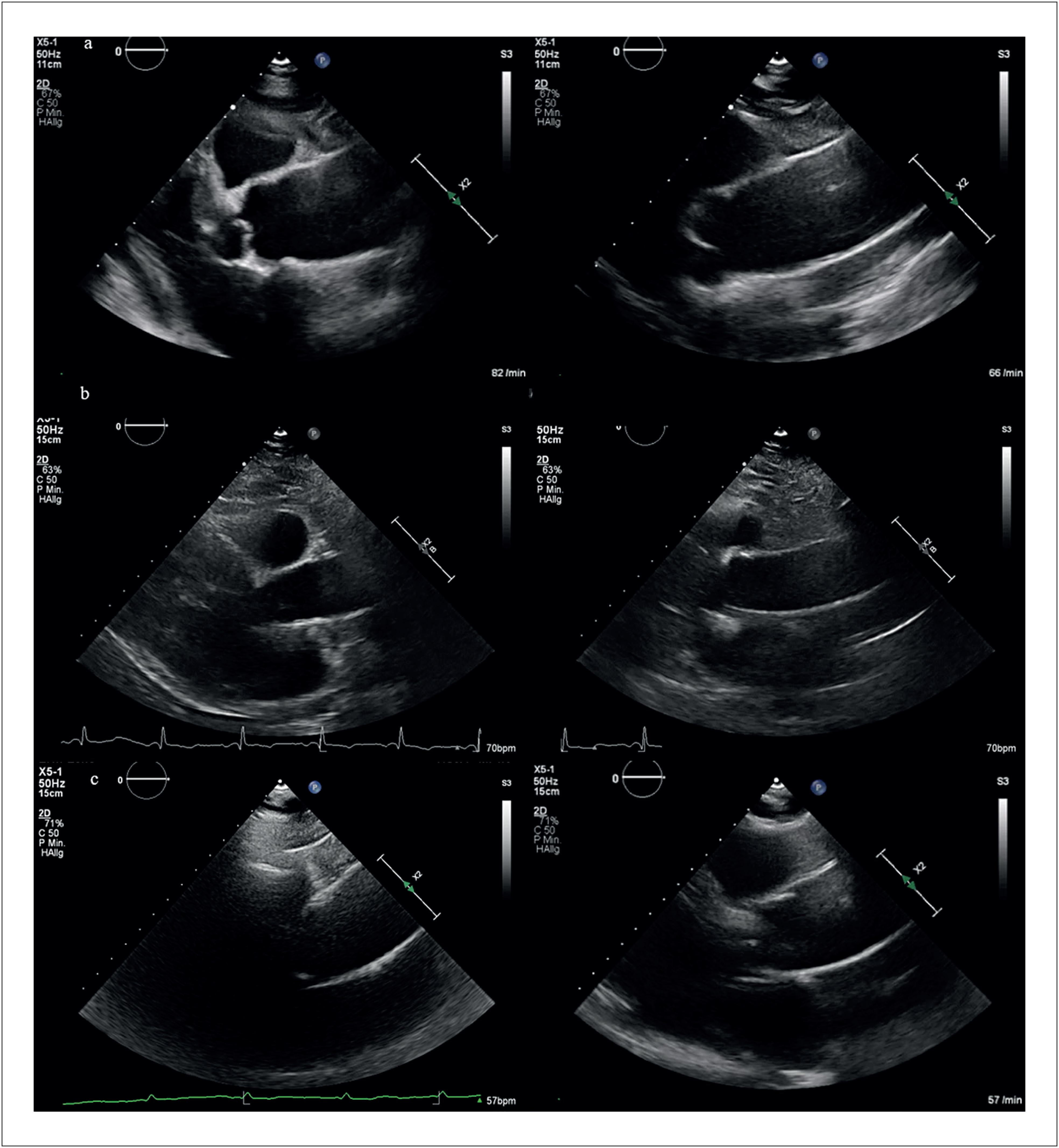
Table 1 summarizes the baseline clinical and echocardiographic characteristics. Figure 1 shows representative examples of imaging the ascending aorta using different TTE views.

Visualization of the ascending aorta in three patients (A, B, and C) using the uPLAX view (left panels) and the mPLAX (right panels).
Results
Visualization of the ascending aorta
The visualized ascending aortic segment was significantly longer using the mPLAX view than using the uPLAX view (60.6 ± 9.3 mm vs 44.3 ± 8.2 mm, respectively). The mean difference (MD) between both techniques was 16.4 mm (95% CI, 15.1-17.6 mm; p < 0.001).
Ascending aortic diameter
The ascending aortic diameter measured in the mPLAX view was significantly greater than that obtained in the uPLAX view (34.9 ± 5.7 mm vs 33.2 ± 5.3 mm, respectively), with a MD of 1.7 mm (95% CI, 1.2-2.3 mm; p < 0.001).
This difference was primarily observed among individuals with hypertension (n = 135). In this subgroup, ascending aortic diameter measured 35.6 ± 5.7 mm in the mPLAX view compared with 33.5 ± 5.4 mm in the uPLAX view, corresponding to a MD of 2.1 mm (95% CI, 1.5-2.7 mm; p < 0.001).
In contrast, among individuals without hypertension (n = 34), ascending aortic diameter was comparable between the two views (32.4 ± 5.0 mm in mPLAX vs 32.1 ± 5.2 mm in uPLAX), with no statistically significant difference (MD, 0.3 mm; 95% CI, −1.3 to 1.9 mm; p = 0.709).
Non-inferiority analysis
The predefined criterion for non-inferiority of the mPLAX view relative to the uPLAX view for visualization of a longer ascending aortic segment was met.
Sex-based analysis
Ascending aortic length and diameter were greater in men than in women using both imaging approaches. However, the incremental gain in visualized aortic length achieved with the mPLAX view compared with the uPLAX view was similar between sexes (16.3 mm [95% CI, 14.8-17.9 mm] in men vs 16.4 mm [95% CI, 14.3-18.5 mm] in women).
Study limitations
This study has several limitations. First, it represents a single-center experience and therefore requires external validation before widespread adoption. Nevertheless, given the simplicity and rapid acquisition of the mPLAX view, we believe that this approach is readily applicable in routine clinical practice.
Second, ascending aortic measurements obtained using the mPLAX view were not compared with reference imaging modalities such as computed tomography. However, in individuals without hypertension, aortic diameters measured using the mPLAX and uPLAX views were highly consistent.
Conclusions
In this study, we proposed a mPLAX focused on optimizing visualization of the ascending aorta. The rightward transducer translation partially removes the left ventricle from the imaging plane, whereas the simultaneous clockwise rotation and slight caudal angulation improve alignment with the ascending aortic long axis by correcting the heart-aorta angle.7
We believe that the mPLAX view may serve as an additional non-inferior TTE window for visualization of a longer segment of the tubular ascending aorta during routine TTE examinations. Because this view can be obtained without changing the patient's position or intercostal space, it is easier and faster to acquire, particularly for general cardiologists and less experienced echocardiographers performing high-volume daily studies.
This approach may be especially useful during routine TTE follow-up of patients with previously documented mid-distal ascending aortic aneurysms identified by computed tomography and adequate parasternal acoustic windows.
-
Sources of Funding
There were no external funding sources for this study.
-
Study Association
This study is not associated with any thesis or dissertation work.
-
Ethics Approval and Consent to Participate
This retrospective study analyzed fully anonymized echocardiographic data. No interventions were performed, and ethics committee approval was not required under local regulations.
-
Use of Artificial Intelligence
The authors did not use any artificial intelligence tools in the development of this work.
Availability of Research Data
All datasets supporting the results of this study are available upon request from the corresponding author, subject to ethical and confidentiality considerations.
References
-
1 Hartnell GG. Imaging of Aortic Aneurysms and Dissection: CT and MRI. J Thorac Imaging. 2001;16(1):35-46. doi: 10.1097/00005382-200101000-00006.
» https://doi.org/10.1097/00005382-200101000-00006 -
2 Elefteriades JA, Farkas EA. Thoracic Aortic Aneurysm Clinically Pertinent Controversies and Uncertainties. J Am Coll Cardiol. 2010;55(9):841-57. doi: 10.1016/j.jacc.2009.08.084.
» https://doi.org/10.1016/j.jacc.2009.08.084 -
3 Evangelista A, Sitges M, Jondeau G, Nijveldt R, Pepi M, Cuellar H, et al. Multimodality Imaging in Thoracic Aortic Diseases: A Clinical Consensus Statement from the European Association of Cardiovascular Imaging and the European Society of Cardiology Working Group on Aorta and Peripheral Vascular Diseases. Eur Heart J Cardiovasc Imaging. 2023;24(5):e65-e85. doi: 10.1093/ehjci/jead024.
» https://doi.org/10.1093/ehjci/jead024 -
4 Evangelista A, Flachskampf FA, Erbel R, Antonini-Canterin F, Vlachopoulos C, Rocchi G, et al. Echocardiography in Aortic Diseases: EAE Recommendations for Clinical Practice. Eur J Echocardiogr. 2010;11(8):645-58. doi: 10.1093/ejechocard/jeq056.
» https://doi.org/10.1093/ejechocard/jeq056 -
5 Upadhyaya K, Ugonabo I, Satam K, Hull SC. Echocardiographic Evaluation of the Thoracic Aorta: Tips and Pitfalls. Aorta. 2021;9(1):1-8. doi: 10.1055/s-0041-1724005.
» https://doi.org/10.1055/s-0041-1724005 -
6 Goldstein SA, Evangelista A, Abbara S, Arai A, Asch FM, Badano LP, et al. Multimodality Imaging of Diseases of the Thoracic Aorta in Adults: From the American Society of Echocardiography and the European Association of Cardiovascular Imaging: Endorsed by the Society of Cardiovascular Computed Tomography and Society for Cardiovascular Magnetic Resonance. J Am Soc Echocardiogr. 2015;28(2):119-82. doi: 10.1016/j.echo.2014.11.015.
» https://doi.org/10.1016/j.echo.2014.11.015
Edited by
-
Editor responsible for the review:
Marcelo Tavares