Clinical Outcomes Following PM Implantation: A 3-Year Comparative Study of Sick Sinus Syndrome and Atrioventricular Block
Abstract
Background Permanent pacemaker implantation is an established treatment for both sick sinus syndrome (SSS) and atrioventricular block (AVB), yet limited data exist comparing long-term clinical outcomes and cardiac remodeling patterns between these distinct bradyarrhythmic etiologies.
Objectives To compare three-year clinical outcomes and cardiac structural changes between SSS and AVB patients following permanent pacemaker implantation.
Methods This retrospective observational study enrolled 192 adult patients (65 SSS, 127 AVB) who underwent pacemaker implantation between January 2018 and December 2020. Demographic data, echocardiographic parameters, pacemaker-recorded data, and clinical outcomes were assessed at baseline, one month, six months, one year, and three years. Primary outcomes included changes in cardiac structure and function, symptomatic improvement, new-onset atrial fibrillation, rehospitalization, and all-cause mortality.
Results Both groups demonstrated significant declines in left ventricular ejection fraction over three years (SSS: 57.2±6.0% to 54.3±8.3%, p<0.001; AVB: 55.5±7.0% to 53.0±6.8%, p<0.001). Despite similar ventricular dysfunction progression, distinct remodeling patterns emerged: SSS patients exhibited significant increases in left atrial diameter (31.3 to 38.8 mm, p=0.040), left ventricular end-diastolic diameter (45.2 to 51.0 mm, p<0.001), and mitral regurgitation prevalence (26.2% to 64.6%), while AVB patients maintained stable chamber dimensions despite high ventricular pacing burden (>90%). Symptomatic improvement was comparable (SSS: 85.6% vs. AVB: 83.5%, p=0.84), as were new-onset atrial fibrillation (41.5% vs. 37.0%, p=0.54), rehospitalization (50.8% vs. 58.3%, p=0.38), and all-cause mortality (13.8% vs. 11.8%, p=0.69). Mortality predictors differed: mitral regurgitation severity in SSS versus coronary artery disease, beta-blocker therapy, and left atrial volume in AVB patients.
Conclusions Despite similar symptomatic benefits and clinical outcomes, SSS and AVB patients demonstrate distinct cardiac remodeling patterns and different mortality predictors, supporting the need for etiology-specific follow-up strategies in pacemaker recipients.
Keywords
Artificial Pacemaker; Sick Sinus Syndrome; Atrioventricular Block; Atrial Fibrillation
Resumo
Fundamento O implante de marca-passo permanente é um tratamento estabelecido tanto para a síndrome do nó sinusal (SNS) quanto para o bloqueio atrioventricular (BAV), porém existem poucos dados comparando os resultados clínicos a longo prazo e os padrões de remodelamento cardíaco entre essas distintas etiologias bradiarrítmicas.
Objetivos Comparar os resultados clínicos e as alterações estruturais cardíacas em três anos entre pacientes com SNS e BAV após implante de marca-passo permanente.
Métodos Este estudo observacional retrospectivo incluiu 192 pacientes adultos (65 com SNS, 127 com BAV) submetidos a implante de marca-passo entre janeiro de 2018 e dezembro de 2020. Dados demográficos, parâmetros ecocardiográficos, dados registrados pelo marca-passo e desfechos clínicos foram avaliados no início do estudo, após um mês, seis meses, um ano e três anos. Os desfechos primários incluíram alterações na estrutura e função cardíacas, melhora dos sintomas, fibrilação atrial de início recente, reinternação e mortalidade por todas as causas.
Resultados Ambos os grupos demonstraram declínios significativos na fração de ejeção do ventrículo esquerdo ao longo de três anos (SNS: 57,2±6,0% para 54,3±8,3%, p<0,001; BAV: 55,5±7,0% para 53,0±6,8%, p<0,001). Apesar da progressão semelhante da disfunção ventricular, padrões distintos de remodelamento emergiram: os pacientes com SNS apresentaram aumentos significativos no diâmetro do átrio esquerdo (31,3 para 38,8 mm, p=0,040), no diâmetro diastólico final do ventrículo esquerdo (45,2 para 51,0 mm, p<0,001) e na prevalência de regurgitação mitral (26,2% para 64,6%), enquanto os pacientes com BAV mantiveram dimensões ventriculares estáveis apesar da alta carga de estimulação ventricular (>90%). A melhora dos sintomas foi comparável (SNS: 85,6% vs. BAV: 83,5%, p=0,84), assim como o surgimento de fibrilação atrial (41,5% vs. 37,0%, p=0,54), a reinternação (50,8% vs. 58,3%, p=0,38) e a mortalidade por todas as causas (13,8% vs. 11,8%, p=0,69). Os preditores de mortalidade diferiram: gravidade da regurgitação mitral em pacientes com SNS versus doença arterial coronariana, terapia com betabloqueadores e volume do átrio esquerdo em pacientes com BAV.
Conclusões Apesar dos benefícios sintomáticos e resultados clínicos semelhantes, pacientes com SNS e BAV demonstram padrões distintos de remodelamento cardíaco e diferentes preditores de mortalidade, o que reforça a necessidade de estratégias de acompanhamento específicas para cada etiologia em pacientes com marca-passo.
Palavras-chave
Marca-Passo Artificial; Síndrome do Nó Sinusal; Bloqueio Atrioventricular; Fibrilação Atrial
Introduction
Bradyarrhythmias, including sick sinus syndrome (SSS) and atrioventricular block (AVB), are common indications for permanent pacemaker implantation.1 While pacemakers effectively restore heart rate and prevent syncope, their long-term effects on cardiac structure, function, and clinical outcomes remain a subject of ongoing investigation.2 Understanding the differences in disease progression and response to pacing therapy between SSS and AVB patients is essential for optimizing patient management and improving prognostic outcomes.3
SSS is characterized by sinus node dysfunction, leading to inappropriate bradycardia and symptoms such as dizziness, syncope, and fatigue.4 In contrast, AVB results from impaired conduction between the atria and ventricles, potentially leading to severe bradycardia and hemodynamic instability.5 Although pacemaker therapy effectively alleviates symptoms in both conditions, concerns persist regarding its impact on cardiac remodelling, valvular function, and the development of heart failure over time.6
Previous studies have suggested that right ventricular pacing may contribute to adverse cardiac remodelling, including left ventricular dysfunction and atrial enlargement, potentially increaszing the risk of atrial fibrillation (AF) and heart failure.7 However, whether these effects differ significantly between SSS and AVB patients remains unclear.8 Furthermore, the clinical implications of pacemaker therapy, including rehospitalization rates, mortality, and symptomatic improvement, require further exploration.9
This study aims to compare the long-term clinical outcomes of patients undergoing pacemaker implantation for SSS versus AVB over a three-year follow-up period. By analyzing echocardiographic changes, pacemaker-recorded data, and clinical endpoints, we seek to provide a comprehensive assessment of the impact of pacing therapy on these two distinct patient populations. The findings from this study will contribute to a better understanding of the prognostic implications of pacemaker implantation and inform future strategies for patient management and therapy optimization.
Methods
Study design and population
This study was a retrospective, observational cohort study conducted to compare the long-term clinical outcomes of patients undergoing permanent pacemaker implantation for SSS or AVB. The study population included adult patients who underwent pacemaker implantation at our institution between January 2018 and December 2020. Inclusion criteria consisted of a confirmed diagnosis of either SSS or AVB as the primary indication for pacemaker implantation, age ≥18 years, and availability of baseline and follow-up clinical and echocardiographic data. Exclusion criteria included prior pacemaker implantation, concomitant severe valvular disease requiring intervention, advanced heart failure (New York Heart Association [NYHA] class IV), or a history of congenital heart disease.
Data collection and follow-up
Demographic, clinical, laboratory, and echocardiographic data were collected from medical records. Baseline characteristics included age, sex, body mass index (BMI), comorbidities (hypertension, diabetes mellitus, coronary artery disease, and smoking status), and medication use (beta-blockers, ACE inhibitors/angiotensin receptor blockers [ARBs], mineralocorticoid receptor antagonists [MRAs], loop diuretics, and non-dihydropyridine calcium channel blockers). Laboratory parameters included serum sodium (Na), potassium (K), estimated glomerular filtration rate (eGFR), and hemoglobin levels.
Echocardiographic assessment
All patients underwent transthoracic echocardiography at baseline and follow-up intervals (first month, sixth month, first year, and third year). Echocardiographic examinations were performed using a commercially available ultrasound system (Philips EPIQ 7) by experienced cardiologists following the American Society of Echocardiography (ASE) guidelines. Key parameters were measured as follows:
-
Left Ventricular Ejection Fraction (EF%): Assessed using the biplane Simpson’s method from the apical four-chamber and two-chamber views.
-
Left Atrial Diameter (LAD): Measured in the parasternal long-axis view at end-systole.
-
Left Atrial Volume Index (LAVI): Calculated using the biplane area-length method and indexed to body surface area.
-
Left Ventricular End-Diastolic Diameter (LVEDD): Measured at the level of the mitral valve leaflet tips in the parasternal long-axis view.
-
Pulmonary Artery Systolic Pressure (PASP): Estimated using the peak tricuspid regurgitation velocity and right atrial pressure estimation.
-
Mitral Regurgitation (MR) Severity: Assessed using color Doppler imaging and graded as mild, moderate, or severe based on the regurgitant jet area and vena contracta width.
Pacemaker data collection and interpretation
Pacemaker programming data were obtained from device interrogation reports at each follow-up visit. The interrogation was performed using the Medtronic pacemaker programmer and involved assessment of:
-
Mean Lower Rate Limit: The programmed minimum heart rate at which the pacemaker delivers pacing support.
-
Atrial Pacing Percentage: The proportion of time atrial pacing is delivered, indicating the pacemaker's reliance on artificial pacing to maintain rhythm.
-
Atrial Premature Beat Percentage: The percentage of premature atrial contractions detected, which may indicate underlying atrial arrhythmia.
-
Atrial High-Rate Episodes (AHREs): Episodes of rapid atrial activity detected by the pacemaker, defined as atrial rates exceeding a predefined threshold (e.g., 180 beats per minute), which may suggest AF or atrial tachyarrhythmia.
-
Pacemaker Programming Protocol: All pacemakers were programmed according to contemporary guidelines with optimization for physiologic pacing. The pacing mode was DDD/DDDR in all patients, with a lower rate limit of 60 beats per minute. In SSS patients, algorithms to minimize ventricular pacing were enabled when available, including extended AV delay (up to 250-300 ms) and AV hysteresis features to promote intrinsic AV conduction. However, in AVB patients, such algorithms were not utilized, given the intrinsic conduction disease, and standard AV delays (120-180 ms) were programmed. Rate-responsive pacing was activated in patients with chronotropic incompetence. Mode-switch algorithms for atrial tachyarrhythmias were enabled in all devices. Despite these programming strategies, the ventricular pacing burden in AVB patients remained high (>90%) due to the nature of their conduction disorder, whereas SSS patients maintained predominantly intrinsic ventricular activation.
Outcome measures
The primary outcome was the longitudinal change in echocardiographic parameters over the three-year follow-up period. Secondary outcomes included:
-
Symptomatic improvement, assessed using the European Heart Rhythm Association (EHRA) symptom score: The EHRA score is a classification system that evaluates the severity of symptoms related to arrhythmia, ranging from EHRA I (no symptoms) to EHRA IV (severe symptoms affecting daily activities).
-
Incidence of new-onset atrial fibrillation (AF): Defined as the first documented episode of AF detected by either routine electrocardiography, Holter monitoring, or pacemaker interrogation, in a patient with no prior history of AF.
-
Rehospitalization rates during the follow-up period.
-
All-cause mortality.
Statistical analysis
Continuous variables were expressed as mean ± standard deviation (SD) and compared between groups using the independent samples t-test. Categorical variables were presented as frequencies and percentages and analyzed using the chi-square or Fisher's exact test. Repeated measures analysis of variance (ANOVA) was used to assess changes in echocardiographic parameters over time. Cox proportional hazards regression analysis was performed to identify predictors of rehospitalization and mortality. Kaplan-Meier survival curves were generated for rehospitalization and mortality outcomes, and comparisons between groups were conducted using the Log-Rank test. A two-sided p-value <0.05 was considered statistically significant.
Ethical considerations
This study was conducted in accordance with the principles of the Declaration of Helsinki and was approved by the institutional ethics committee. Given the retrospective nature of the study, informed consent was waived by the ethics board. Patient confidentiality was maintained by de-identifying data before analysis.
Result
The study included a total of 192 patients, with 65 (33.9%) undergoing pacemaker implantation for SSS and 127 (66.1%) for AVB. The mean age of the cohort was 66.3 ± 7.2 years, and 89 patients (46.4%) were male. Hypertension was the most prevalent comorbidity, affecting 93 patients (48.4%).
The two groups were compared in terms of demographic, echocardiographic, and clinical characteristics. No significant differences were observed in age, sex, or BMI. The prevalence of hypertension, diabetes mellitus, coronary artery disease, and active smoking was similar between the groups; however, heart failure was more frequent in AVB patients than in those with SSS (17.3% vs. 10.8%, p=0.03). The use of ACE inhibitors/ARBs and mineralocorticoid receptor antagonists (MRAs) did not differ between groups. However, beta-blockers (69.2% vs. 13.4%, p<0.001) and non-dihydropyridine calcium channel blockers (12.3% vs. 7.1%, p=0.01) were more commonly prescribed in SSS patients. Conversely, loop diuretics were used more frequently in the AVB group (20.5% vs. 9.2%, p=0.05). Serum sodium (Na), potassium (K), estimated glomerular filtration rate (eGFR), and hemoglobin levels were comparable between the two groups. On echocardiographic assessment, LAD was significantly larger in AVB patients (34.6 ± 6.5 mm vs. 31.3 ± 6.9 mm, p=0.02), whereas no other significant differences were observed in the remaining parameters (Table 1).
The two patient groups were compared using pacemaker-recorded data. The mean lower rate limit at baseline did not differ significantly between groups (60.5 ± 2.5 vs. 61.2 ± 3.6, p = 0.20). As expected, atrial pacing percentage was consistently higher in the SSS group at all follow-up intervals (first month: 84.5±19.1% vs. 18.1±14.5%; third year: 88.4±20.3% vs. 17.3±21.3%; p < 0.001), while ventricular pacing percentage was markedly higher in AVB patients (first month: 92.5±6.3% vs. 12.3±8.5%; third year: 95.3±4.8% vs. 9.8±7.6%; p < 0.001). The minimal ventricular pacing in SSS patients reflects preserved AV conduction in this population, whereas the high ventricular pacing burden in AVB patients was inherent to their conduction disorder. Atrial premature beat percentage and atrial high-rate episode frequency were comparable between the groups throughout the follow-up period (Table 2).
Longitudinal changes in echocardiographic parameters were assessed from pre-implantation to 3-year follow-up in both patient subgroups. Among patients with SSS, we observed significant deterioration in multiple cardiac parameters. Left ventricular systolic function declined, with EF decreasing from 57.2 ± 6.0% at baseline to 54.3 ± 8.3% at 3 years (p < 0.001). These patients exhibited progressive left atrial remodeling, demonstrated by increases in LAD from 31.3 ± 6.9 mm to 38.8 ± 7.9 mm (p = 0.040) and LAVI from 46.1 ± 10.8 mL/m2 to 53.2 ± 10.4 mL/m2 (p < 0.001). Furthermore, left ventricular dimensions expanded significantly, with LVEDD increasing from 45.2 ± 3.5 mm to 51.0 ± 3.5 mm (p < 0.001). Valvular function also worsened, with the prevalence of mild or greater MR increasing substantially from 26.2% (17/65 patients) to 58.4% (38/65 patients; p = 0.001). Additionally, PASP rose from 26.9 ± 6.2 mmHg to 31.6 ± 7.0 mmHg (p=0.025). Patients with AVB similarly demonstrated a significant reduction in EF% from 55.5 ± 7.0% to 53.0 ± 6.8% (p < 0.001). However, in contrast to the SSS group, AVB patients maintained stable left atrial dimensions (LAD and LAVI) and LVEDD throughout the follow-up period. Both groups showed comparable progression of valvular dysfunction, with AVB patients exhibiting an increase in mild or worse MR from 30.7% (39/127 patients) to 57.4% (73/127 patients; p < 0.001) and a rise in PASP from 25.8 ± 7.0 mmHg to 30.8 ± 8.9 mmHg (p = 0.019) (Table 3).
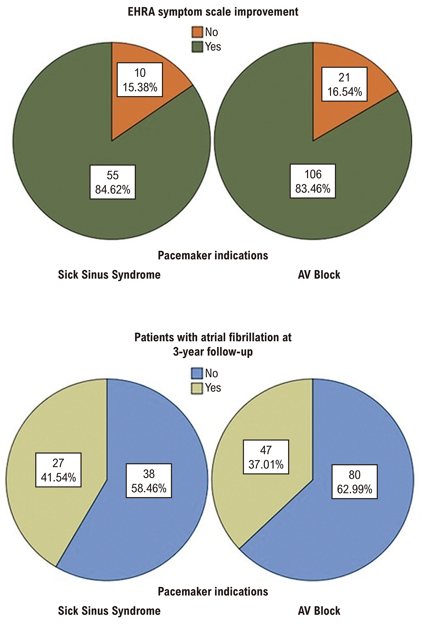
At the 3-year follow-up assessment, clinical outcomes were evaluated according to EHRA symptom scores. The analysis revealed that 83.9% of patients (161 of 192) experienced symptomatic improvement following pacemaker implantation. When stratified by indication, 85.6% of patients with SSS (55 of 65) and 83.5% of those with AVB (106 of 127) demonstrated clinical benefit, with no statistically significant difference between groups (p=0.84; odds ratio [OR] 0.92, 95% confidence interval [CI] 0.40–2.09). Subsequent analysis of patient characteristics associated with symptomatic improvement yielded distinct patterns for each group. Among patients with SSS, no significant predictors of treatment response were identified. However, in the AVB cohort, several factors showed significant inverse correlations with symptomatic benefit: female sex (p=0.039), pre-existing coronary artery disease (p=0.006), increased LAD (p=0.05), and greater LVEDD (p=0.042). During the 3-year observation period, new-onset AF was documented in 38.5% of the study population (74 of 192). The incidence was comparable between etiological groups, occurring in 41.5% of SSS patients (27 of 65) versus 37.0% of AVB patients (47 of 127) (p=0.54; OR 0.83, 95% CI 0.45–1.52). Examination of potential risk factors for atrial fibrillation development revealed no significant associations in the SSS group. Conversely, among patients with AVB, three parameters emerged as significant predictors of AF occurrence: advanced age (p=0.045), larger LAD (p=0.003), and increased LAVI (p=0.046) (Figure 1).
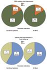
– Comparison of EHRA symptom scale and development of atrial fibrillation between patient groups at 3-year follow-up.
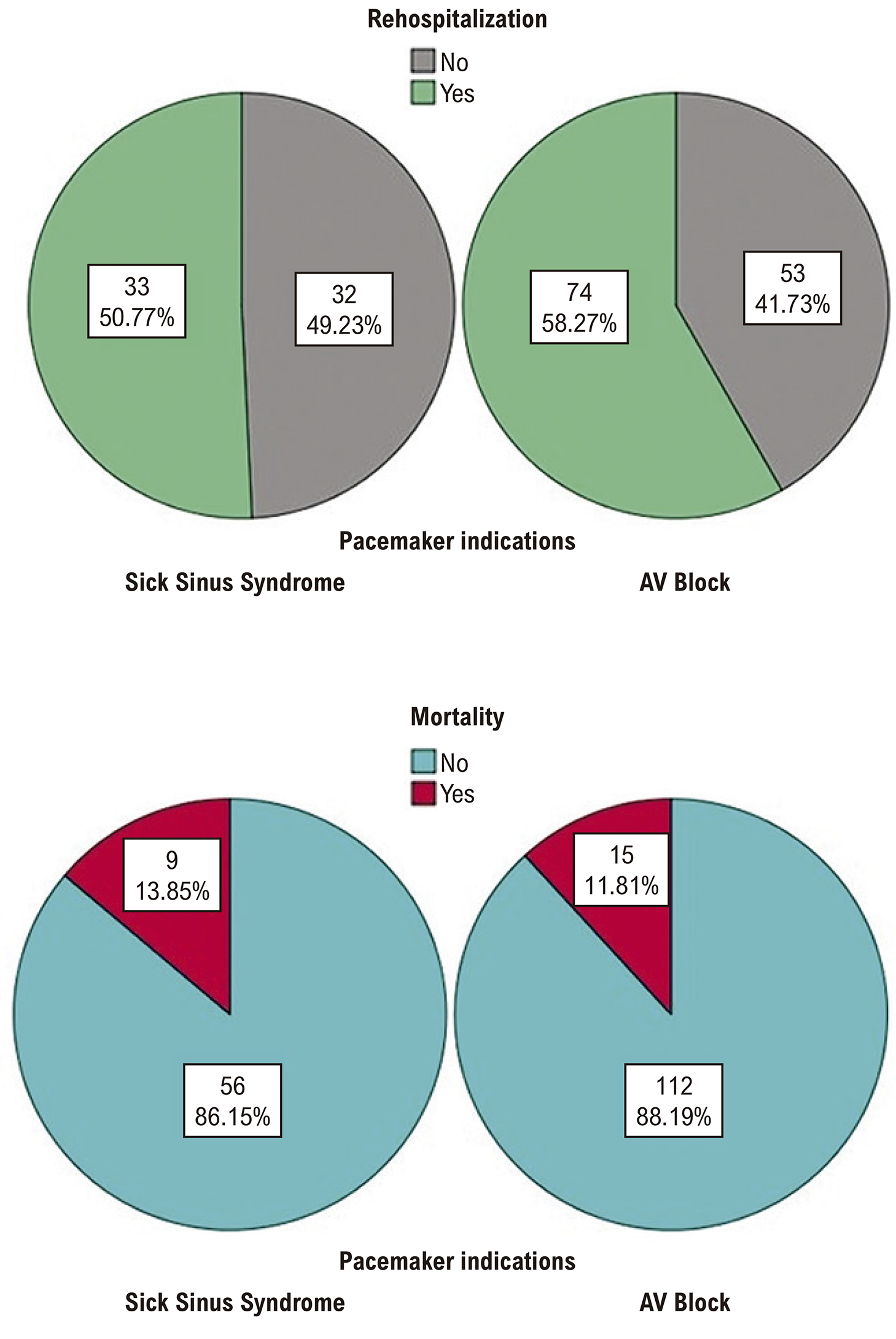
At the 3-year follow-up evaluation, we assessed rehospitalization and mortality outcomes across the study cohort. Among the 192 enrolled patients, 107 (55.7%) required rehospitalization during the observation period. When stratified by pacemaker indication, rehospitalization rates were comparable between groups, occurring in 50.8% of SSS patients (33/65) versus 58.3% of AVB patients (74/127) (p=0.38; odds ratio [OR] 1.20, 95% confidence interval [CI] 0.80-1.81).
Predictive analysis of rehospitalization risk factors revealed distinct patterns between etiologies. For SSS patients, only elevated PASP demonstrated a significant association with rehospitalization (p=0.04). In contrast, the AVB cohort showed significant inverse relationships between rehospitalization and both female sex (p=0.037) and hypertension diagnosis (p=0.013). All-cause mortality was documented in 12.5% of patients (24/192), with no significant difference between groups (SSS: 13.8% [9/65] vs AVB: 11.8% [15/127]; p=0.69; OR 0.84, 95% CI 0.37-1.93). Mortality predictors differed substantially between groups. Among SSS patients, only the severity of MR showed significant correlation with mortality risk (p=0.031). Conversely, AVB patients demonstrated significant mortality associations with three independent factors: pre-existing coronary artery disease (p=0.009), beta-blocker therapy (p=0.005), and elevated LAVI (p=0.001) (Figure 2).
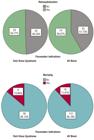
– Comparison of rehospitalization and mortality events between patient groups at 3-year follow-up.
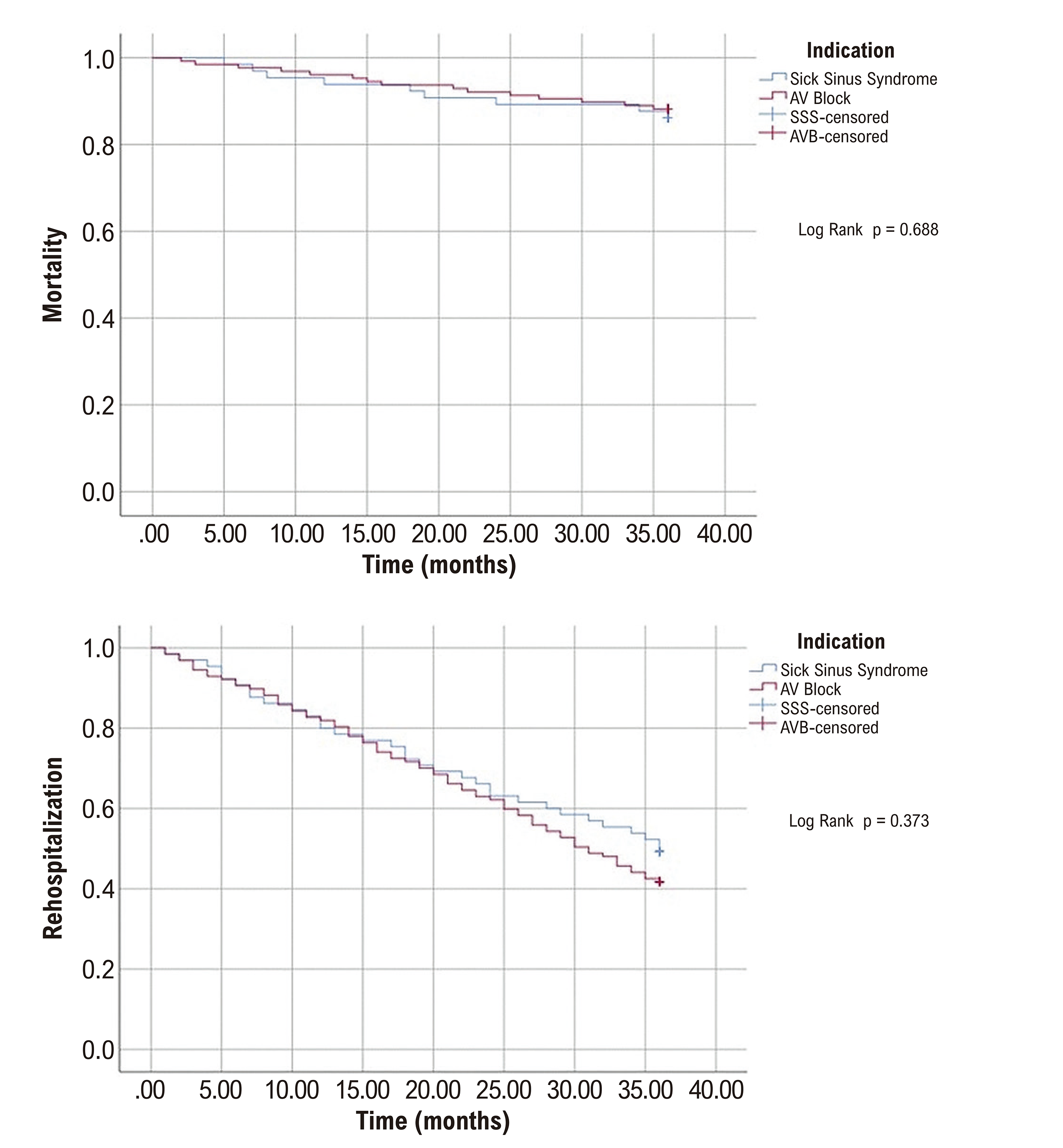
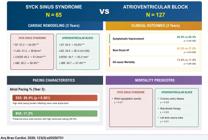
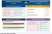
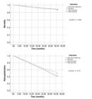
During the 3-year follow-up, the groups were compared using Cox regression and survival analysis for rehospitalization and mortality outcomes. No significant difference was observed in mortality between groups (Log Rank p=0.688). However, survival analysis for rehospitalization revealed a trend toward lower rehospitalization rates over time in sick sinus syndrome patients, though this did not reach statistical significance (Log-Rank p=0.373) (Figure 3). Key comparative findings between the SSS and AVB groups regarding cardiac remodeling, clinical outcomes, pacing characteristics, and mortality predictors are summarized in the Central Illustration.
Discussion
This study provides a comprehensive comparison of the long-term clinical outcomes between patients with SSS and AVB following permanent pacemaker implantation. Our findings demonstrate that while pacemaker therapy provides symptomatic improvement in the majority of patients from both groups, important differences exist in cardiac remodeling patterns and predictors of adverse outcomes.
The observed higher atrial pacing percentage in SSS patients is consistent with underlying sinus node dysfunction, which necessitates more frequent atrial pacing support. 4,10 This finding aligns with previous studies that have demonstrated the dependence of SSS patients on atrial pacing for maintaining adequate heart rates.11 In contrast, AVB patients typically maintain intrinsic sinus node function but require ventricular pacing due to impaired AV conduction.5
The substantially different ventricular pacing burdens between groups (>90% in AVB vs. <12% in SSS patients) provide crucial context for interpreting our remodeling findings. The high ventricular pacing percentage in AVB patients (averaging 93-95% throughout follow-up) likely contributed to the adverse effects observed in both groups, though the patterns differed. While both groups experienced declining EF, the progressive left atrial and ventricular dilation observed predominantly in SSS patients despite minimal ventricular pacing suggests that sinus node dysfunction itself may predispose to adverse remodeling independent of pacing burden. Conversely, the stable chamber dimensions in AVB patients despite near-universal ventricular pacing challenges the notion that RV pacing universally causes chamber dilation, suggesting other factors, such as intrinsic atrial disease in SSS, may play important roles. These findings align with recent observations that the detrimental effects of RV pacing may be more pronounced in patients with underlying atrial pathology.
A particularly notable finding was the differential pattern of cardiac remodeling between the two groups. SSS patients exhibited significant increases in left atrial dimensions and LVEDD over the follow-up period, suggesting progressive adverse cardiac remodeling. This observation supports earlier studies suggesting that right ventricular pacing might contribute to ventricular dyssynchrony and subsequent adverse remodeling.7,12 The maintenance of stable left atrial dimensions in AVB patients, despite comparable decreases in EF, suggests potential differences in the pathophysiological response to pacing therapy between these two conditions.
The substantial incidence of new-onset AF in both groups (41.5% in SSS vs. 37.0% in AVB) highlights the close relationship between bradyarrhythmia and AF development.13 While the difference between groups was not statistically significant, the identification of advanced age and increased left atrial dimensions as predictors of AF in AVB patients aligns with established risk factors for this arrhythmia.14 The high incidence of AF raises important questions about the potential benefits of prophylactic anticoagulation in high-risk patients with pacemakers, particularly those with increased left atrial dimensions.15
Regarding symptomatic improvement, our finding that approximately 84% of patients experienced clinical benefit following pacemaker implantation confirms the efficacy of pacing therapy for symptom relief in bradyarrhythmias.16 The absence of significant between-group differences in symptomatic improvement suggests that the underlying etiology of bradyarrhythmia may not substantially influence the subjective response to pacing therapy. However, the identification of female sex, coronary artery disease, and increased cardiac dimensions as negative predictors of symptomatic improvement in AVB patients warrants further investigation and may have implications for patient selection and counseling.17
The rehospitalization rate of approximately 56% over three years indicates a substantial healthcare burden associated with pacemaker patients. Although the trend toward lower rehospitalization rates in SSS patients did not reach statistical significance, the different predictors of rehospitalization between groups suggest potentially distinct pathophysiological mechanisms contributing to clinical decompensation.18 The association between elevated PASP and rehospitalization in SSS patients points to the potential importance of right ventricular function and pulmonary hemodynamics in this population.19
Mortality outcomes were comparable between groups, affecting approximately 12% of patients over three years. However, the distinct predictors of mortality—MR severity in SSS patients versus coronary artery disease, beta-blocker therapy, and increased left atrial volume in AVB patients—suggest different pathways contributing to adverse outcomes.20,21 The association between beta-blocker therapy and mortality in AVB patients is particularly intriguing and may reflect either the severity of underlying cardiac disease necessitating beta-blocker use or potentially adverse interactions between pharmacological therapy and pacing.22
The progressive increase in MR observed in both groups deserves special attention. This finding is consistent with previous studies suggesting that right ventricular pacing can induce papillary muscle dyssynchrony and subsequent functional MR.23 The correlation between MR severity and mortality in SSS patients emphasizes the potential prognostic importance of valvular function in this population.24
This study provides several important advances beyond existing literature. First, we demonstrate for the first time that SSS patients exhibit progressive cardiac remodeling despite minimal ventricular pacing (<12%), contradicting the prevailing assumption that adverse remodeling is primarily pacing-induced. This finding suggests an intrinsic pathophysiological process in SSS that warrants recognition as an independent risk factor for structural deterioration, not previously emphasized in major pacemaker trials such as MOST or DAVID. Second, our observation that AVB patients maintain stable chamber dimensions despite >90% ventricular pacing challenges the universality of pacing-induced cardiomyopathy and suggests protective factors or compensatory mechanisms that merit further investigation. Third, we identify etiology-specific mortality predictors that have not been previously described: while MR predicts outcomes in SSS (potentially related to progressive atrial dysfunction), the predictive value of LAVI in AVB patients suggests different mechanistic pathways to adverse outcomes. These distinct risk profiles provide a rationale for differentiated surveillance strategies based on underlying bradyarrhythmia etiology—a precision medicine approach not currently reflected in guideline recommendations, which treat all pacemaker patients similarly regardless of indication. Finally, the high incidence of new-onset AF (41.5% in SSS, 37.0% in AVB) substantially exceeds rates reported in earlier trials, likely reflecting both improved detection through continuous device monitoring and our longer follow-up duration, emphasizing the need for systematic atrial arrhythmia surveillance in all pacemaker recipients.
Our study has several limitations that should be acknowledged. First, its retrospective design limits causal inference and may introduce selection bias. Second, we did not systematically assess ventricular dyssynchrony, which might have provided mechanistic insights into the observed cardiac remodeling patterns. Third, the single-center nature of our study may limit generalizability to other populations. Finally, we did not analyze the impact of different pacing modes or settings, which might influence long-term outcomes.
Despite these limitations, our findings have important clinical implications. The different patterns of cardiac remodeling and predictors of adverse outcomes between SSS and AVB patients suggest that individualized follow-up strategies based on the underlying etiology of bradyarrhythmia may be beneficial. SSS patients may benefit from closer monitoring of cardiac dimensions and optimization of pacing settings to minimize ventricular pacing burden.25 In AVB patients, particular attention to left atrial parameters and management of underlying coronary artery disease might improve long-term outcomes.26
Future research should explore whether alternative pacing strategies, such as His bundle pacing or cardiac resynchronization therapy, might prevent the observed structural changes and improve outcomes in specific patient subgroups.27,28 Additionally, prospective studies with longer follow-up periods are needed to better understand the long-term impact of pacing therapy on cardiac structure and function, particularly in relation to the development of heart failure and valvular dysfunction.29
Conclusion
This three-year comparative study demonstrates that while permanent pacemaker implantation provides comparable symptomatic improvement in both SSS and AVB patients, the two conditions exhibit fundamentally different pathophysiological responses to pacing therapy. Despite minimal ventricular pacing burden, SSS patients developed progressive adverse cardiac remodeling with significant left atrial and ventricular chamber dilation, suggesting that sinus node dysfunction itself, independent of pacing, predisposes to structural deterioration. Conversely, AVB patients maintained stable chamber dimensions despite near-universal ventricular pacing (>90%), challenging conventional assumptions about the uniformly deleterious effects of right ventricular pacing.
The distinct mortality predictors between groups, MR severity in SSS versus coronary artery disease, and left atrial volume in AVB underscore the need for etiology-specific risk stratification and tailored follow-up strategies. These findings support individualized management approaches: closer echocardiographic surveillance for structural changes in SSS patients, and enhanced attention to left atrial parameters and coronary disease management in AVB patients. Future research should investigate whether alternative pacing strategies can mitigate the intrinsic remodeling tendency observed in SSS patients and validate risk-based surveillance protocols for each patient population.
References
-
1 Kusumoto FM, Schoenfeld MH, Barrett C, Edgerton JR, Ellenbogen KA, Gold MR, et al. 2018 ACC/AHA/HRS Guideline on the Evaluation and Management of Patients with Bradycardia and Cardiac Conduction Delay: Executive Summary: A Report of the American College of Cardiology/American Heart Association Task Force on Clinical Practice Guidelines, and the Heart Rhythm Society. J Am Coll Cardiol. 2019;74(7):932-87. doi: 10.1016/j.jacc.2018.10.043.
» https://doi.org/10.1016/j.jacc.2018.10.043 - 2 Sweeney MO, Hellkamp AS, Ellenbogen KA, et al. Adverse effect of ventricular pacing on heart failure and atrial fibrillation among patients with normal baseline QRS duration in a clinical trial of pacemaker therapy for sinus node dysfunction. Circulation. 2003;107(23):2932-2937.
-
3 Sweeney MO, Hellkamp AS, Ellenbogen KA, Greenspon AJ, Freedman RA, Lee KL, et al. Adverse Effect of Ventricular Pacing on Heart Failure and Atrial Fibrillation among Patients with Normal Baseline QRS Duration in a Clinical Trial of Pacemaker Therapy for Sinus Node Dysfunction. Circulation. 2003;107(23):2932-7. doi: 10.1161/01.CIR.0000072769.17295.B1.
» https://doi.org/10.1161/01.CIR.0000072769.17295.B1 -
4 Brignole M, Moya A, Lange FJ, Deharo JC, Elliott PM, Fanciulli A, et al. 2018 ESC Guidelines for the Diagnosis and Management of Syncope. Eur Heart J. 2018;39(21):1883-948. doi: 10.1093/eurheartj/ehy037.
» https://doi.org/10.1093/eurheartj/ehy037 -
5 Epstein AE, Dimarco JP, Ellenbogen KA, Estes NA 3rd, Freedman RA, Gettes LS, et al. ACC/AHA/HRS 2008 Guidelines for Device-Based Therapy of Cardiac Rhythm Abnormalities. Heart Rhythm. 2008;5(6):e1-62. doi: 10.1016/j.hrthm.2008.04.014.
» https://doi.org/10.1016/j.hrthm.2008.04.014 -
6 Tops LF, Schalij MJ, Bax JJ. The Effects of Right Ventricular Apical Pacing on Ventricular Function and Dyssynchrony Implications for Therapy. J Am Coll Cardiol. 2009;54(9):764-76. doi: 10.1016/j.jacc.2009.06.006.
» https://doi.org/10.1016/j.jacc.2009.06.006 -
7 Yu CM, Hayes DL. Cardiac Resynchronization Therapy: State of the Art 2013. Eur Heart J. 2013;34(19):1396-403. doi: 10.1093/eurheartj/ehs454.
» https://doi.org/10.1093/eurheartj/ehs454 - 8 Cheng YJ, Liu ZH, Yao FJ, et al. Post-pacemaker implant QRS duration and heart failure admission in patients with sick sinus syndrome and complete atrioventricular block. ESC Heart Failure. 2019;6(5):1005-1013.
-
9 Gillis AM, Pürerfellner H, Israel CW, Sunthorn H, Kacet S, Anelli-Monti M, et al. Reducing Unnecessary Right Ventricular Pacing with the Managed Ventricular Pacing Mode in Patients with Sinus Node Disease and AV Block. Pacing Clin Electrophysiol. 2006;29(7):697-705. doi: 10.1111/j.1540-8159.2006.00422.x.
» https://doi.org/10.1111/j.1540-8159.2006.00422.x -
10 Brignole M, Auricchio A, Baron-Esquivias G, Bordachar P, Boriani G, Breithardt OA, et al. 2013 ESC Guidelines on Cardiac Pacing and Cardiac Resynchronization Therapy: The Task Force on Cardiac Pacing and Resynchronization Therapy of the European Society of Cardiology (ESC). Developed in Collaboration with the European Heart Rhythm Association (EHRA). Eur Heart J. 2013;34(29):2281-329. doi: 10.1093/eurheartj/eht150.
» https://doi.org/10.1093/eurheartj/eht150 -
11 Andersen HR, Nielsen JC, Thomsen PE, Thuesen L, Mortensen PT, Vesterlund T, et al. Long-Term Follow-Up of Patients from a Randomised Trial of Atrial versus Ventricular Pacing for Sick-Sinus Syndrome. Lancet. 1997;350(9086):1210-6. doi: 10.1016/S0140-6736(97)03425-9.
» https://doi.org/10.1016/S0140-6736(97)03425-9 -
12 Sweeney MO, Prinzen FW. A New Paradigm for Physiologic Ventricular Pacing. J Am Coll Cardiol. 2006;47(2):282-8. doi: 10.1016/j.jacc.2005.09.029.
» https://doi.org/10.1016/j.jacc.2005.09.029 -
13 Healey JS, Connolly SJ, Gold MR, Israel CW, van Gelder IC, Capucci A, et al. Subclinical Atrial Fibrillation and the Risk of Stroke. N Engl J Med. 2012;366(2):120-9. doi: 10.1056/NEJMoa1105575.
» https://doi.org/10.1056/NEJMoa1105575 -
14 Hirsh BJ, Copeland-Halperin RS, Halperin JL. Fibrotic Atrial Cardiomyopathy, Atrial Fibrillation, and Thromboembolism: Mechanistic Links and Clinical Inferences. J Am Coll Cardiol. 2015;65(20):2239-51. doi: 10.1016/j.jacc.2015.03.557.
» https://doi.org/10.1016/j.jacc.2015.03.557 -
15 Kirchhof P, Benussi S, Kotecha D, Ahlsson A, Atar D, Casadei B, et al. 2016 ESC Guidelines for the Management of Atrial Fibrillation Developed in Collaboration with EACTS. Eur Heart J. 2016;37(38):2893-962. doi: 10.1093/eurheartj/ehw210.
» https://doi.org/10.1093/eurheartj/ehw210 -
16 Lamas GA, Orav EJ, Stambler BS, Ellenbogen KA, Sgarbossa EB, Huang SK, et al. Quality of Life and Clinical Outcomes in Elderly Patients Treated with Ventricular Pacing as Compared with Dual-Chamber Pacing. Pacemaker Selection in the Elderly Investigators. N Engl J Med. 1998;338(16):1097-104. doi: 10.1056/NEJM199804163381602.
» https://doi.org/10.1056/NEJM199804163381602 -
17 Fleischmann KE, Orav EJ, Lamas GA, Mangione CM, Schron E, Lee KL, et al. Pacemaker Implantation and Quality of Life in the Mode Selection Trial (MOST). Heart Rhythm. 2006;3(6):653-9. doi: 10.1016/j.hrthm.2006.02.1031.
» https://doi.org/10.1016/j.hrthm.2006.02.1031 -
18 Poole JE, Gleva MJ, Mela T, Chung MK, Uslan DZ, Borge R, et al. Complication Rates Associated with Pacemaker or Implantable Cardioverter-Defibrillator Generator Replacements and Upgrade Procedures: Results from the REPLACE Registry. Circulation. 2010;122(16):1553-61. doi: 10.1161/CIRCULATIONAHA.110.976076.
» https://doi.org/10.1161/CIRCULATIONAHA.110.976076 -
19 Rosenhek R, Iung B, Tornos P, Antunes MJ, Prendergast BD, Otto CM, et al. ESC Working Group on Valvular Heart Disease Position Paper: Assessing the Risk of Interventions in Patients with Valvular Heart Disease. Eur Heart J. 2012;33(7):822-8. doi: 10.1093/eurheartj/ehr061.
» https://doi.org/10.1093/eurheartj/ehr061 -
20 Barsheshet A, Moss AJ, McNitt S, Jons C, Glikson M, Klein HU, et al. Long-Term Implications of Cumulative Right Ventricular Pacing among Patients with an Implantable Cardioverter-Defibrillator. Heart Rhythm. 2011;8(2):212-8. doi: 10.1016/j.hrthm.2010.10.035.
» https://doi.org/10.1016/j.hrthm.2010.10.035 -
21 Ricci RP, Morichelli L, Santini M. Remote Control of Implanted Devices Through Home Monitoring Technology Improves Detection and Clinical Management of Atrial Fibrillation. Europace. 2009;11(1):54-61. doi: 10.1093/europace/eun303.
» https://doi.org/10.1093/europace/eun303 -
22 Chatterjee NA, Upadhyay GA, Ellenbogen KA, McAlister FA, Choudhry NK, Singh JP. Atrioventricular Nodal Ablation in Atrial Fibrillation: A Meta-Analysis and Systematic Review. Circ Arrhythm Electrophysiol. 2012;5(1):68-76. doi: 10.1161/CIRCEP.111.967810.
» https://doi.org/10.1161/CIRCEP.111.967810 -
23 Spartera M, Galderisi M, Mele D, Cameli M, D'Andrea A, Rossi A, et al. Role of Cardiac Dyssynchrony and Resynchronization Therapy in Functional Mitral Regurgitation. Eur Heart J Cardiovasc Imaging. 2016;17(5):471-80. doi: 10.1093/ehjci/jev352.
» https://doi.org/10.1093/ehjci/jev352 -
24 Goldenberg I, Kutyifa V, Klein HU, Cannom DS, Brown MW, Dan A, et al. Survival with Cardiac-Resynchronization Therapy in Mild Heart Failure. N Engl J Med. 2014;370(18):1694-701. doi: 10.1056/NEJMoa1401426.
» https://doi.org/10.1056/NEJMoa1401426 -
25 Leyva F, Nisam S, Auricchio A. 20 Years of Cardiac Resynchronization Therapy. J Am Coll Cardiol. 2014;64(10):1047-58. doi: 10.1016/j.jacc.2014.06.1178.
» https://doi.org/10.1016/j.jacc.2014.06.1178 -
26 Vijayaraman P, Naperkowski A, Subzposh FA, Abdelrahman M, Sharma PS, Oren JW, et al. Permanent His-Bundle Pacing: Long-Term Lead Performance and Clinical Outcomes. Heart Rhythm. 2018;15(5):696-702. doi: 10.1016/j.hrthm.2017.12.022.
» https://doi.org/10.1016/j.hrthm.2017.12.022 -
27 Abdelrahman M, Subzposh FA, Beer D, Durr B, Naperkowski A, Sun H, et al. Clinical Outcomes of His Bundle Pacing Compared to Right Ventricular Pacing. J Am Coll Cardiol. 2018;71(20):2319-30. doi: 10.1016/j.jacc.2018.02.048.
» https://doi.org/10.1016/j.jacc.2018.02.048 -
28 Moss AJ, Hall WJ, Cannom DS, Klein H, Brown MW, Daubert JP, et al. Cardiac-Resynchronization Therapy for the Prevention of Heart-Failure Events. N Engl J Med. 2009;361(14):1329-38. doi: 10.1056/NEJMoa0906431.
» https://doi.org/10.1056/NEJMoa0906431 -
29 Glikson M, Nielsen JC, Kronborg MB, Michowitz Y, Auricchio A, Barbash IM, et al. 2021 ESC Guidelines on Cardiac Pacing and Cardiac Resynchronization Therapy. Europace. 2022;24(1):71-164. doi: 10.1093/europace/euab232.
» https://doi.org/10.1093/europace/euab232
-
Study Association:
This study is not associated with any thesis or dissertation work.
-
Ethics approval and consent to participate:
This study was approved by the Ethics Committee of the Non-Interventional Health Sciences Research at Istanbul Yeni Yüzyıl University under the protocol number 2024/07-1289. All the procedures in this study were in accordance with the 1975 Helsinki Declaration, updated in 2013. Informed consent was obtained from all participants included in the study.
-
Use of Artificial Intelligence:
The authors did not use any artificial intelligence tools in the development of this work.
-
Data Availability Statement:
All datasets supporting the results of this study are available upon request from the corresponding author
-
Sources of Funding:
There were no external funding sources for this study.
Edited by
-
Editor responsible for the review:
Mauricio Scanavacca
All datasets supporting the results of this study are available upon request from the corresponding author

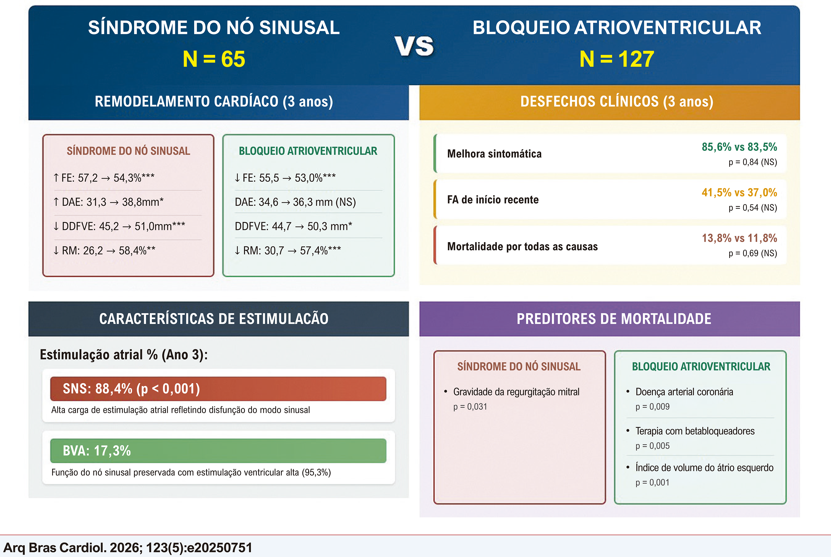 Significância estatística: * p < 0,05; ** p < 0,01; *** p < 0,001; NS indica insignificância (p ≥ 0,05). Todas as comparações baseadas na linha de base vs. medições de acompanhamento de 3 anos. Código de cores: Caixas vermelhas/rosas: Resultados do grupo SNS; Caixas verdes: Resultados do grupo BAV; Seção laranja: Resultados clínicos; Seção roxa: Preditor de morbilidade. AVB: bloqueio atrioventricular; EF: Fração de ejeção; DDFVE: Diâmetro diastólico final do ventrículo esquerdo; RM: Regurgitação mitral; AF: fibrilação atrial; NS: insignificante. Setas: ↓ indica diminuição do valor basal para o acompanhamento de 3 anos; ↑ indica aumento do valor basal para o acompanhamento de 3 anos.
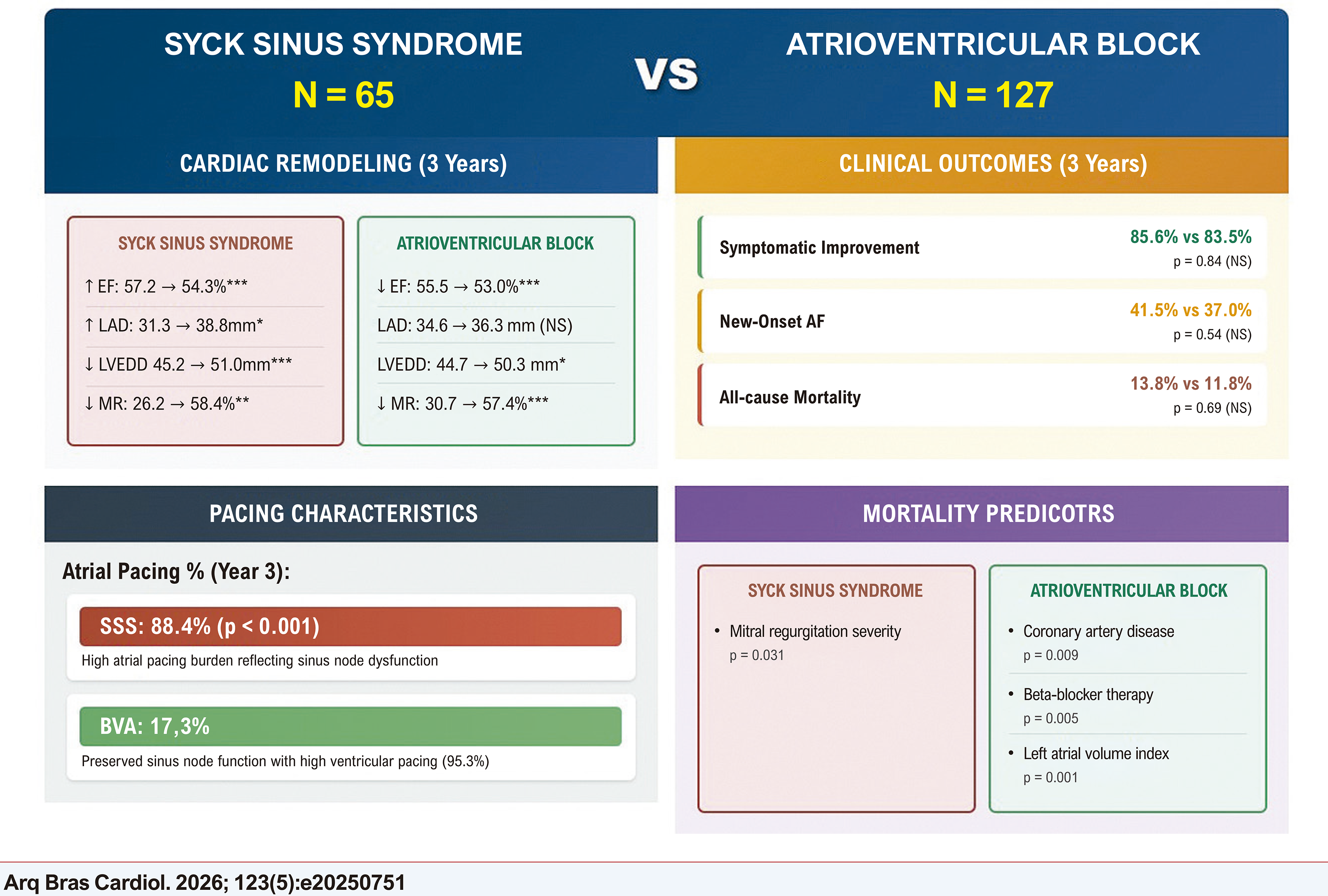 Statistical significance: * p < 0.05; ** p < 0.01; *** p < 0.001; NS indicates not significant (p ≥ 0.05). All comparisons based on baseline vs. 3-year follow-up measurements. Color coding: Red/pink boxes: SSS group findings; Green boxes: AVB group findings; Orange section: Clinical outcomes; Purple section: Mortality predictors. SSS: Sick Sinus Syndrome; AVB: Atrioventricular Block; EF: Ejection Fraction; LAD: Left Atrial Diameter; LVEDD: Left Ventricular End-Diastolic Diameter; MR: Mitral Regurgitation; AF: Atrial Fibrilation; NS: Not Significant. Arrows: ↓ indicates decrease from baseline to 3-year follow-up; ↑ indicates increase from baseline to 3-year follow-up.