Artificial Intelligence-Derived ECG-Age as a Predictor of Mortality and Cardiovascular Events: A Systematic Review and Meta-Analysis
Abstract
Background Artificial intelligence (AI)-derived electrocardiographic age (ECG-age) and the difference between ECG-age and chronological age (delta-age) are emerging biomarkers of cardiovascular aging and adverse outcomes, but their prognostic value remains unclear.
Objectives We performed a systematic review and meta-analysis to evaluate associations between AI-derived ECG-age or delta-age and clinical outcomes.
Methods Nine databases were searched through May 1, 2025. Eligible studies evaluated mortality or cardiovascular events and reported measures of association. Pooled hazard ratios (pHRs) with 95% confidence intervals (CIs) were calculated via fixed- or random-effects models, with statistical significance set at p < 0.05. The protocol was registered in PROSPERO (CRD420251042467).
Results Ten studies (2021–2025) with over 550,000 participants from East Asia, the Americas, and the UK were included. Most used convolutional neural networks to estimate ECG-age; delta-age was calculated as ECG-age minus chronological age. Elevated delta-age was associated with increased all-cause mortality (pHR = 1.83, 95% CI: 1.45–2.32) and cardiovascular mortality (pHR = 2.63, 95% CI: 1.93–3.58). Three studies reported increased risk of atrial fibrillation (pHR = 1.96, 95% CI: 1.43–2.69), although data were limited and heterogeneous. Descriptive analyses suggested that greater delta-age predicts incident heart failure and stroke.
Conclusions AI-derived ECG-age, particularly delta-age, is associated with all-cause and cardiovascular mortality, supporting its role as a noninvasive biomarker of cardiovascular aging. Evidence for atrial fibrillation is suggestive but limited. Standardized algorithms, robust external validation, and prospective multicenter studies are needed to confirm clinical utility and integrate ECG-age into risk stratification, even in asymptomatic individuals.
Keywords:
Electrocardiography; Artificial Intelligence; Deep Learning; Machine Learning; Prognosis

Resumo
Fundamento A idade eletrocardiográfica (idade-ECG) estimada pore inteligência artificial (IA) e a diferença entre a idade-ECG e a idade cronológica (delta-idade) são biomarcadores emergentes do envelhecimento cardiovascular e de desfechos adversos, mas seu valor prognóstico permanece incerto.
Objetivos Realizamos uma revisão sistemática e metanálise para avaliar as associações entre a idade-ECG estimada por IA ou a delta-idade e os desfechos clínicos.
Métodos Nove bases de dados foram pesquisadas até 1º de maio de 2025. Os estudos elegíveis avaliaram mortalidade ou eventos cardiovasculares e relataram medidas de associação. As hazard ratios agrupadas (pHRs) com intervalos de confiança (IC) de 95% foram calculadas por meio de modelos de efeitos fixos ou aleatórios, com significância estatística definida em p < 0,05. O protocolo foi registrado no PROSPERO (CRD420251042467).
Resultados Dez estudos (2021–2025) com mais de 550.000 participantes do Leste Asiático, das Américas e do Reino Unido foram incluídos. A maioria utilizou redes neurais convolucionais para estimar a idade-ECG; a delta-idade foi calculada como a idade-ECG menos idade cronológica. Uma delta-idade elevada foi associada ao aumento da mortalidade por todas as causas (pHR = 1,83, IC 95%: 1,45–2,32) e da mortalidade cardiovascular (pHR = 2,63, IC 95%: 1,93–3,58). Três estudos relataram aumento do risco de fibrilação atrial (pHR = 1,96, IC 95%: 1,43–2,69), embora os dados fossem limitados e heterogêneos. Análises descritivas sugeriram que uma delta-idade maior prediz a incidência de insuficiência cardíaca e acidente vascular cerebral.
Conclusões A idade-ECG estimada por IA, particularmente a delta-idade, está associada à mortalidade por todas as causas e à mortalidade cardiovascular, reforçando seu papel como um biomarcador não invasivo do envelhecimento cardiovascular. As evidências para fibrilação atrial são sugestivas, mas limitadas. Algoritmos padronizados, validação externa robusta e estudos prospectivos multicêntricos são necessários para confirmar a utilidade clínica e integrar a idade-ECG à estratificação de risco, mesmo em indivíduos assintomáticos.
Palavras-chave:
Eletrocardiografia; Inteligência Artificial; Aprendizagem Profunda; Aprendizado de Máquina; Prognóstico

Introduction
The electrocardiogram (ECG) is a vital diagnostic tool across diverse clinical scenarios.1,2 It is well established that the electrical signals of the heart are modulated by numerous physiological factors related to both the cardiovascular system and broader biological mechanisms.3-5 As such, not only can cardiovascular diseases significantly influence ECG signals, but also lifestyle,6 dietary patterns,7 and chronic diseases.8 Its application extends beyond the identification of cardiac disorders, and additional latent information based on ECG data can be harnessed to provide a broader assessment of cardiovascular health and aging.9,10
The integration of computer-assisted interpretation into clinical ECGs has gained prominence,11 and ECG-age, an age estimate derived from 12-lead ECGs using artificial intelligence (AI), has emerged as a potential biomarker of cardiovascular aging and a measurable indicator of global cardiovascular health.12-15Previous studies have shown a strong correlation between ECG-age and chronological age in healthy individuals, while an elevated ECG-age relative to chronological age has been associated with higher cardiovascular risk.16 This discrepancy is thought to reflect accelerated cardiovascular or biological aging, resulting from the cumulative impact of comorbidities, subclinical disease, and lifestyle factors. This suggests that ECG-age could serve as an integrative marker of subclinical disease burden and overall cardiovascular health.
The difference between ECG-age and chronological age, known as delta-age, has been proposed as a predictor of adverse outcomes,16 including mortality and major adverse cardiovascular events (MACEs). However, its clinical impact has not yet been fully established.
A recent systematic review17 evaluated seventeen original studies that employed AI-based algorithms to assess delta-age. The analysis revealed that hypertension and diabetes mellitus were the most prevalent factors contributing to elevated delta-age, and myocardial infarction and heart failure (HF) had the most significant impacts. Although a significant association was observed between increased delta-age and both all-cause mortality and cardiovascular mortality, the meta-analysis was limited to six and three studies for these respective outcomes.
Therefore, the primary objective of this review was to assess the prognostic value of AI-derived ECG-age and delta-age in predicting all-cause and cardiovascular mortality. Secondary objectives included evaluating their associations with other adverse cardiovascular outcomes, such as atrial fibrillation, heart failure, and stroke, incorporating updated data and analyses from recent studies. The main findings and clinical implications of this review are summarized in the Central Illustration.
Methods
Search strategy and selection criteria
This review followed the Preferred Reporting Items for Systematic Reviews and Meta-Analyses (PRISMA) guidelines. The study protocol was registered in PROSPERO (CRD420251042467).
Nine databases (LILACS, Scielo, MEDLINE, ScienceDirect, EMBASE, CENTRAL, CINAHL, Web of Science, and Scopus) were systematically searched from inception to May 1, 2025, for primary development or validation studies of predictive models based on AI-determined ECG-age that assessed the associations between ECG-age or delta-age and clinical outcomes. The search strategy included the following MeSH terms: "electrocardiographic age" OR "AI ECG-heart age" OR "AI ECG age" OR "ECG-age". Studies were eligible if they reported model performance metrics (e.g., AUC, sensitivity, specificity, accuracy, and calibration) or measures of association (e.g., hazard ratio [HR], odds ratio [OR], and risk ratio [RR]). The primary outcomes were all-cause mortality, cardiovascular mortality, and adverse cardiovascular events, including myocardial infarction, atherosclerotic cardiovascular disease, stroke, heart failure, and atrial fibrillation.
The follow-up periods varied across studies, with a predominance of retrospective cohort designs. Systematic reviews, meta-analyses, editorials, letters, opinion pieces, conference abstracts without complete data, and purely methodological studies without application to clinical outcomes were excluded.
The retrieved articles were uploaded into Rayyan,18 and duplicates were removed. Two reviewers independently screened the articles and extracted the data. Any discrepancies were resolved through discussion with a third reviewer.
Data collection process and data items
Data from all studies meeting the inclusion criteria were extracted into a table in accordance with the CHARMS checklist. The extracted data included the study design, study period, total study population, prognostic analysis population, population characteristics, inclusion and exclusion criteria, data source, type of ECG used, and kind of IA algorithm. Additional variables collected included the correlation between ECG-age and chronological age, data split method, validation type, delta-age cutoff, outcomes assessed, number of events per group, HR of events per group, covariates adjusted for, and risk of bias assessment. For studies that reported multiple HRs with different covariate adjustments, we extracted the most fully adjusted HR.
Quality assessment
We assessed the risk of bias in the included studies via the Newcastle-Ottawa Scale (NOS) for cohort studies. This scale comprises three domains–Selection, Comparability, and Outcome–each containing specific items with multiple answer options, in which those reflecting higher methodological quality are awarded stars. Because several studies have conducted various analyses in different populations, each population was assessed separately. A study was classified as “good” if it received three or four stars in the selection domain, one or two stars in the comparability domain, and two or three stars in the Outcome domain. A “fair” classification requires the same number of stars in the comparability and outcome domains but only two stars in the selection domain.
Data synthesis and meta-analysis
The pooled hazard ratio (HR) with its 95% confidence interval (CI) was calculated to assess the associations between elevated delta-age and mortality outcomes, as well as atrial fibrillation. An HR greater than 1 was interpreted as a significantly greater risk among patients with higher delta-age, assuming that the 95% CI did not cross 1. For all analyses, statistical significance was set at p < 0.05.
To quantify heterogeneity, we calculated Cochran’s Q, the I2 statistic, and the τ2 statistic. A random-effects meta-analysis with Hartung–Knapp adjustment was conducted in the analysis of all-cause mortality and atrial fibrillation, due to substantial heterogeneity among studies (I2 > 25%). However, the fixed-effects model was applied in the cardiovascular mortality analysis because no significant heterogeneity was detected (I2 = 0.0%). Publication bias was assessed using funnel plot inspection and Begg’s test. Additionally, the trim-and-fill method was used to address potentially missing studies. Owing to the limited number of included studies (k = 3), Begg’s test could not be used to assess atrial fibrillation and cardiovascular mortality.
All the statistical analyses were performed using R software, version 4.5.0 (R Foundation for Statistical Computing, Vienna, Austria).
Results
Study selection
The study selection process is shown in the PRISMA flow diagram (Figure 1). We identified 1249 records across nine databases and 486 duplicates. After removing duplicates, 763 articles were screened for inclusion. Of these, 752 were excluded based on title or abstract, primarily because they did not assess ECG-age. The remaining 11 articles underwent full-text analysis. One was excluded from the study design. Ten studies13,15,19-26 were included in the qualitative and quantitative analyses.

– PRISMA flow diagram. Study selection process, illustrating the identification, screening, and inclusion of studies in the systematic review.
Characteristics of included studies
All included studies were published between 2021 and 2025 and were based on ECG recordings from retrospective cohorts. Some were conducted in East Asian populations,13,19-22 whereas others included data from Brazil,16 the United States,23,24 and the United Kingdom.25 The number of participants included in the prognostic analysis varied substantially across studies, ranging from approximately 2,00026 individuals to more than 200,000.16 Collectively, the ten studies encompassed a total prognostic analysis population of nearly 558,000 participants, which was reduced to 336,026 after accounting for overlapping datasets used in more than one study. The participants were predominantly adults, with ages ranging from 18 years to over 68 years. Most studies analyzed general population cohorts or individuals undergoing cardiovascular risk screening. Tables 1 and 2 summarize the study characteristics.
Most studies have used deep neural networks (DNNs) trained on ECG signals to predict ECG-age, with the difference between the AI-estimated age and the actual chronological age–delta-age–subsequently calculated and interpreted as accelerated cardiovascular aging. Delta-age was analyzed both as a continuous variable and in categorical form, most frequently defined as being more than eight years above chronological age. While most analyses have been based on ECG signals,13,16,20,21,23-26 one study used 12-lead ECG images as input,22 and another employed XML data extracted from 12-lead ECGs.19 Many of these analyses utilized Attia’s12 CNN23,25 or Ribeiro’s27 AI model,16,20,24,26 and the majority of investigations relied on large, well-established cohorts that were used to train those CNNs, particularly the CODE dataset in Brazil and the Mayo Clinic dataset in the United States, although some developed and validated models using independent national datasets.13,19,21,22 The validation procedures also varied, ranging from internal validation within the same dataset to analyses in independent cohorts, although external validation was not always performed with fully distinct populations.
The risk of bias assessment was performed via the Newcastle-Ottawa Scale (NOS) tool, which awards stars on the basis of predefined criteria, resulting in a total score ranging from 0 to 9. Studies with higher methodological quality receive more stars. In our assessment, the mean score was 8.5 stars. Most studies were classified as “good.” Only one study had a single analysis rated as “fair.” The detailed results of the risk of bias assessment are presented in Table 3.
All-cause mortality
Seven studies13,15,19,21,23,24,26 reported the HR for higher delta-age for all-cause mortality, with eleven analyses considering that some studies evaluated more than one population subgroup.
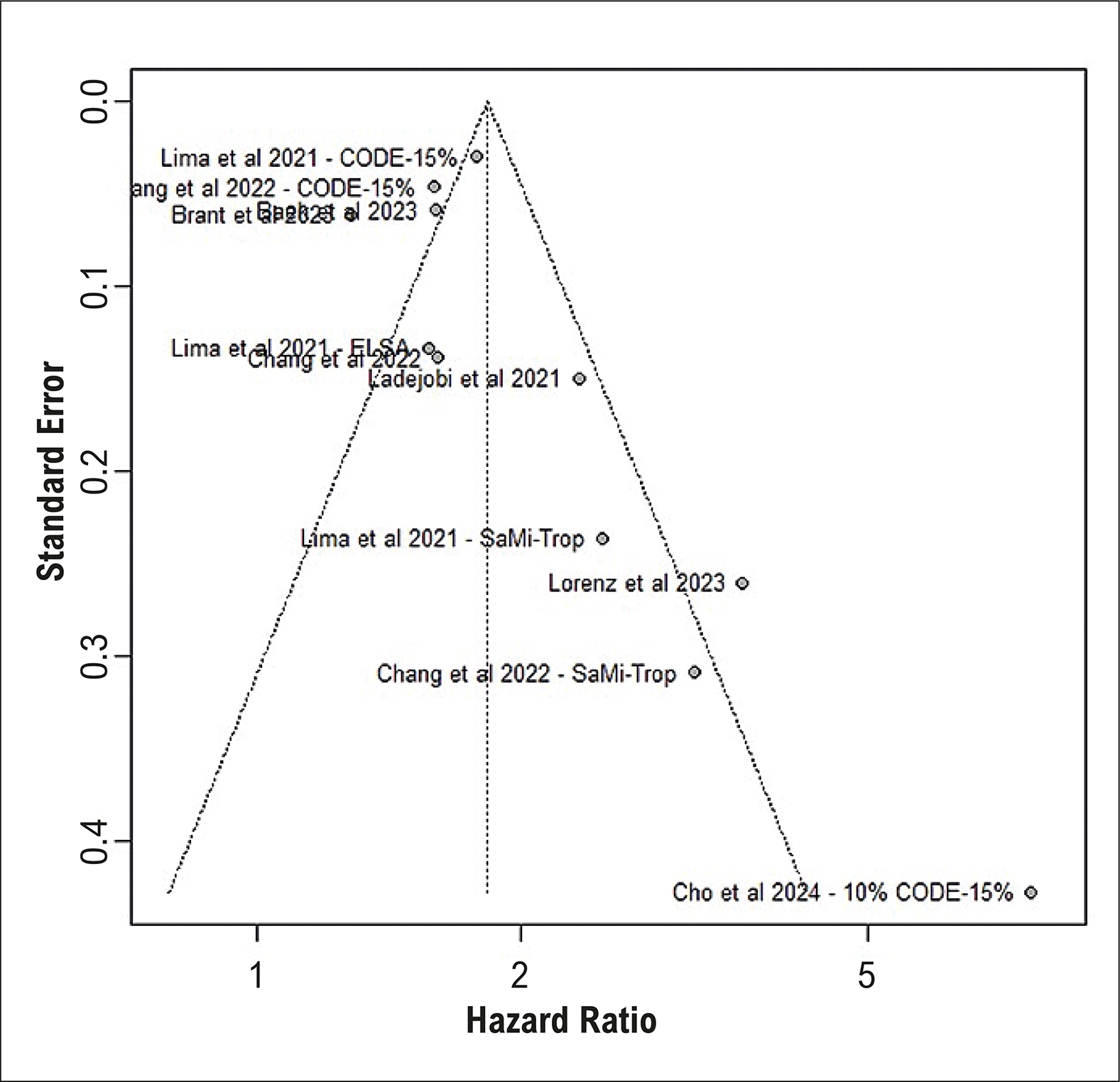
The pooled estimate for all-cause mortality was HR = 1.83 (95% CI: 1.45-2.32), indicating that a significantly increased risk was associated with high delta-age (Figure 2). The included studies were significantly heterogeneous, with I2 = 82,9%. We assessed for potential publication bias using a funnel plot and statistical tests. The visual inspection of the funnel plot suggested asymmetry, indicating a potential small-study effect (Figure 3). Statistical analysis further supported this observation, as Begg's rank correlation test indicated significant evidence of publication bias (p = 0.0290). The trim-and-fill method imputed three missing studies on the right side of the funnel plot. After imputing the three missing studies, the adjusted pooled HR was 1.678 (95% CI: 1.26-2.23). While this adjusted effect size was slightly smaller than the original, the finding remained statistically significant. This suggests that while publication bias may have led to an overestimation of the effect, the analysis still revealed a significant positive association.

– Pooled hazard ratios (HRs) and 95% confidence intervals (CIs) for all-cause mortality in higher delta-age groups.

Cardiovascular mortality
Three studies13,18,23 reported the hazard ratio (HR) for the association between higher delta-age and cardiovascular mortality. The pooled estimate was HR = 2.63 (95% CI: 1.93-3.5), indicating that a significantly increased risk was associated with increased delta-age (Figure 4). No heterogeneity was identified among these studies (I2=0.0%). Owing to the low number of studies (k = 3), Begg's test was not performed. A trim and fill analysis was conducted to address potential publication bias, which imputed two studies. After the inclusion of these studies, the pooled HR decreased to 2.2 (95% CI: 1.67-2.89), but the results remained statistically significant, suggesting that the overall positive association is robust.

– Pooled hazard ratios (HRs) and 95% confidence intervals (CIs) for cardiovascular mortality in higher delta-age groups.
Atrial fibrillation
Similarly, three studies13,20,23 were included in the meta-analysis that reported the pooled hazard ratio (HR) for the association between greater delta-age and atrial fibrillation. The pooled estimate was HR = 1.96 (95% CI: 1.43-2.69), indicating that a significantly increased risk was associated with greater delta-age (Figure 5). However, this analysis revealed high heterogeneity (I2 = 91.1%). Due to the small number of studies (k=3), Begg’s test was not performed to assess publication bias. A subsequent trim-and-fill analysis showed that no studies were missing, confirming that all eligible studies were included in the meta-analysis. Notably, when the Hartung–Knapp adjustment was applied, the confidence interval widened and included the null (HR = 1.96, 95% CI: 0.99–3.88), reflecting the uncertainty related to the limited number of studies.

– Pooled hazard ratios (HRs) and 95% confidence intervals (CIs) for new-onset atrial fibrillation in higher delta-age groups.
Heart failure
Greater delta-age has also been associated with increased risk of HF. Brant et al.20 found that participants whose ECG-age exceeded their chronological age by 9 years had a 75% higher risk of developing HF (HR 1.75; 95% CI: 1.45–2.12). Chang et al.13 reported an even stronger association with newly onset HF (HR 2.79; 95% CI: 2.25–3.45). These studies consistently demonstrate that elevated delta-age is associated with a higher risk of incident HF.
Stroke
Two studies13,24 analyzed the predictive value of delta-age for stroke. Chang et al.13 found that individuals with accelerated ECG-age had a significantly increased risk of stroke (HR 1.65; 95% CI: 1.42–1.92). This finding was corroborated by Leung et al.,24 who reported a 42% higher risk of incident stroke among participants with greater delta-age (HR 1.42; 95% CI: 1.12–1.80). These studies demonstrate that higher delta-age is consistently associated with increased risk of stroke across different cohorts.
Discussion
Our systematic review and meta-analysis revealed a strong correlation between ECG-derived age (ECG-age) and the risk of clinically significant outcomes, including all-cause mortality, cardiovascular mortality, and atrial fibrillation. Specifically, a greater delta-age (the difference between a patient’s ECG-age and his chronological age) was consistently associated with a higher risk of these outcomes.
According to the studies included, increased ECG-age was generally defined as a predicted age exceeding the model’s mean absolute error (MAE), which typically ranged between 6 and 9 years (Table 1). This threshold represented the delta-age above which adverse outcomes were more frequently observed, reinforcing the potential of ECG-age as an indicator of accelerated cardiovascular aging. However, no specific or standardized delta-age cutoff has been established for each outcome, as studies adopted different definitions and reporting strategies.
With respect to all-cause mortality, all eleven analyses reviewed revealed a positive correlation between greater delta-age and increased risk. The pooled estimates indicated a significantly higher risk among individuals with elevated delta-age. The increased risk ranged widely from 28% to 600%, most likely reflecting variations in the subgroups analyzed. For example, Cho et al.21 reported a particularly high hazard ratio, possibly because they analyzed only a small subgroup from the CODE study. The significant heterogeneity observed among studies can be attributed to several factors. First, the study populations varied significantly in terms of race and ethnicity, which influences ECGs. Second, the presence of various comorbidities, such as Chagas cardiomyopathy, in the patients from the SaMi-Trop study likely played a role. Finally, the use of different machine learning algorithms, each with unique training populations and specific settings, may have also contributed to this variation.
Our statistical analyses revealed evidence of publication bias. This finding may reflect the small-study effect, where smaller studies tend to show larger effect sizes. After performing a trim-and-fill analysis to account for potential bias, the adjusted pooled effect remained statistically significant. Thus, while publication bias may have slightly inflated the effect size, the overall conclusion remains unchanged: the analysis is robust and suggests a clear increase in the risk of all-cause mortality in patients with a higher ECG-age.
For cardiovascular mortality, the meta-analysis demonstrated a robust and statistically significant effect. Our analyses also revealed a strong positive correlation, with the increased risk for this event ranging from 120% to 249%. Unlike the all-cause mortality analysis, there was no heterogeneity among the included studies. The trim-and-fill sensitivity analysis slightly attenuated the pooled estimate but did not alter the significance of the results, confirming the robustness of the association between higher delta-age and cardiovascular mortality risk.
For atrial fibrillation, the meta-analysis using the conventional random-effects model showed that individuals with a greater delta-age had almost twice the risk of atrial fibrillation. However, estimates varied depending on the statistical method applied, reflecting the inherent uncertainty associated with the small number of studies. High heterogeneity was observed, indicating that the effect of ECG-age on atrial fibrillation risk varies across studies. The disparity in effect sizes is likely explained by differences in study populations: one study23 analyzed 9,877 participants from the Framingham Heart Study, another13 included 30,469 Taiwanese adults aged 20–80 years without pre-existing cardiovascular disease, and the largest study20 analyzed over 121,000 South Korean patients aged 20–90 years. These differences in patient demographics, baseline health, and geographic location likely contributed to the high heterogeneity observed. These findings suggest a potential relationship between accelerated ECG-age and AF risk, possibly mediated by subclinical atrial remodeling, age-related fibrosis, or cumulative cardiovascular risk burden. However, the evidence is currently insufficient to draw definitive conclusions, and these findings should be interpreted with caution. Additional large, prospectively designed studies with diverse populations are needed to clarify the prognostic value of ECG-age for AF.
The association between delta-age and heart failure suggests ECG-age may reflect early subclinical cardiac remodeling or functional impairment not captured by traditional risk factors. Although only a few studies analyzed this outcome13,20 and a formal meta-analysis was not possible, consistent results across cohorts suggest that ECG-age may be a promising non-invasive marker of HF risk. However, these results warrant cautious interpretation due to the small number of studies available.
Similarly, the association between accelerated ECG-age and incident stroke highlights its potential as a marker of subclinical vascular aging. Given the small number of studies.13,24 this synthesis was descriptive rather than quantitative. The observed trends suggest that ECG-age captures pathophysiological processes that predispose to cerebrovascular events independently of traditional risk factors. Because this outcome was evaluated in only a few studies, the observed association should be viewed as preliminary and confirmed in future research.
Various comorbidities and lifestyle habits can compromise physiological function, leading to a disparity between a person's chronological and biological age. Recent research7-9 has consistently shown that a wide range of lifestyle factors and diseases can cause abnormalities on an ECG. For example, conditions such as hypertension, type 2 diabetes, obesity, smoking, and sedentarism are known to affect ECG measurements. Given this, ECG-age is a simple, noninvasive, and cost-effective tool for cardiovascular risk stratification, particularly in asymptomatic individuals, enabling more precise preventive and prognostic decisions. While several studies have concluded that a greater delta-age is associated with atherosclerotic cardiovascular disease, abnormal peripheral endothelial dysfunction, and increased mortality, our systematic review and meta-analysis highlight the predictive power of these data. Our findings revealed that an increased delta-ECG is consistently associated with higher mortality and cardiovascular risk. A promising application of this tool would be the stratification of apparently healthy or asymptomatic patients, identifying those at greater risk for these events. This concept is further supported by studies such as the one by Mossavarali et al.17 which demonstrated that chronic diseases, genetic mutations, and structural cardiovascular diseases are associated with an increased ECG-age. This suggests that ECG-age can become a valuable tool for providing a more precise assessment of overall cardiovascular health, even in patients who already have a high cardiovascular risk.
When interpreting these findings, some methodological considerations are important. The repeated use of the same large datasets, often with different algorithms applied to the same population, may limit the independence of the results and the incremental value of each new approach. Furthermore, external validation was not uniformly rigorous, as several studies relied on cohorts from the same institution or with overlapping datasets, while truly independent validation across distinct populations and healthcare systems was less frequent. External validation was performed in five studies,13,16,21,22,26 using datasets from diverse cohorts, such as community-based populations (CODE, Brazil), clinical populations with cardiovascular disease (SaMi-Trop, Brazil), health check-up participants (Severance Health Check-up, South Korea), and large biobanks (UK Biobank, USA/UK). Given that ECG characteristics may vary according to ethnicity, age distribution, comorbidities, and recording standards, the lack of consistent, rigorous external validation raises concerns about the generalizability of ECG-age. To ensure reproducibility and mitigate potential biases, future investigations should prioritize validation in diverse, multicenter, and international cohorts, ideally including both community-based populations and high-risk clinical groups, with transparent reporting of data preprocessing, model calibration, and performance metrics. Such efforts are essential to establish ECG-age as a reliable biomarker across varied clinical and demographic contexts.
In addition to these methodological limitations, another important aspect relates to explainability and interpretability. While most of the included studies did not provide in-depth analyses in this regard, Cho et al. in 202521applied saliency maps to their AI-based ECG-age model, segmenting the ECG into PQ, QRS, ST, and TP intervals. Their results showed that the PQ segment consistently presented the highest saliency values across five independent validation datasets. This finding indicates that the model focused on physiologically meaningful regions of the ECG rather than random artifacts. By identifying which waveform components most strongly drive age prediction, such analyses enhance transparency and help bridge the gap between AI modeling and clinical interpretation. However, beyond this isolated effort, explainability remains largely underexplored in the current literature, and future studies should systematically incorporate and standardize interpretability frameworks in ECG-age research.
Future research should focus on prospective, longitudinal studies to establish the real-world clinical impact of ECG-age. Such trials are essential to determine whether ECG-age provides incremental prognostic value over established risk scores, improves risk stratification, or guides preventive interventions in daily practice. Prospective multicenter studies embedded within healthcare systems will be key to evaluating whether the use of ECG-age can lead to meaningful improvements in patient outcomes. These efforts are crucial to move from proof-of-concept to clinical implementation, paving the way for AI-derived ECG biomarkers to be incorporated into routine cardiovascular risk assessment and management.
Conclusions
This systematic review and meta-analysis demonstrated that AI-derived ECG-age, particularly greater delta-age, is strongly associated with both all-cause and cardiovascular mortality, supporting its role as a biomarker of cardiovascular aging. Evidence for atrial fibrillation is suggestive but inconclusive, highlighting the need for further studies. These findings reinforce the potential of ECG-age as a noninvasive, simple, and cost-effective biomarker to enhance cardiovascular risk stratification, even among asymptomatic individuals. To strengthen clinical utility and guide implementation in cardiovascular risk stratification, standardization of algorithms, robust external validation, and prospective multicenter studies are needed, as are assessments of the incremental value of this approach in combination with traditional risk scores. In parallel, improving model explainability will be essential to ensure transparency and foster clinical trust in AI-based ECG biomarkers.
References
-
1 Kligfield P, Gettes LS, Bailey JJ, Childers R, Deal BJ, Hancock EW, et al. Recommendations for the Standardization and Interpretation of the Electrocardiogram: Part I: The Electrocardiogram and its Technology: A Scientific Statement from the American Heart Association Electrocardiography and Arrhythmias Committee, Council on Clinical Cardiology; the American College of Cardiology Foundation; and the Heart Rhythm Society: Endorsed by the International Society for Computerized Electrocardiology. Circulation. 2007;115(10):1306-24. doi: 10.1161/CIRCULATIONAHA.106.180200.
» https://doi.org/10.1161/CIRCULATIONAHA.106.180200 -
2 Rautaharju PM, Surawicz B, Gettes LS, Bailey JJ, Childers R, Deal BJ, et al. AHA/ACCF/HRS Recommendations for the Standardization and Interpretation of the Electrocardiogram: Part IV: The ST Segment, T and U Waves, and the QT Interval: A Scientific Statement from the American Heart Association Electrocardiography and Arrhythmias Committee, Council on Clinical Cardiology; the American College of Cardiology Foundation; and the Heart Rhythm Society. Endorsed by the International Society for Computerized Electrocardiology. J Am Coll Cardiol. 2009;53(11):982-91. doi: 10.1016/j.jacc.2008.12.014.
» https://doi.org/10.1016/j.jacc.2008.12.014 -
3 Bachman S, Sparrow D, Smith LK. Effect of Aging on the Electrocardiogram. Am J Cardiol. 1981;48(3):513-6. doi: 10.1016/0002-9149(81)90081-3.
» https://doi.org/10.1016/0002-9149(81)90081-3 -
4 Liao YL, Liu KA, Dyer A, Schoenberger JA, Shekelle RB, Colette P, et al. Major and Minor Electrocardiographic Abnormalities and Risk of Death from Coronary Heart Disease, Cardiovascular Diseases and All Causes in Men and Women. J Am Coll Cardiol. 1988;12(6):1494-500. doi: 10.1016/s0735-1097(88)80016-0.
» https://doi.org/10.1016/s0735-1097(88)80016-0 -
5 Tsuji H, Larson MG, Venditti FJ Jr, Manders ES, Evans JC, Feldman CL, et al. Impact of Reduced Heart Rate Variability on Risk for Cardiac Events. The Framingham Heart Study. Circulation. 1996;94(11):2850-5. doi: 10.1161/01.cir.94.11.2850.
» https://doi.org/10.1161/01.cir.94.11.2850 -
6 Kang M, Shin S, Zhang G, Jung J, Kim YT. Mental Stress Classification Based on a Support Vector Machine and Naive Bayes Using Electrocardiogram Signals. Sensors. 2021;21(23):7916. doi: 10.3390/s21237916.
» https://doi.org/10.3390/s21237916 -
7 Widerlöv E, Jostell KG, Claesson L, Odlind B, Keisu M, Freyschuss U. Influence of Food Intake on Electrocardiograms of Healthy Male Volunteers. Eur J Clin Pharmacol. 1999;55(9):619-24. doi: 10.1007/s002280050682.
» https://doi.org/10.1007/s002280050682 -
8 Sahranavard T, Alimi R, Arabkhazaei J, Nasrabadi M, Dana SMMA, Gholami Y, et al. Association of Major and Minor ECG Abnormalities with Traditional Cardiovascular Risk Factors in the General Population: A Large Scale Study. Sci Rep. 2024;14(1):11289. doi: 10.1038/s41598-024-62142-8.
» https://doi.org/10.1038/s41598-024-62142-8 -
9 Ansari MY, Qaraqe M, Charafeddine F, Serpedin E, Righetti R, Qaraqe K. Estimating Age and Gender from Electrocardiogram Signals: A Comprehensive Review of the Past Decade. Artif Intell Med. 2023;146:102690. doi: 10.1016/j.artmed.2023.102690.
» https://doi.org/10.1016/j.artmed.2023.102690 -
10 Moraes DN, Nascimento BR, Pires MC, Paixão GMM, MacFarlane PW, Ribeiro ALP. Prognostic Value of Resting Heart Rate and Heart Rate Variability in the 12-lead Electrocardiogram: Mortality Data from the CODE Database. Am J Cardiol. 2025;248:23-31. doi: 10.1016/j.amjcard.2025.03.038.
» https://doi.org/10.1016/j.amjcard.2025.03.038 -
11 Paixão GMM, Santos BC, Araujo RM, Ribeiro MH, Moraes JL, Ribeiro AL. Machine Learning in Medicine: Review and Applicability. Arq Bras Cardiol. 2022;118(1):95-102. doi: 10.36660/abc.20200596.
» https://doi.org/10.36660/abc.20200596 -
12 Attia ZI, Friedman PA, Noseworthy PA, Lopez-Jimenez F, Ladewig DJ, Satam G, et al. Age and Sex Estimation Using Artificial Intelligence from Standard 12-Lead ECGs. Circ Arrhythm Electrophysiol. 2019;12(9):e007284. doi: 10.1161/CIRCEP.119.007284.
» https://doi.org/10.1161/CIRCEP.119.007284 -
13 Chang CH, Lin CS, Luo YS, Lee YT, Lin C. Electrocardiogram-Based Heart Age Estimation by a Deep Learning Model Provides More Information on the Incidence of Cardiovascular Disorders. Front Cardiovasc Med. 2022;9:754909. doi: 10.3389/fcvm.2022.754909.
» https://doi.org/10.3389/fcvm.2022.754909 -
14 Liu CM, Kuo MJ, Kuo CY, Wu IC, Chen PF, Hsu WT, et al. Reclassification of the Conventional Risk Assessment for Aging-Related Diseases by Electrocardiogram-Enabled Biological Age. NPJ Aging. 2025;11(1):7. doi: 10.1038/s41514-025-00198-0.
» https://doi.org/10.1038/s41514-025-00198-0 -
15 Medina-Inojosa J, Ladejobi A, Attia Z, Shelly-Cohen M, Gersh B, Noseworthy P, et al. The Association of Artificial Intelligence-Enabled Electrocardiogram-Derived Age (Physiologic Age) with Atherosclerotic Cardiovascular Events in the Community. Eur Heart J. 2020 Nov;41(Suppl 2):ehaa946.2905. doi:10.1093/ehjci/ehaa946.2905.
» https://doi.org/10.1093/ehjci/ehaa946.2905 -
16 Lima EM, Ribeiro AH, Paixão GMM, Ribeiro MH, Pinto-Filho MM, Gomes PR, et al. Deep Neural Network-Estimated Electrocardiographic Age as a Mortality Predictor. Nat Commun. 2021;12(1):5117. doi: 10.1038/s41467-021-25351-7.
» https://doi.org/10.1038/s41467-021-25351-7 -
17 Mossavarali S, Vaezi A, Gholami Z, Molaei A, Yekaninejad MS, Asselbergs FW, et al. Determinants of Artificial Intelligence Electrocardiogram-Derived Age and Its Association with Cardiovascular Events and Mortality: A Systematic Review and Meta-Analysis. NPJ Digit Med. 2025;8(1):322. doi: 10.1038/s41746-025-01727-7.
» https://doi.org/10.1038/s41746-025-01727-7 -
18 Ouzzani M, Hammady H, Fedorowicz Z, Elmagarmid A. Rayyan-a Web and Mobile App for Systematic Reviews. Syst Rev. 2016;5(1):210. doi: 10.1186/s13643-016-0384-4.
» https://doi.org/10.1186/s13643-016-0384-4 -
19 Baek YS, Lee DH, Jo Y, Lee SC, Choi W, Kim DH. Artificial Intelligence-Estimated Biological Heart Age Using a 12-Lead Electrocardiogram Predicts Mortality and Cardiovascular Outcomes. Front Cardiovasc Med. 2023;10:1137892. doi: 10.3389/fcvm.2023.1137892.
» https://doi.org/10.3389/fcvm.2023.1137892 -
20 Brant LCC, Ribeiro AH, Pinto-Filho MM, Kornej J, Preis SR, Fetterman JL, et al. Association between Electrocardiographic Age and Cardiovascular Events in Community Settings: The Framingham Heart Study. Circ Cardiovasc Qual Outcomes. 2023;16(7):e009821. doi: 10.1161/CIRCOUTCOMES.122.009821.
» https://doi.org/10.1161/CIRCOUTCOMES.122.009821 -
21 Cho S, Eom S, Kim D, Kim TH, Uhm JS, Pak HN, et al. Artificial Intelligence-Derived Electrocardiographic Aging and Risk of Atrial Fibrillation: A Multi-National Study. Eur Heart J. 2025;46(9):839-52. doi: 10.1093/eurheartj/ehae790.
» https://doi.org/10.1093/eurheartj/ehae790 -
22 Cho Y, Kim JS, Kim J, Yoon YE, Jung SY. Image-Based ECG Analyzing Deep-Learning Algorithm to Predict Biological Age and Mortality Risks: Interethnic Validation. J Cardiovasc Med. 2024;25(11):781-8. doi: 10.2459/JCM.0000000000001670.
» https://doi.org/10.2459/JCM.0000000000001670 -
23 Ladejobi AO, Medina-Inojosa JR, Cohen MS, Attia ZI, Scott CG, LeBrasseur NK, et al. The 12-Lead Electrocardiogram as a Biomarker of Biological Age. Eur Heart J Digit Health. 2021;2(3):379-89. doi: 10.1093/ehjdh/ztab043.
» https://doi.org/10.1093/ehjdh/ztab043 -
24 Leung R, Wang B, Gottbrecht M, Doerr A, Marya N, Soni A, et al. Association between Deep Neural Network-Derived Electrocardiographic-Age and Incident Stroke. Front Cardiovasc Med. 2024;11:1368094. doi: 10.3389/fcvm.2024.1368094.
» https://doi.org/10.3389/fcvm.2024.1368094 -
25 Lorenz EC, Zaniletti I, Johnson BK, Petterson TM, Kremers WK, Schinstock CA, et al. Physiological Age by Artificial Intelligence-Enhanced Electrocardiograms as a Novel Risk Factor of Mortality in Kidney Transplant Candidates. Transplantation. 2023;107(6):1365-72. doi: 10.1097/TP.0000000000004504.
» https://doi.org/10.1097/TP.0000000000004504 -
26 Park H, Kwon OS, Shim J, Kim D, Park JW, Kim YG, et al. Artificial Intelligence Estimated Electrocardiographic Age as a Recurrence Predictor after Atrial Fibrillation Catheter Ablation. NPJ Digit Med. 2024;7(1):234. doi: 10.1038/s41746-024-01234-1.
» https://doi.org/10.1038/s41746-024-01234-1 -
27 Ribeiro AH, Ribeiro MH, Paixão GMM, Oliveira DM, Gomes PR, Canazart JA, et al. Automatic Diagnosis of the 12-Lead ECG Using a Deep Neural Network. Nat Commun. 2020;11(1):1760. doi: 10.1038/s41467-020-15432-4.
» https://doi.org/10.1038/s41467-020-15432-4
-
Study Association:
This article is part of the thesis of master submitted by Isadora Cristine Reis Sguizzato Bozzi, from Universidade Federal de Minas Gerais.
-
Ethics Approval and Consent to Participate:
This article does not contain any studies with human participants or animals performed by any of the authors.
-
Use of Artificial Intelligence:
During the preparation of this work, the author(s) used Grammarly for to assist with English language revision and improve the clarity and fluency of the manuscript. After using this tool/service, the author(s) reviewed and edited the content as needed and take full responsibility for the content of the published article.
-
Availability of Research Data:
The underlying content of the research text is contained within the manuscript.
-
Sources of Funding:
There were no external funding sources for this study.
Edited by
-
Editor responsible for the review:
Marcio Bittencourt
The underlying content of the research text is contained within the manuscript.