Nanoemulsions with cannabidiol reduced autistic-like behaviors and reversed decreased hippocampus viable cells and cerebral cortex neuronal death in a prenatal valproic acid rat model
Abstract
The highly lipophilic nature and low aqueous solubility of cannabidiol (CBD) limit its oral bioavailability, resulting in poor intestinal absorption. To overcome these limitations, we proposed the production of a nanoemulsion with CBD to be included in the therapeutic treatment of autism spectrum disorder. The current study aimed to evaluate the effect of CBD-rich corn oil nanoemulsion treatment in male rats born to females exposed to valproic acid (VPA) during pregnancy on autistic-like behaviors and hippocampal histology. Offspring rats were treated orally twice daily with CBD nanoemulsions at different doses (1 and 2 mg/animal). The endpoints evaluated were anxiety, grooming time, exploratory activity, sociability, the social preference index, and hippocampal and cerebral cortex histology. All formulations were characterized as nanoemulsions and showed a reduced vesicle size (107.6 - 72.6 nm), low PDI (0.290-0.432), negative zeta potential (-40.6 mv), and good stability. Prenatal exposure to VPA increased anxiety and grooming time, and reduced exploratory activity, sociability, and the social preference index in the animals. Furthermore, VPA-exposed animals exhibited elevated neuronal death and a reduction in viable cells in the hippocampus. In conclusion, CBD nanoemulsion treatment reversed autistic-like behaviors, potentially by protecting against hippocampal neuronal death.
The highly lipophilic nature and low aqueous solubility of cannabidiol (CBD) limit its oral bioavailability, resulting in poor intestinal absorption. To overcome these limitations, we proposed the production of a nanoemulsion with CBD to be included in the therapeutic treatment of autism spectrum disorder. The current study aimed to evaluate the effect of CBD-rich corn oil nanoemulsion treatment in male rats born to females exposed to valproic acid (VPA) during pregnancy on autistic-like behaviors and hippocampal histology. Offspring rats were treated orally twice daily with CBD nanoemulsions at different doses (1 and 2 mg/animal). The endpoints evaluated were anxiety, grooming time, exploratory activity, sociability, the social preference index, and hippocampal and cerebral cortex histology. All formulations were characterized as nanoemulsions and showed a reduced vesicle size (107.6 - 72.6 nm), low PDI (0.290-0.432), negative zeta potential (-40.6 mv), and good stability. Prenatal exposure to VPA increased anxiety and grooming time, and reduced exploratory activity, sociability, and the social preference index in the animals. Furthermore, VPA-exposed animals exhibited elevated neuronal death and a reduction in viable cells in the hippocampus. In conclusion, CBD nanoemulsion treatment reversed autistic-like behaviors, potentially by protecting against hippocampal neuronal death.
Key words
Nanotechnology; bioavailability; cannabinoids; autism; treatment
INTRODUCTION
Autism spectrum disorder (ASD) is a heterogeneous neurodevelopmental disorder characterized by communication and social interaction alterations associated with restricted and repetitive behavior (Association 2013). Comorbidities like seizures, self-injury, aggressive behavior, depression, sleep disorders, and anxiety are also commonly observed (Besag Vasey 2020). ASD has severe economic consequences since it is a chronic condition that generates disability, with associated societal costs. However, there is no specific drug therapy available for children with autism and established treatments include psychosocial and educational interventions, which aim to improve social and communication skills (Lord et al. 2020). The pharmacotherapy used is symptomatic and can have severe side effects, as well as not being effective (Turner 2020).
Recent evidence points to the involvement of the hippocampus in autism (Banker et al. 2021). The hippocampus participates in several functions that are impaired in autism, among them social interaction and memory. The modulation of social behaviors involves projections from the basolateral complex of the amygdala to the ventral hippocampus (Felix-Ortiz Tye 2014). Furthermore, a plethora of pathological processes seem to be involved in the etiopathogenesis of autism, including genetic, epigenetic, and environmental factors (Lai et al. 2017). Among the environmental factors, prenatal exposure to valproic acid (VPA) during pregnancy has been demonstrated to increase the risk of autism in children (Nicolini Fahnestock 2018). In addition, rodents prenatally exposed to VPA display behavioral changes that reproduce this condition in humans (Taleb et al. 2021, Tartaglione et al. 2019). It has been speculated that VPA exposure causes an imbalance in epigenetic regulation by indirectly increasing histone deacetylase expression, generating behavior changes in the model, and programmed cell death in the neo-cortex (Mehra et al. 2022). Consequently, prenatal administration of valproic acid has been accepted and used as an experimental model of autism in rats, as it reproduces autistic characteristics in rodents that are similar to those in humans (Bossu Roux 2019).
It has been reported that endocannabinoid system (ECS) dysfunction is also associated with autism. The ECS is composed mainly of cannabinoid receptors 1 and 2 (CB1 and CB2), endocannabinoids 2-arachidonoylglycerol (2-AG) and N-arachidonoylethanolamine (anandamide or AEA), and enzymes involved in endocannabinoid synthesis and degradation (Araujo et al. 2019). It has already been demonstrated that inhibition of AEA degradation decreases synaptic dysfunction in rats prenatally exposed to VPA and ameliorates behavioral changes (Kerr et al. 2016, Wu et al. 2020). Furthermore, increased AEA signaling reverses the behavioral changes displayed by VPA-exposed animals (Melancia et al. 2018). A recent report showed that inhibition of diacylglycerol lipase alpha (DGL-α), an enzyme that catalyzes the biosynthesis of 2-AG, induces autism and associated comorbidities in adult mice (Fyke et al. 2021b). Furthermore, it was observed that CB1 knockout mice display impairments in communication and social interaction (Fyke et al. 2021a). Folkes et al. (Folkes et al. 2020) showed that activation of the basolateral amygdala-nucleus accumbens glutamatergic circuit harms social interaction in mice and that 2-AG endocannabinoid signaling blocks this effect.
A preclinical study that used the autism model with VPA rescued behavioral changes in female rats exposed to VPA (Thornton et al. 2021). In another preclinical study, phytocannabinoid cannabidivarin restored hippocampal endocannabinoid signaling and neuroinflammation induced by prenatal VPA exposure, reducing behavioral changes associated with ASD (Zamberletti et al. 2019). In addition, phytocannabinoids showed therapeutic effects in the ECS of patients with ASD (Carbone et al. 2021, Pedrazzi et al. 2022). Therefore, the ECS may be a promising therapeutic target in the treatment of ASD, and phytocannabinoids emerge as a possible strategy for this purpose (Dias-de Freitas et al. 2022).
Phytocannabinoids are cannabis-derived compounds that are widely studied due to their pharmacological properties. Among them, cannabidiol (CBD) stands out as the largest active component of the plant, presenting therapeutic properties mainly in diseases of the central nervous system (Poleg et al. 2019). The CBD action involves the modulation of the ECS, predominantly through inhibition of intracellular reuptake and degradation of the endocannabinoid AEA (Bisogno et al. 2001). The components of ECS act as regulators of the immune system and immune response, as well as of oxidative stress. CBD has anti-inflammatory and antioxidant properties that appear to be associated with ECS modulation (Atalay et al. 2019). Considering the role of CBD in the modulation of the ECS, it could be a viable option to ameliorate the autism phenotype.
Biological barriers limit bioavailability in approximately 90% of therapeutic agents (Cabrera-Perez Pham-The 2018). The intestinal absorption capacity of drugs is influenced by their physicochemical properties, the absorption capacity of epithelial cells, and intestinal permeability (Lundquist Artursson 2016). Cannabinoids are highly lipophilic molecules with low aqueous solubility and very low oral bioavailability (Bruni et al. 2018). Low water solubility leads to poor intestinal absorption. Therefore, it is important to use mechanisms that improve the intestinal absorption capacity of lipophilic drugs (Choudhury et al. 2019), and nanotechnology could overcome all these limitations.
Currently, nanotechnology has been employed as a strategy to enable therapeutic agents to cross membranes, since it favors changes in physicochemical properties and allows the formation of nanometric systems (Naqvi et al. 2020). Among the nanotechnology products, we highlight nanoemulsions, which are colloidal dispersions of oil-in-water (O/W) or water-in-oil (W/O), formulated using emulsifiers, since they are amphiphilic molecules (McClements 2021). Oil-in-water emulsions are produced by small oil droplets dispersed in water, with a hydrophobic core formed by the oil molecules and a hydrophilic membrane containing emulsifier molecules (Choi McClements 2020). This system increases the solubility of poorly soluble substances. The droplets have dimensions of between 1 and 200 nanometers and their small size enhances penetrability into membranes (Thakkar et al. 2015). Systemic bioavailability and kinetic stability of drugs increase when nanoemulsion is administered orally, increasing the therapeutic potency (Singh et al. 2017).
A study carried out by Esposito et al. (2015) highlighted the efficiency of cannabinoid antagonists encapsulated in nanostructured lipid carriers administered to rats, improving the solubility of these compounds. Another study demonstrated the therapeutic efficacy of cannabidiol lipid nanoparticles in diabetic Parkinson’s rats (Lapmanee et al. 2024). In addition, Nakano et al. (2019) showed that CBD nanoemulsions administered to rats successfully improved CBD absorption compared to conventional CBD oil formulations.
Therefore, the present study aimed to evaluate the effect of CBD-rich corn oil nanoemulsion treatment in autistic-like behaviors and the hippocampus in a rat model prenatally exposed to VPA. First, we developed the nanoemulsion characterized by macroscopic physics analysis, stability tests, and physicochemical properties, such as measuring droplet size, the polydispersity index, and zeta potential. Subsequently, we analyzed the therapeutic effect of these nanoemulsions on autistic-like behaviors such as anxiety, exploratory activity, repetitive behavior, sociability, and social preference, and on the numerical density of apoptotic neurons in various hippocampal subfields.
MATERIALS AND METHODS
Chemical analysis of CBD-rich corn oil
CBD-enriched corn oil (CBDO) (Extract CBD 22-06-2020, CAS number 13956-29-1, lot# CL001) was kindly donated by Abrace Esperança (Paraíba, Brazil), a non-profit institution authorized by the Brazilian courts to cultivate and produce Cannabis sativa derivatives in the form of oils and ointments. According to Abrace, the oil was obtained by the alcoholic extraction method. In the current study, CBDO was used at concentrations of 5, 10, and 20 mg/mL. The chemical analysis was carried out by gas chromatography coupled with mass spectrometry (GC-MS). The analysis was performed in Agilent Technologies 8890 equipment, a 7693A series auto-injector, coupled to a selective mass detector (5977B inert model) with electron impact ionization (70 eV), and a quad-type analyzer. CBDO (5, 10, and 20 mg/mL) was filtered through a membrane with 0.22 µm PTFE pores. Briefly, 100 µL of sample in the oily vehicle were diluted and homogenized in 900 µL of chloroform (Vetec, CAS number 67663, lot# DCBD 0246V). In the final samples, 35 µL of caffeine (CAF) (Cayman Chemical Company, CAS number 58082, lot# 05557523) was added as an internal standard at a concentration of 0.014 mg/mL. CBD was identified by their spectra in comparison with the authentic pattern (Cerilliant, CAS number 67561, lot# FE10071912) contained in the equipment. Analyses were carried out at the federal police laboratory (Recife, Pernambuco, Brazil).
Preparation of nanoemulsions
The experiments were assayed at the Universidade Federal Rural de Pernambuco, Brazil. The preparation of nanoemulsions was carried out according to Santos Magnabosco et al. (Santos-Magnabosco et al. 2022) by the high-energy emulsification method. Briefly, oil-in-water (O/W) nanoemulsions were produced using an oil phase containing 10% (w/w) sorbitan monooleate 80 (Span 80, Sigma, CAS number 1338438, lot# MKCK7756, EHL = 4.3) as a nonionic surfactant, and 10% (w/w) CBDO (at the concentrations of 5, 10, and 20 mg/mL) or 10% (w/w) corn oil (without CBD) homogenized using a magnetic stirrer at 7 g for 5 min at room temperature (25 ± 2 °C).
At the same time, the aqueous phase was prepared using 10% (w/w) polysorbate 80 (Tween 80, Neon, lot# 56538, EHL 15.0) as a nonionic surfactant and 70% (w/w) distilled water homogenizer at 7 g for 5 min. In addition, 40 mg of propylparaben (Dinâmica, lot# 106924) in alcoholic solution, an antimicrobial preservative, was added to the aqueous phase and mixed at 7 g for 5 min for total ethanol volatilization. Lastly, the oily phase was dripped using a burette into the aqueous phase under magnetic stirring at 7 g at room temperature (27 °C), and the mixture was then stirred continuously at 60 g for 20 hours to obtain the nanometric size.
Macroscopic physics analysis and stability tests
Organoleptic characteristics (dispersion phase: homogeneous or heterogeneous; aspect: presence or absence of lumps; smell: present or absent; consistency: fluid, slightly thick, thick, or very thick; opacity: translucent, partially opaque, or opaque; and color: present or absent) of nanoemulsions were evaluated immediately after preparation according to guidelines from ANVISA (2004) and Santos-Magnabosco et al. (2022). For stability analysis, nanoemulsions were stored at room temperature (27 °C) and monitored for 2, 7, 15, and 30 days for the new assessment of all organoleptic characteristics. Centrifugation (at 1210 g for 30 minutes - Centrifuge Kacil CE-01) and thermal stress analysis (cooling: 5 ± 2 °C; freezing: −20 ± 2 °C; and heating: 40 and 50 ± 2 °C) were also carried out. Following centrifugation, the appearance-dispersion phase of the formulations was evaluated, and after thermal stress, the macroscopic physical analyses of each formulation were performed again. For the animal experiments, nanoemulsions were prepared biweekly, kept at room temperature, and protected from light.
Physicochemical analysis
The droplet size (nm), polydispersity index (PDI), and zeta potential (ζ – mV) of CBDO nanoemulsion (CBDON) at concentrations of 5, 10, and 20 mg/mL (CBDON0.5, CBDON1, and CBDON2, respectively) and empty nanoemulsion (corn oil) were performed using the standard proton correlation spectroscopy (PCS) technique, set at 90° to 25°C, using a Zetasizer Nano ZS (Malvern Instruments UK) (Cadena et al. 2013, Santos-Magnabosco et al. 2022). Analysis was carried out in triplicate.
Animals
The animal protocols were approved by the Ethics Committee on the Use of Animals of Universidade Federal Rural de Pernambuco - UFRPE (License number #2881210120). Animals were supplied by the Departamento de Morfologia e Fisiologia Animal at the same University. Adult Female Wistar rats (n = 15) were placed in collective polypropylene cages (5 per cage), at 22 ± 2 °C, and a 12-hour light/dark cycle. Water and food were provided ad libitum. Females were mated overnight. Day 0.5 of pregnancy was considered when spermatozoa were found in the vaginal smear. On day 12.5, pregnant females (n = 12) received a single dose of valproic acid (Sigma, CAS number 99661, lot# 0567447-3) intraperitoneally (500 mg/kg in 0.9% saline at a concentration of 250 mg/mL), and the remaining animals (n = 3) received the same dose of saline via the same route (Control group). Male rats born from valproic acid-treated females were randomly divided into three groups (n = 16 per group x 3 = 48): VPA group, VPA group treated with 1 mL of CBDON1 (1 mg/animal ≈ 3.35 mg/Kg), and VPA group treated with 1 mL of CBDON2 (2 mg/animal ≈ 6.80 mg/Kg). Offspring from females who received saline intraperitoneally formed the control group (n = 16). The control and VPA groups were treated with empty nanoemulsions (corn oil) at a dose of 1 mL/animal. The nanoemulsions were administered by gavage twice a day for 60 days. Only CBDON1 and CBDON2 were used for the treatment of animals because they presented better physical-chemical characteristics compared to CBDON0.5 (results). We did not include a group treated only with CBD-enriched oil because it was not possible to administer it to animals by gavage due to the high viscosity of the oil.
Behavioral Analysis
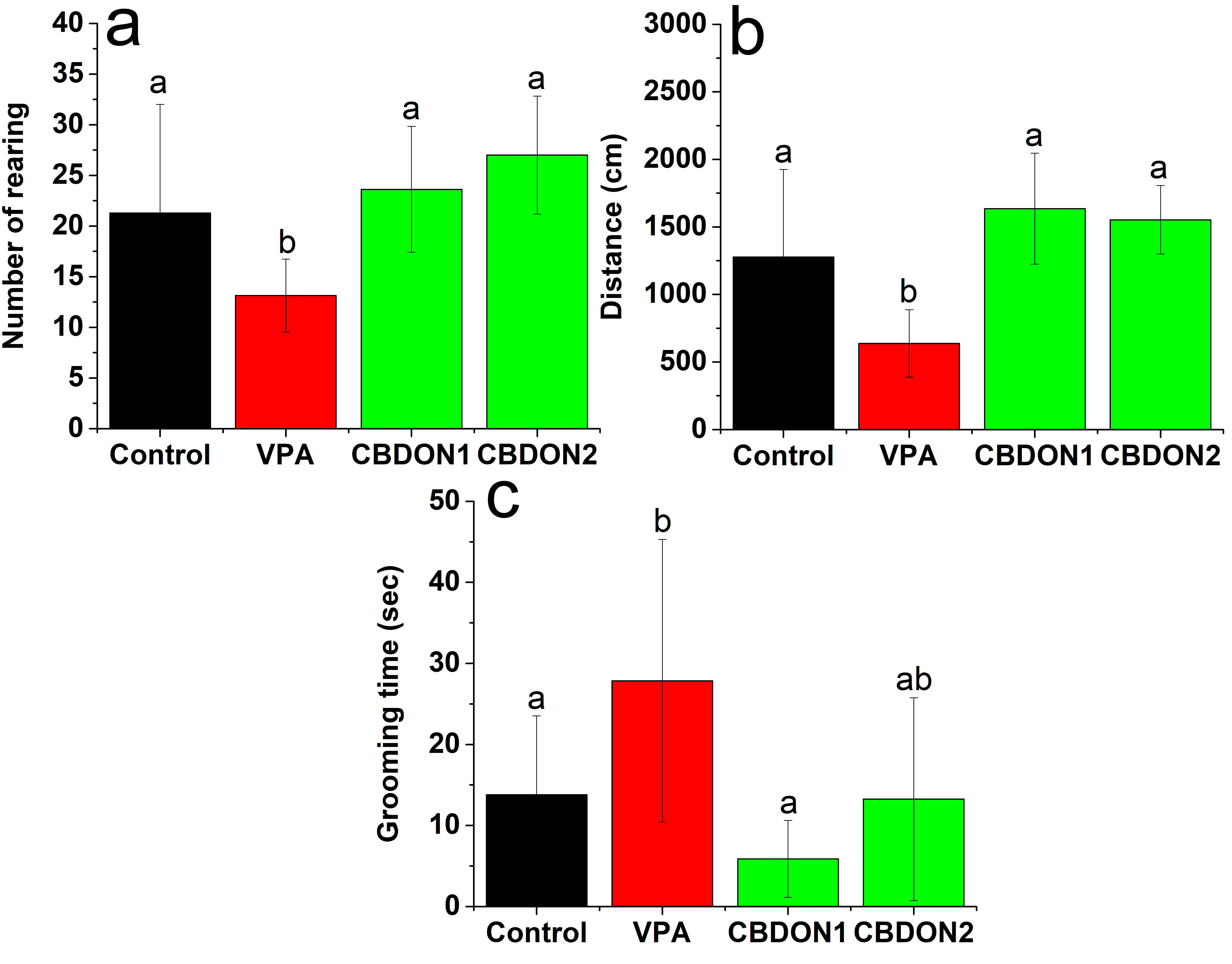
The experimenters handled the animals daily for one week before the beginning of the behavioral tests. All behavioral tests were performed between 6:00 p.m. and 4:00 a.m. after 30 days of treatment. The testing room was maintained with low-intensity lighting using a 40-watt red lamp in all tests to maintain animal welfare. Materials (maze and boxes) were cleaned between animals with 20% ethanol solution and dried with a paper towel. The schedule of animal experiments is summarized in Figure 1.

Schematic representation of the study schedule. GD, gestational day; PND, postnatal day; EPMT, elevated plus-maze test; ACT, activity cage test; TCT, Three-Chamber Social Interaction Test.
Elevated Plus-Maze Test
The elevated plus-maze test (EPM) was performed as previously described by Kraeuter et al. (Kraeuter et al. 2019) to assess anxiety-like behavior. The EPM was made of white polyvinyl chloride (PVC) with two open arms (36 x 6 x 1 cm3) and two closed arms (36 x 6 x 15 cm3) that crossed perpendicularly and was mounted on a raised platform 60 cm from the floor. On postnatal day (PND) 61 the offspring rats (16 x 4 groups) were placed individually in the center of the platform (intersection of the open and closed arms) facing the closed arms and allowed to explore the maze for 5 minutes. During the test period, the following parameters were evaluated: the number of entries into the open and closed arms and the time spent in each arm. These measures were recorded and scored manually. Low anxiety behavior was defined when animals explored the open arms for longer, and high anxiety behavior was defined when animals spent more time in the closed arms.

Chromatographic profile of CBD analyte at concentrations of 5 mg/ml (a), 10 mg/ml (b), 20 mg/ml (c), and the internal caffeine (CAF) pattern (0.014 mg/ml).
Activity Cage Test
An activity cage was used to evaluate the locomotor activity and repetitive behavior of rats, as previously described by Zamberletti et al. (Zamberletti et al. 2019). Additionally, we assessed rearing-up behavior. Briefly, the test was carried out in a wooden box (66 x 57 x 40 cm3) with a black floor. To encourage the exploration of the animals, the walls had 2 cm holes equally distributed in two rows. On PND 62, each rat was placed in the box and allowed to explore the environment freely for 5 min. Behavior was recorded using a cellphone camera positioned above the box. The total distance covered, number of rearing-up actions, and grooming times were evaluated manually. The rearing-up behavior was defined as elevation on the two hind legs, while grooming behavior was defined as rubbing the body with paws or mouth and rubbing the head with paws.
Three-Chamber Social Interaction Test
The three-chamber social test was carried out to evaluate the animal’s sociability and interest in social novelty according to Zamberletti et al. (Zamberletti et al. 2019). The experiment was performed in a social test apparatus (105 x 60 x 35 cm3), internally divided into three similar compartments (35 x 60 x 35 cm3) which allowed free access between them. Rats usually prefer to spend more time with other rats and are more interested in a novel intruder than a familiar one. The test consisted of three phases before starting the experiment. First, each rat was habituated, exploring the box freely for 5 min. The next phase started immediately after the habituation period and evaluated the social approach. A wire cage containing an unfamiliar rat was placed randomly in the right or left side chamber. The unfamiliar rat was randomly selected and belonged to the same lineage, age, and sex as the experimental groups. An identical empty wire cage was placed in the contralateral chamber. The test was performed on PND 63-65 and consisted of placing the animals individually and randomly in the central compartment and letting them freely explore all chambers for 10 min. Finally, the third phase provided a measure of preference for social novelty. For this, a new unfamiliar rat was placed inside a previously empty wire cage, and the rat being tested was allowed to explore the chambers for 10 min. All sessions were recorded, and the time spent in each chamber was measured with a stopwatch. The sociability index (SI) and social preference index (SPI) were calculated as the ratio of time spent exploring the first unfamiliar rat (second phase) or the second unfamiliar rat (third phase), and the total time of exploration x 100.
Brain collection and processing of histological material
On PDN 91 the animals were deeply anesthetized with intraperitoneal administration of ketamine (60 mg/kg) and xylazine (20 mg/kg) and were euthanized by decapitation. Their brains were quickly removed, fixed by immersion in 10% formaldehyde in 10 mM phosphate buffer pH 7.3 solution, and stored for 48 hours. Next, coronal cuts of approximately 2 mm were made in each brain and placed in histological cassettes. The material was dehydrated in an increasing series of ethanol and included in paraffin. Semi-serial sections of 4 μm were obtained using a rotating microtome (RM 2255, Leica Biosystems, Nussloch, Germany), respecting an interval of at least 39 μm between sections. Histological preparations were stained with hematoxylin and eosin.
Histological analysis of the hippocampus and cerebral cortex
The hippocampus and cortical region close to it were photomicrographed using a digital image capture system coupled to a Leica DM500® microscope (Leica Microsystems, Heerbrugg, Switzerland). In determining the volumetric density of the hippocampus, quantifications were performed of all living neurons and the necrotic neurons constituting the hippocampus in its different layers CA1, CA2, CA3, hilum, and dentate gyrus (DG) of all animals in the different experimental groups.
To measure the diameter, photomicrographs were randomly taken in 70 fields per experimental group, in the different layers of the hippocampus, at 400x magnification, and analyzed using the ImageJ Software (version 1.53k, 2019, National Institutes of Health, MD).
The volumetric data of the cellular composition of the cortex cerebral region over the hippocampus were measured by capturing images and inserting a micrometric graticule with 441 points of intersection at 400x magnification. Glial cells, living neurons, necrotic neurons, neuropils, and blood vessels were counted in 10 fields randomly, totaling 4410 points in each animal in the different experimental groups. After obtaining these data, the relations between glial cells/living neurons and glial cells/necrotic neurons were determined.
Statistical analysis
Statistical analyses were performed using OriginPro 2015 academic software (Origin Lab. Northampton, MA USA). Data were analyzed by one-way ANOVA followed by Tukey’s test. Differences were considered significant when p 0.05.
RESULTS
Chromatographic profile of CBDO
Chromatograms of the CBDO at 5, 10, and 20 mg/mL are shown in Figures 2a-c, respectively. CBDs were identified by their spectra in comparison with the authentic pattern contained in the equipment. It was observed that the chemical compositions of the CBDO at 5, 10, and 20 mg/mL were 0.56% (≈ 0.5%), 0.93% (≈ 1.0%), and 1.94% (≈ 2.0%) CBD, respectively.
Nanoemulsions had good macroscopic characteristics, stability, and physicochemical properties
The nanoemulsions were characterized according to dispersion phase (homogeneous or heterogeneous); aspect (presence or absence of lumps); smell (present or absent); consistency (fluid, slightly thick, thick, or very thick); opacity (translucent, partially opaque, or opaque); and color (present or absent). During the period evaluated, results revealed that all nanoemulsions were homogeneous, with no lumps and no odor. The formulations had a fluid consistency, but the nanoemulsion produced with oil rich in CBD at a concentration of 2% was slightly thick. Regarding opacity, all nanoemulsions were opaque due to addiction to propylparaben preservative. Regarding color, formulations showed a slightly yellowish tone as the concentration of CBD-rich oil increased. To evaluate stability, on the day of preparation and 2, 7, 15, and 30 days after preparation, nanoemulsions were submitted to centrifugation and thermal stress, remaining for 24 hours under cooling (5 ± 2 °C), freezing (-20 ± 2 °C), and heating (40 and 50 ± 2 °C). Subsequently, the formulations were kept at room temperature (25 ± 2 °C) for approximately one hour, and organoleptic characteristics were assessed again. Any macroscopic physical change was observed in nanoemulsions after centrifuging, cooling, freezing, and heating (creaming or phase separation), indicating good stability.
Concerning the physicochemical characteristics (Table I), the nanoemulsion containing corn oil without CBD had a droplet size of 87.5 ± 1.0 nm, while CBDON0.5, CBDON1, and CBDON2 had droplet sizes of 107.6 ± 1.1 nm, 90.4 ± 1.3 nm, and 72.6 ± 0.4 nm, respectively. All CBD formulations were characterized as nanoemulsions, as they had vesicles with a droplet size smaller than 200 nm (McClements Öztürk 2021). PDI values are used to describe the degree of uniformity of vesicle size distribution, also known as the heterogeneity index. A PDI less than 0.05 indicates a highly monodisperse, i.e., homogeneous, pattern, while a value greater than 0.7 shows that the sample has a very broad, heterogeneous particle size distribution (Danaei et al. 2018). The PDI values of CBDON0.5, CBDON1, and CBDON2 were 0.432, 0.360, and 0.290, respectively, while the nanoemulsion without CBD had a PDI of 0.327. Thus, CBDON1 and CBDON2 were chosen for administration to animals and subsequent behavioral assessment since they had the lowest PDIs. These values indicate that the formulations are homogeneous, as they have a PDI lower than 0.7. Zeta potential measurements were used to determine the stability of colloidal systems. The formulation containing only corn oil showed a zeta potential of -40.6 ± 0.1 mv. CBDON0.5, CBDON1, and CBDON2 showed zeta potentials of -30.2 ± 1.3 mv, -26.0 ± 3.6 mv, and -24.9 ± 0.1, respectively. All formulations can easily be incorporated into syrups.
Influence evaluations of cannabidiols according to vesicle size, polydispersity index (PDI), and zeta potential ζ (mv) of oil-in-water (O/A) nanoemulsions of CBD and corn oil nanoemulsion without CBD (empty - E).
Anxiety behavior was alleviated by CBDON treatment
Anxiety behavior was assessed by an elevated plus maze test. Time spent in the closed arms was significantly higher in the VPA group compared to the control group (F3.28 = 6.22; p 0.005), showing an anxiety behavior that usually occurs in autistic individuals and valproic-acid-induced autism. In turn, the control group had a higher percentage of time spent in the open arms (F3.28 = 4.8; p 0.05), behavior considered normal in this test. CBDON1 treatment significantly reduced the length of stay in the closed arms, as shown in Figure 3b, and rescued the decrease in time spent in the open arms (Figure 3a), suggesting an anxiety reduction observed in the VPA group. The VPA group presented a reduced number of entries in the open arms (F3.28 = 5.48; p 0.05) and closed arms (F3.28 = 5.88; p 0.05) in relation to the control group, however, CBDON1 treatment attenuated this effect (Figure 3c, d).

Effect of prenatal exposure to VPA (VPA group) and CBD-enriched Cannabis oil nanoemulsion (CBDON) treatment at the concentrations of 1 mg/animal (CBDON1) and 2 mg/animal (CBDON2) on anxiety in autistic rats. a, b: Time spent in the open and closed arms, respectively, in an elevated plus-maze test. c, d: Number of entries in the open and closed arms, respectively, in an elevated plus-maze test. Each experimental group was compared with the control (a) and VPA groups (b) by one-way ANOVA followed by Tukey’s test and considered significant when p 0.05. (a) Tukey post hoc test against control group (VPA p 0.05, CBDON1 p = 0.99, CBDON2 p = 0.92) and against VPA group (CBDON1 p 0.05, CBDON2 p = 0.07). (b) Tukey post hoc test against control group (VPA p 0.005, CBDON1 p = 0.70, CBDON2 p = 0.28) and against VPA group (CBDON1 p 0.05, CBDON2 p = 0.11). (c) Tukey post hoc test against control group (VPA p 0.005, CBDON1 p = 0.51, CBDON2 p = 0.91) and against VPA group (CBDON1 p 0.05, CBDON2 p = 0.07). (d) Tukey post hoc test against the control group (VPA p 0.05, CBDON1 p = 0.67, CBDON2 p = 0.99) and against the VPA group (CBDON1 p 0.005, CBDON2 p 0.05).
Exploratory activity was rescued, and stereotyped behavior was reduced by CBDON treatment
The VPA group presented a significant reduction in total distance covered (F3.28 = 9.18; p 0.05) and number of rearing-up actions (F3.40 = 7.23; p 0.05) when compared to the control group. CBDON1 and CBDON2 treatment significantly attenuated the decrease in total distance traveled and rearing-up behavior (Figure 4a, b) in the VPA group. The self-grooming evaluation displayed increased time of repetitive self-grooming in the VPA group compared with control rats (F3.40 = 5.99; p 0.05). CBDON1 administration significantly reduced the time spent in repetitive behavior in the VPA group (Figure 4c).

Effect of prenatal exposure to VPA (VPA group) and CBD-enriched Cannabis oil nanoemulsion treatment at the concentrations of 1 mg/animal (CBDON1) and 2 mg/animal (CBDON2) on exploratory activity and stereotype behavior (grooming) in autistic rats. a: Number of rearing-up actions in the open field test. b: Total distance traveled in the open field test. c: Grooming time in the open field test. Each experimental group was compared with the control (a) and VPA groups (b) by one-way ANOVA followed by Tukey’s test and considered significant when p 0.05. (a) Tukey post hoc test against the control group (VPA p 0.05, CBDON1 p = 0.88, CBDON2 p = 0.31) and against the VPA group (CBDON1 p 0.05, CBDON2 p 0.05). (b) Tukey post hoc test against the control group (VPA p 0.01, CBDON1 p = 0.34, CBDON2 p = 0.56) and against the VPA group (CBDON1 p 0.05, CBDON2 p 0.05). (c) Tukey post hoc test against control group (VPA p 0.05, CBDON1 p = 0.50, CBDON2 p = 0.99) and against VPA group (CBDON1 p 0.005, CBDON2 p = 0.06).
The sociability index and social preference index were improved by CBDON treatment
In the first phase (sociability) in the three-chamber apparatus, the control group exhibited a preference for the unfamiliar rat compared to the empty cage, demonstrating normal sociability. The VPA group spent more time in the empty cage (F3.40 = 6.01; p 0.005) and less time with the unfamiliar rat (F3.40 = 8.61; p 0.05) compared to the control group, reflecting social avoidance. CBDON1 and CBDON2 treatment significantly attenuated the decrease in time spent with unfamiliar rats observed in the VPA-exposed group as well as reduced time spent in the empty cage (Figure 5a, b). The VPA group exhibited a decrease in the sociability index compared to the control group. However, this parameter was significantly reversed by CBDON1 and CBDON2 treatment (Figure 5c). In the second phase (social preference), a preference of the control group for the unfamiliar rat was observed (F3.39 = 9.83; p 0.05) when compared to familiar rats (F3.39 = 7.83; p 0.05). However, the VPA group spent significantly more time with the familiar rat than the unfamiliar rat, showing a reduced social preference (Figure 5d, e). CBDON treatment in both doses rescued the social preference in the VPA group. The VPA group exhibited a reduction in the social preference index when compared to the control group, which was attenuated by the CBDON2 treatment (F3.40 = 7.38; p 0.05) (Figure 5f).

Effect of prenatal exposure to VPA (VPA group) and CBD-enriched Cannabis oil nanoemulsion treatment at the concentrations of 1 mg/animal (CBDON1) and 2 mg/animal (CBDON2) on the sociability index and social preference index of autistic rats. a, b: Time spent with stranger rat and object on three chamber tests. c, d: Time spent with novel rat (unknown) and familiar rat (stranger) in three chamber tests. e, f: Sociability and social preference index. Each experimental group was compared with the control (a) and VPA groups (b) by one-way ANOVA followed by Tukey’s test and considered significant when p 0.05. (a) Tukey post hoc test against the control group (VPA p 0.05, CBDON1 p = 0.75, CBDON2 p = 0.47) and against the VPA group (CBDON1 p 0.05, CBDON2 p 0.05). (b) Tukey post hoc test against the control group (VPA p 0.005, CBDON1 p = 0.97, CBDON2 p = 0.94) and against the VPA group (CBDON1 p 0.05, CBDON2 p 0.05). (c) Tukey post hoc test against the control group (VPA p 0.05, CBDON1 p = 0.75, CBDON2 p = 0.47) and against the VPA group (CBDON1 p 0.05, CBDON2 p 0.05). (d) Tukey post hoc test against the control group (VPA p 0.05, CBDON1 p = 0.24, CBDON2 p = 0.37) and against the VPA group (CBDON1 p 0.05, CBDON2 p 0.05). (e) Tukey post hoc test against the control group (VPA p 0.005, CBDON1 p = 0.63, CBDON2 p = 0.72) and against the VPA group (CBDON1 p = 0.12, CBDON2 p = 0.09). (f) Tukey post hoc test against the control group (VPA p 0.05, CBDON1 p = 0.51, CBDON2 p = 0.67) and against the VPA group (CBDON1 p = 0.06, CBDON2 p 0.05).
CBDON treatment protected cells in the hippocampus and cortex cerebral region against prenatal exposure to VPA
The VPA group presented a decrease in cell viability compared to control animals, and treatment with CBDON2 attenuated this effect (Table II). At the same time, an increase in cell mortality was observed in animals that received valproic acid in the prenatal period. CBDON treatment at both doses reduced valproic acid-induced neuronal death (Table III). The cellular composition of the cortical region can be observed in Table V. The animals exposed to valproic acid showed a decrease in the number of viable neurons and an increase in the number of red neurons (apoptotic). Consequently, there was an increase and a reduction in the glial/VN and glial/RN ratio, respectively. The number of blood vessels was raised in the VPA group, whereas we did not observe these alterations in the CBDON treatment at either dose. The measurements of the hippocampal layers CA1, CA2, CA3, and dentate gyrus are shown in Table IV. The VPA group presented a reduction in all hippocampal layers compared to control animals, and CBDON at both doses recovered the diameter of the hippocampal regions (figure 6a-h).

Representative photomicrographs from the hippocampus of control, VPA, CBDON1, and CBDON2 groups. a and b: Photomicrograph of the hippocampal structure of the control group, which can be seen in the absence of neuronal death in all structures CA1, CA2, CA3, CA4, hilum (H), dentate gyrus (DG), internal molecular layer (IML), in addition to of the stratum pyramidale (SP), stratum oriens (SO) and stratum radiatum (SR). Figures 6c and 6d: VPA group, in which an intense reduction in the thickness of the CA1 cell layer can be observed and, in some areas, there is an absence of neuron bodies. Note intense necrosis of neuron bodies (arrow), mainly in the hilum area, dentate gyrus, and CA1. Figures 6e and 6f: CBDON1 group, maintenance of hippocampus structures, and intense reduction of neuronal necrosis can be observed (arrow). g and h: CBDON2 group, note the maintenance of structures, as well as the presence of rare necrotic neurons (arrow). Magnification: x 400 (a, c, e, g) and x 1000 (b, d, f, h).
Quantification of the population of viable neurons (VN) in the areas of the hippocampus (CA1, CA2, CA3, hilus, and dentate gyrus) of control, VPA, CBDON1, and CBDON2 groups. Each experimental group was compared with control (a) and VPA group (b) by one-way ANOVA (CA1 F3, 24 = 7.16 and CA2 F3, 24 = 6.42) followed by Tukey’s test and considered significant when p < 0.05.
Quantification of the population of red neurons (RN) in the hippocampus (CA1, CA2, CA3, hilus, and dentate gyrus) of control, VPA, CBDON1, and CBDON2 groups. Each experimental group was compared with control (a) and VPA group (b) by one-way ANOVA (CA3 F3, 24 = 3.057) followed by Tukey’s test and considered significant when p < 0.05.
Diameter (µm) of the neuronal population in the hippocampus (CA1, CA2, CA3, and dentate gyrus) of control, VPA, CBDON1, and CBDON2 groups. Each experimental group was compared with control (a) and VPA group (b) by one-way ANOVA (CA1 F3, 308 = 13.148; CA2 F3, 308 = 39.608; CA3 F3, 308 = 52.252; and, Dentate gyrus F3, 308 = 6.206) followed by Tukey’s test and considered significant when p < 0.05.
Corticocerebral regions of control, VPA, CBDON1, and CBDON2 groups. Each experimental group (number) was compared with control (a) and VPA group (b) by one-way ANOVA (VN F3, 24 = 3.154; RN F3, 24 = 36.110; VESSEL F3, 24 = 3.889; and GLIAL/RN F3, 24 = 11.172) followed by Tukey’s test and considered significant when p < 0.05.
The research reported in this manuscript is part of a patent (BR1020210268301) in the course of the process.
DISCUSSION
In the present study, we investigated the effects of CBDON administration on autistic-like behaviors in a prenatal valproic acid rat model. Previously, we focused on the preparation of CBD-rich oil nanoemulsion to enhance oil bioavailability and reduce the dose administered to animals. Nanoemulsions are colloidal systems that may potentially increase water solubility, as well as the bioavailability of lipophilic bioactive compounds. Despite the benefits, nanoemulsions can suffer instability phenomena (McClements 2021). To assess nanoemulsion stability, mechanical and thermal tests were carried out, and macroscopic characteristics were evaluated for 30 days. Exposure to high temperatures and centrifugation forces enhances the probability of collision generating aggregation and increasing the coalescence rate (Dantas et al. 2021). Nevertheless, the nanoemulsions were macroscopically stable after the tests, preserving their appearance and homogeneity.
Other parameters were used to evaluate the stability of nanoemulsions, such as droplet diameter, the polydispersity index, and the zeta potential. CBDON showed a vesicle size larger than the nanoemulsion without CBD. Moreover, it was observed that CBDON2 had a smaller vesicle size than CBDON1. On the other hand, CBDON0.5 had a droplet size larger than CBDON1. These results demonstrate that the presence of CBD modifies the size of the nanoemulsion and that increasing the concentration of CBD decreases the size of the vesicle. The size of the vesicle is important because it influences the physicochemical and functional characteristics of the nanoemulsion (Choi McClements 2020). Mikulcova et al. (2017) reported that the reduced droplet size is related to greater stability and nanoemulsions with large vesicles (such as 590 – 780 nm) quickly lose stability. According to Stokes’ law and Derjaguin-Landau-Verwey-Overbeek (DLVO) theory, smaller particle size may result in a high electrostatic repulsion, preventing emulsion coalescence and increasing the repulsion force between vesicles (Meng et al. 2019). Our results corroborate this theory since they showed that particle size is directly related to nanoemulsion stability.
The polydispersity index is an indicator of the association of the particles; a small PDI value indicates homogeneity and a high PDI value indicates a heterogeneous and polydispersed distribution (Danaei et al. 2018). The nanoemulsions had low PDI values, which indicates a stable and uniform distribution. In addition, the PDI value was inversely proportional to the CBD concentration, and CBDON1 and CBDON2 demonstrated lower PDI values than CBDON0.5, indicating the best monodispersity. Zeta potential is an important physicochemical characteristic of formulations. These parameters can determine vesicle surface charge, which may be cationic, anionic, or neutral. Negative values indicate a greater interaction of repulsion forces between vesicles, which ensures stability (Smith et al. 2017). The zeta potential obtained in all formulations showed negative values, indicating good stability. Additionally, it was observed that the presence of CBD modified the zeta potential of nanoemulsions, so the higher concentration of CBD produced a lower zeta potential. Anionic surfactants were used in the development of nanoemulsions and negative potential zeta may be related to the presence of anions in the surfactants (Shen et al. 2021). CBDON2 was the most stable nanoemulsion based on the droplet size, PDI, and zeta potential value.
Currently, the use of CBD in individuals with autism is receiving attention. Pre-clinical and clinical studies have shown the efficacy and safety of CBD in the core symptoms of autism (Bar-Lev Schleider et al. 2019, Poleg et al. 2019). For example, Manduca et al. (2024) demonstrated that CBD rescued the cognitive deficits exhibited by Fmr1 - Δexon 8 mice, a validated animal model for ASD. Another preclinical study showed the general anxiolytic and prosocial efficacy of a broad-spectrum hemp oil rich in CBD administered to BTBR mice, a strain with ASD-like behaviors (Staben et al. 2023). Furthermore, CBD was effective in attenuating autism-like behaviors and comorbid behaviors associated with ASD in a mouse model of idiopathic ASD (Shrader et al. 2024). Clinical studies have also demonstrated the therapeutic effects of whole-plant CBD extract in core symptoms of ASD as well as social impairments (Parrella et al. 2023). Moreover, clinical studies are being assayed to study the efficacy of CBD to ameliorates behavioral changes in neurodevelopmental disorders (Muller et al. 2024). Based on this, we have important findings to discuss in our paper based on the therapeutic effects of CBDON.
In the current work, to determine the effects of CBDON behavioral changes after 30 days of treatment, we assessed anxiety, locomotion, exploratory activity, repetitive behavior, sociability, and social preference. We used the prenatal VPA rat model since rodents prenatally exposed to this drug develop behavioral phenotypes of the human condition. This model is a valid and reliable tool to investigate the behavior and other features of autism (Bossu Roux 2019). Prenatal exposure to VPA has been shown to increase anxiety and stereotyped movements, as well as to reduce exploratory activity, sociability, and social preference. Several animals and human studies have evidenced that exposure to VPA in intrauterine life results in behavioral changes, such as increased repetitive behavior (Schneider Przewlocki 2005) and anxiety (Schneider et al. 2007) along with a decrease in social interaction and exploratory activity (Wang et al. 2019) corroborating our present data.
CBDON in both doses significantly attenuated alterations induced by VPA in behavioral parameters in rats. In the elevated plus-maze test, we observed an increase in the anxiety of animals exposed to VPA. This behavior consists of aversion to open spaces, which is observed through the animal spends more time in the closed arms of the maze, a behavior reproduced by the VPA group. The CBDON1 treatment was able to reduce anxiety signs induced by prenatal exposure to VPA. Exploratory activity was evaluated by the number of rearing-up actions and total distance covered. A reduction was observed in the number of rearing-up actions and total distance covered in the VPA group. It has been reported that exploratory activity is reduced in prenatal exposure to VPA, in accordance with our study (Uccelli et al. 2021).
Grooming behavior is innate in rodents. It consists of movements that involve licking the forelegs, belly, and back in a stereotyped anteroposterior progression. Excessive repetitive self-grooming in VPA rats may be analogous to repetitive motor stereotypies and self-stimulation, a phenotype commonly observed in autism (Keller et al. 2021). Studies have shown an increase in self-grooming behavior in VPA rats (McKinnell et al. 2021, Mohammadi et al. 2020). Our results also show similar results. This parameter was rescued by CBDON1 and CBDON2 treatment. The VPA group showed reduced sociability and social preference index, in other words, VPA treatment impaired social behavior. CBDON1 and CBDON2 treatments also reversed this effect.
Beneficial effects of CBD on behavior are related to both the route of administration and the dose used, and oral administration was reported to be more effective (Hlozek et al. 2017). However, studies in animal models have shown effective CBD responses in reversing behavior changes with higher doses, ranging from 20 mg/kg to 120 mg/kg (Deiana et al. 2012, Kaplan et al. 2017). Thus, the use of nanoemulsion may have increased the effectiveness of CBD from 5 to 17 times, since positive results were observed using very small doses (3.35, and ≈ 6.80 mg/Kg for CBDON1 and CBDON2, respectively). In this context, nanoemulsion is a great option to overcome the need to administer CBD at high doses.
In the search for possible correlates of the effects observed at the behavioral level, we performed histological analysis in the hippocampus of the experimental groups. We found that VPA generates an increase in cell death in the CA3 hippocampal region and reduces the number of viable neurons in regions CA1, CA2, CA3, and the dentate gyrus. Although our study does not have data to evidence these findings, they may be related to the neuroinflammation and microglial activation generated by VPA, since these events contribute to neuronal dysfunction and death (Codagnone et al. 2015). When overactivated in response to neuronal damage and genetic or environmental factors, microglia cause widespread damage to neighboring neurons. Indeed, reactive microglia kill neurons by producing neurotoxic factors and proinflammatory molecules (Galvani et al. 2021). Additionally, our study showed an increase in the number of blood vessels in the cortical region of the VPA group. Neuroinflammation and microglial activation promote angiogenesis, which could justify these findings (Huang et al. 2020). Associated with this, the occurrence of neurotoxic effects related to oxidative stress in prenatal exposure to VPA was previously reported. The excess of oxidative stress leads to DNA damage and neuronal apoptosis (Taleb et al. 2021). In contrast, studies have shown a beneficial effect of VPA on reducing neuronal death and neuroinflammation, which does not corroborate our results (Chen et al. 2018).
It is important to point out that there is evidence of hippocampal structural abnormalities in autistic individuals (Banker et al. 2021). Moreover, the dentate gyrus and CA1/CA2/CA3 regions are involved in the circuitry underlying social memory (Bertoni et al. 2021, Hitti Siegelbaum 2014). Furthermore, the amygdala has long been linked to social interaction and shares reciprocal connections with the ventral hippocampus, and there are abnormal patterns of hippocampal and amygdala development in autistic patients (Felix-Ortiz Tye 2014). Therefore, the behavioral changes observed in our study may be related to the lesions found in the hippocampus.
CBDON in both doses has been found to reduce the death of neurons and increase the number of viable cells in the hippocampus and cortical region. It has been demonstrated that CBD has a neuroprotective role in various neurological conditions. This neuroprotective role may be due to one of the pharmacological effects of CBD, the reduction in brain oxidative stress and inflammation (Atalay et al. 2019). The decrease in the number of cortical blood vessels may be related to a probable reduction in angiogenesis as a consequence of a probable reduction in neuroinflammation.
Thus, CBDON showed beneficial effects on autistic-like behaviors, as well as reducing hippocampal cell death in prenatal VPA exposure rats. Given that prenatal VPA exposure generates neuroinflammation and oxidative stress, we assume that the anti-inflammatory and antioxidant activity of CBD could justify these results. The development of a nanoemulsion to overcome the lipophilic characteristics of CBD showed promising results, as the dose used could be decreased.
CONCLUSIONS
It was concluded that CBDON demonstrates great potential in attenuating autistic-like behaviors in rats. This effect may be related to the protection against neuronal death in the hippocampus, also resulting from the treatment with the nanoemulsion. Although nanoemulsions at both doses showed favorable responses and increased the effect of CBD, CBDON2 was shown to be more efficient, and it is possible to incorporate syrups to attenuate all evaluated parameters. However, further research should be directed to exploring the related pharmacokinetics, pharmacodynamics, and toxicity. These findings bring new insights into the treatment of autism, and nanotechnology appears to represent a promising perspective that may bring CBD closer to clinical use.
ACKNOWLEDGMENTS
The authors would like to thank the Universidade Federal Rural de Pernambuco and all the members involved in this research. The authors would also like to thank the Coordenação de Aperfeiçoamento de Pessoal de Nível Superior (CAPES) for the scholarship to Mariana de França Oliveira da Silva (Grant 88882.436160/2019-01), and Conselho Nacional de Desenvolvimento Científico e Tecnológico (CNPq) for the scholarship to Cláudia Kathariny da Silva Farias (Grant 408879/2018-1), the productivity grant to Pabyton G. Cadena (306947/2020-0), and financial support (Grant 408879/2018-1).The authors would like to thank the Universidade Federal Rural de Pernambuco and all the members involved in this research. The authors would also like to thank the Coordenação de Aperfeiçoamento de Pessoal de Nível Superior (CAPES) for the scholarship to Mariana de França Oliveira da Silva (Grant 88882.436160/2019-01), and Conselho Nacional de Desenvolvimento Científico e Tecnológico (CNPq) for the scholarship to Cláudia Kathariny da Silva Farias (Grant 408879/2018-1), the productivity grant to Pabyton G. Cadena (306947/2020-0), and financial support (Grant 408879/2018-1).
REFERENCES
- ANVISA - AGÊNCIA NACIONAL DE VIGILÂNCIA SANITÁRIA. 2004. Guia de estabilidade de produtos cosméticos, Brasilia, Brazil.
- ARAUJO DJ, TJOA K SAIJO K. 2019. The Endocannabinoid System as a Window Into Microglial Biology and Its Relationship to Autism. Front Cell Neurosci 13: 424.
- ASSOCIATION AP. 2013. Diagnostic and Statistical Manual of Mental Disorders, 5th ed. American Psychiatric Publishing, Arlington, VA.
- ATALAY S, JAROCKA-KARPOWICZ I SKRZYDLEWSKA E. 2019. Antioxidative and Anti-Inflammatory Properties of Cannabidiol. Antioxidants 9: 21.
- BANKER SM, GU X, SCHILLER D FOSS-FEIG JH. 2021. Hippocampal contributions to social and cognitive deficits in autism spectrum disorder. Trends Neurosci 44(10): 793-807.
- BAR-LEV SCHLEIDER L, MECHOULAM R, SABAN N, MEIRI G NOVACK V. 2019. Real life Experience of Medical Cannabis Treatment in Autism: Analysis of Safety and Efficacy. Sci Rep 9(1): 200.
- BERTONI A ET AL. 2021. Oxytocin administration in neonates shapes hippocampal circuitry and restores social behavior in a mouse model of autism. Mol Psychiatry 26(12): 7582-7595.
- BESAG FMC VASEY MJ. 2020. Seizures and Epilepsy in Autism Spectrum Disorder. Child Adolesc Psychiatr Clin N Am 29(3): 483-500.
- BISOGNO T, HANUŠ L, DE PETROCELLIS L, TCHILIBON S, PONDE DE, BRANDI I, MORIELLO AS, DAVIS JB, MECHOULAM R DI MARZO V. 2001. Molecular targets for cannabidiol and its synthetic analogues: Effect on vanilloid VR1 receptors and on the cellular uptake and enzymatic hydrolysis of anandamide. Br J Pharmacol 134(4): 845-852.
- BOSSU JL ROUX S. 2019. The valproate model of autism. Med Sci (Paris) 35(3): 236-243.
- BRUNI N, DELLA PEPA C, OLIARO-BOSSO S, PESSIONE E, GASTALDI D DOSIO F. 2018. Cannabinoid Delivery Systems for Pain and Inflammation Treatment. Molecules 23: 2478.
- CABRERA-PEREZ MA PHAM-THE H. 2018. Computational modeling of human oral bioavailability: what will be next? Expert Opin Drug Discov 13(6): 509-521.
- CADENA PG, PEREIRA MA, CORDEIRO RB, CAVALCANTI IM, BARROS NETO B, PIMENTEL MDOC, LIMA FILHO JL, SILVA VL SANTOS-MAGALHAES NS. 2013. Nanoencapsulation of quercetin and resveratrol into elastic liposomes. Biochim Biophys Acta 1828(2): 309-316.
- CARBONE E, MANDUCA A, CACCHIONE C, VICARI S TREZZA V. 2021. Healing autism spectrum disorder with cannabinoids: a neuroinflammatory story. Neurosci Biobehav Rev 121: 128-143.
- CHEN JY, CHU LW, CHENG KI, HSIEH SL, JUAN YS WU BN. 2018. Valproate reduces neuroinflammation and neuronal death in a rat chronic constriction injury model. Sci Rep 8(1): 16457.
- CHOI SJ MCCLEMENTS DJ. 2020. Nanoemulsions as delivery systems for lipophilic nutraceuticals: strategies for improving their formulation, stability, functionality and bioavailability. Food Sci Biotechnol 29(2): 149-168.
- CHOUDHURY H, GORAIN B, PANDEY M, KHURANA RK KESHARWANI P. 2019. Strategizing biodegradable polymeric nanoparticles to cross the biological barriers for cancer targeting. Int J Pharm 565: 509-522.
- CODAGNONE MG, PODESTA MF, UCCELLI NA REINES A. 2015. Differential Local Connectivity and Neuroinflammation Profiles in the Medial Prefrontal Cortex and Hippocampus in the Valproic Acid Rat Model of Autism. Dev Neurosci 37(3): 215-231.
- DANAEI M, DEHGHANKHOLD M, ATAEI S, HASANZADEH DF, JAVANMARD R, DOKHANI A, KHORASANI S MOZAFARI MR. 2018. Impact of Particle Size and Polydispersity Index on the Clinical Applications of Lipidic Nanocarrier Systems. Pharmaceutics 10: 57.
- DANTAS AGB, DE SOUZA RL, DE ALMEIDA AR, XAVIER JUNIOR FH, PITTA M, REGO M OLIVEIRA EE. 2021. Development, Characterization, and Immunomodulatory Evaluation of Carvacrol-loaded Nanoemulsion. Molecules 26: 3899.
- DEIANA S ET AL. 2012. Plasma and brain pharmacokinetic profile of cannabidiol (CBD), cannabidivarine (CBDV), Delta(9)-tetrahydrocannabivarin (THCV) and cannabigerol (CBG) in rats and mice following oral and intraperitoneal administration and CBD action on obsessive-compulsive behaviour. Psychopharmacology (Berl) 219(3): 859-873.
- DIAS-DE FREITAS F, PIMENTA S, SOARES S, GONZAGA D, VAZ-MATOS I PRIOR C. 2022. The role of cannabinoids in neurodevelopmental disorders of children and adolescents. Rev Neurol 75(7): 189-197.
- ESPOSITO E ET AL. 2015. Cannabinoid antagonist in nanostructured lipid carriers (NLCs): design, characterization and in vivo study. Mater Sci Eng C Mater Biol Appl 48: 328-336.
- FELIX-ORTIZ AC TYE KM. 2014. Amygdala inputs to the ventral hippocampus bidirectionally modulate social behavior. J Neurosci 34(2): 586-595.
- FOLKES OM ET L. 2020. An endocannabinoid-regulated basolateral amygdala-nucleus accumbens circuit modulates sociability. J Clin Invest 130(4): 1728-1742.
- FYKE W, ALARCON JM, VELINOV M CHADMAN KK. 2021a. Pharmacological inhibition of the primary endocannabinoid producing enzyme, DGL-alpha, induces autism spectrum disorder-like and co-morbid phenotypes in adult C57BL/J mice. Autism Res 14(7): 1375-1389.
- FYKE W, PREMOLI M, ECHEVERRY AV, LOPEZ-MORENO JA, LEMAIRE-MAYO V, CRUSIO WE, MARSICANO G, WOHR M PIETROPAOLO S. 2021b. Communication and social interaction in the cannabinoid-type 1 receptor null mouse: Implications for autism spectrum disorder. Autism Res 14(9): 1854-1872.
- GALVANI G, MOTTOLESE N, GENNACCARO L, LOI M, MEDICI G, TASSINARI M, FUCHS C, CIANI E TRAZZI S. 2021. Inhibition of microglia overactivation restores neuronal survival in a mouse model of CDKL5 deficiency disorder. J Neuroinflammation 18(1): 155.
- HITTI FL SIEGELBAUM SA. 2014. The hippocampal CA2 region is essential for social memory. Nature 508: 88-92.
- HLOZEK T ET AL. 2017. Pharmacokinetic and behavioural profile of THC, CBD, and THC+CBD combination after pulmonary, oral, and subcutaneous administration in rats and confirmation of conversion in vivo of CBD to THC. Eur Neuropsychopharmacol 27(12): 1223-1237.
- HUANG Y, CHEN S, LUO Y HAN Z. 2020. Crosstalk between Inflammation and the BBB in Stroke. Curr Neuropharmacol 18(12): 1227-1236.
- KAPLAN JS, STELLA N, CATTERALL WA WESTENBROEK RE. 2017. Cannabidiol attenuates seizures and social deficits in a mouse model of Dravet syndrome. Proc Natl Acad Sci USA 114(42): 11229-11234.
- KELLER R, COSTA T, IMPERIALE D, BIANCO A, RONDINI E, HASSIOTIS A BERTELLI MO. 2021. Stereotypies in the Autism Spectrum Disorder: Can We Rely on an Ethological Model? Brain Sciences 11(6): 762.
- KERR DM, GILMARTIN A ROCHE M. 2016. Pharmacological inhibition of fatty acid amide hydrolase attenuates social behavioural deficits in male rats prenatally exposed to valproic acid. Pharmacol Res 113: 228-235.
- KRAEUTER AK, GUEST PC SARNYAI Z. 2019. The Elevated Plus Maze Test for Measuring Anxiety-Like Behavior in Rodents. Methods Mol Biol 1916: 69-74.
- LAI MC, LERCH JP, FLORIS DL, RUIGROK AN, POHL A, LOMBARDO MV BARON-COHEN S. 2017. Imaging sex/gender and autism in the brain: Etiological implications. J Neurosci Res 95: 380-397.
- LAPMANEE S, BHUBHANIL S, WONGCHITRAT P, CHAROENPHON N, INCHAN A, NGERNSUTIVORAKUL T, DECHBUMROONG P, KHONGKOW M NAMDEE K. 2024. Assessing the Safety and Therapeutic Efficacy of Cannabidiol Lipid Nanoparticles in Alleviating Metabolic and Memory Impairments and Hippocampal Histopathological Changes in Diabetic Parkinson’s Rats. Pharmaceutics 16: 514.
- LORD C ET AL. 2020. Autism spectrum disorder. Nat Rev Dis Primers 6(1).
- LUNDQUIST P ARTURSSON P. 2016. Oral absorption of peptides and nanoparticles across the human intestine: Opportunities, limitations and studies in human tissues. Adv Drug Deliv Rev 106: 256-276.
- MANDUCA A ET AL. 2024. Cannabidiol and positive effects on object recognition memory in an in vivo model of Fragile X Syndrome: Obligatory role of hippocampal GPR55 receptors. Pharmacol Res 203: 107176.
- MCCLEMENTS DJ. 2021. Advances in edible nanoemulsions: Digestion, bioavailability, and potential toxicity. Prog Lipid Res 81: 101081.
- MCCLEMENTS, DJ ÖZTÜRK B. 2021. Utilization of Nanotechnology to Improve the Handling, Storage and Biocompatibility of Bioactive Lipids in Food Applications. Foods 10(2): 365.
- MCKINNELL ZE, MAZE T, RAMOS A, CHALLANS B PLAKKE B. 2021. Valproic acid treated female Long-Evans rats are impaired on attentional set-shifting. Behav Brain Res 397: 112966.
- MEHRA S, UL AHSAN A, SETH E CHOPRA M. 2022. Critical Evaluation of Valproic Acid-Induced Rodent Models of Autism: Current and Future Perspectives. J Mol Neurosci 72(6): 1259-1273.
- MELANCIA F, SCHIAVI S, SERVADIO M, CARTOCCI V, CAMPOLONGO P, PALMERY M, PALLOTTINI V TREZZA V. 2018. Sex-specific autistic endophenotypes induced by prenatal exposure to valproic acid involve anandamide signalling. Br J Pharmacol 175(18): 3699-3712.
- MENG Q, LONG P, ZHOU J, HO CT, ZOU X, CHEN B ZHANG L. 2019. Improved absorption of beta-carotene by encapsulation in an oil-in-water nanoemulsion containing tea polyphenols in the aqueous phase. Food Res Int 116: 731-736.
- MIKULCOVA V, KASPARKOVA V, HUMPOLICEK P BUNKOVA L. 2017. Formulation, Characterization and Properties of Hemp Seed Oil and Its Emulsions. Molecules 22: 700.
- MOHAMMADI S, ASADI-SHEKAARI M, BASIRI M, PARVAN M, SHABANI M NOZARI M. 2020. Improvement of autistic-like behaviors in adult rats prenatally exposed to valproic acid through early suppression of NMDA receptor function. Psychopharmacology (Berl) 237(1): 199-208.
- MULLER AR ET AL. 2024. Cannabidiol (Epidyolex(R)) for severe behavioral manifestations in patients with tuberous sclerosis complex, mucopolysaccharidosis type III and fragile X syndrome: protocol for a series of randomized, placebo-controlled N-of-1 trials. BMC Psychiatry 24(1): 23.
- NAKANO Y, TAJIMA M, SUGIYAMA E, SATO VH SATO H. 2019. Development of a Novel Nano-emulsion Formulation to Improve Intestinal Absorption of Cannabidiol. Med Cannabis Cannabinoids 2(1): 35-42.
- NAQVI S, PANGHAL A FLORA SJS. 2020. Nanotechnology: A Promising Approach for Delivery of Neuroprotective Drugs. Front Neurosci 14: 494.
- NICOLINI C FAHNESTOCK M. 2018. The valproic acid-induced rodent model of autism. Exp Neurol 299: 217-227.
- PARRELLA NF, HILL AT, ENTICOTT PG, BARHOUN P, BOWER IS FORD TC. 2023. A systematic review of cannabidiol trials in neurodevelopmental disorders. Pharmacol Biochem Behav 230: 173607.
- PEDRAZZI JFC ET AL. 2022. Cannabidiol for the treatment of autism spectrum disorder: hope or hype? Psychopharmacology (Berl) 239(9): 2713-2734.
- POLEG S, GOLUBCHIK P, OFFEN D WEIZMAN A. 2019. Cannabidiol as a suggested candidate for treatment of autism spectrum disorder. Prog Neuropsychopharmacol Biol Psychiatry 89: 90-96.
- SANTOS-MAGNABOSCO AR ET AL. 2022. Testosterone nanoemulsion produced masculinized Nile tilapia (Oreochromis niloticus). Fish Physiol Biochem 48(6): 1449-1462.
- SCHNEIDER T PRZEWLOCKI R. 2005. Behavioral alterations in rats prenatally exposed to valproic acid: animal model of autism. Neuropsychopharmacology 30(1): 80-89.
- SCHNEIDER T, ZIOLKOWSKA B, GIERYK A, TYMINSKA A PRZEWLOCKI R. 2007. Prenatal exposure to valproic acid disturbs the enkephalinergic system functioning, basal hedonic tone, and emotional responses in an animal model of autism. Psychopharmacology (Berl) 193(4): 547-555.
- SHEN X, WANG J XIN G. 2021. Effect of the Zeta Potential on the Corrosion Resistance of Electroless Nickel and PVDF Composite Layers Using Surfactants. ACS Omega 6(48): 33122-33129.
- SHRADER SH, MELLEN N, CAI J, BARNES GN SONG ZH. 2024. Cannabidiol is a behavioral modulator in BTBR mouse model of idiopathic autism. Front Neurosci 18: 1359810.
- SINGH Y, MEHER JG, RAVAL K, KHAN FA, CHAURASIA M, JAIN NK CHOURASIA MK. 2017. Nanoemulsion: Concepts, development and applications in drug delivery. J Control Release 252: 28-49.
- SMITH MC, CRIST RM, CLOGSTON JD MCNEIL SE. 2017. Zeta potential: a case study of cationic, anionic, and neutral liposomes. Anal Bioanal Chem 409(24): 5779-5787.
- STABEN J ET AL. 2023. Cannabidiol and cannabis-inspired terpene blends have acute prosocial effects in the BTBR mouse model of autism spectrum disorder. Front Neurosci 17: 1185737.
- TALEB A, LIN W, XU X, ZHANG G, ZHOU QG, NAVEED M, MENG F, FUKUNAGA K HAN F. 2021. Emerging mechanisms of valproic acid-induced neurotoxic events in autism and its implications for pharmacological treatment. Biomed Pharmacother 137: 111322.
- TARTAGLIONE AM, SCHIAVI S, CALAMANDREI G TREZZA V. 2019. Prenatal valproate in rodents as a tool to understand the neural underpinnings of social dysfunctions in autism spectrum disorder. Neuropharmacology 159: 107477.
- THAKKAR HP, KHUNT A, DHANDE RD PATEL AA. 2015. Formulation and evaluation of Itraconazole nanoemulsion for enhanced oral bioavailability. J Microencapsul 32(6): 559-569.
- THORNTON AM, HUMPHREY RM, KERR DM, FINN DP ROCHE M. 2021. Increasing Endocannabinoid Tone Alters Anxiety-Like and Stress Coping Behaviour in Female Rats Prenatally Exposed to Valproic Acid. Molecules 26: 3720.
- TURNER M. 2020. The role of drugs in the treatment of autism. Australian Prescriber 43(6): 185-190.
- UCCELLI NA, CODAGNONE MG, TRAETTA ME, LEVANOVICH N, ROSATO SIRI MV, URRUTIA L, FALASCO G, VAZQUEZ S, PASQUINI JM REINES AG. 2021. Neurobiological substrates underlying corpus callosum hypoconnectivity and brain metabolic patterns in the valproic acid rat model of autism spectrum disorder. J Neurochem 159(1): 128-144.
- WANG J, FENG S, LI M, LIU Y, YAN J, TANG Y, DU D CHEN F. 2019. Increased Expression of Kv10.2 in the Hippocampus Attenuates Valproic Acid-Induced Autism-Like Behaviors in Rats. Neurochem Res 44(12): 2796-2808.
- WU HF, LU TY, CHU MC, CHEN PS, LEE CW LIN HC. 2020. Targeting the inhibition of fatty acid amide hydrolase ameliorate the endocannabinoid-mediated synaptic dysfunction in a valproic acid-induced rat model of Autism. Neuropharmacology 162: 107736.
- ZAMBERLETTI E, GABAGLIO M, WOOLLEY-ROBERTS M, BINGHAM S, RUBINO T PAROLARO D. 2019. Cannabidivarin Treatment Ameliorates Autism-Like Behaviors and Restores Hippocampal Endocannabinoid System and Glia Alterations Induced by Prenatal Valproic Acid Exposure in Rats. Front Cell Neurosci 13: 367.