 Characterization of cucumber mosaic virus from Handroanthus heptaphyllus in Brazil
ABSTRACT
In January 2022 and April 2024, plants of Handroanthus heptaphyllus, designated HP22 and HP24, respectively, exhibiing symptoms of mosaic, leaf blistering, and ring spots were observed in the municipality of Piracicaba, São Paulo state, Brazil. Molecular analysis of plant HP22 revealed the presence of cucumber mosaic virus (CMV, genus Cucumovirus), and phylogenetic analysis classified it into subgroup IA. Additionally, CMV was identified using a plate-trapped antigen enzyme-linked immunosorbent assay (PTA-ELISA) in both plants (HP22 and HP24). The virus of HP22 plant was mechanically transmitted to Handroanthus albus, H. heptaphyllus, H. impetiginosus, Tabebuia heptaphylla, and T. roseoalba plants, a transmission that PTA-ELISA confirmed. Furthermore, transmission was observed from Aphis gossypii to H. heptaphyllus and T. heptaphylla plants. Despite the absence of discernible symptoms, the presence of CMV was confirmed through PTA-ELISA. To the best of our knowledge, this represents the first report on the occurrence of CMV in H. heptaphyllus worldwide.
Keywords:
Bignoniaceae;
Cucumovirus
; mechanical; aphid transmission
Handroanthus heptaphyllus (Vell.) Mattos (family Bignoniaceae), commonly known as "pink ipê", is a tree 10-20 m tall, native to South America. It is widely cultivated as an ornamental street plant due to its abundant flowering, showy flowers, and leaf senescence at the same time as flowering (Amaral et al., 2011). Additionally, it is used in the timber industry, for medicinal purposes, and forest restoration (Scarante et al., 2017).
The incidence of plant diseases can have a detrimental impact on the production of H. heptaphyllus, impeding the optimal development and flowering of the plant. In Brazil, only plant diseases caused by fungi and bacteria affecting plants of the genera Handroanthus and Tabebuia (also known as ipê) have been identified and documented (Auer, 2001). To date, no viral infection has been documented in H. heptaphyllus plants.
Cucumber mosaic virus (CMV) is a member of the species Cucumber mosaic virus and belongs to the genus Cucumovirus (family Bromoviridae). CMV is an RNA virus with a vast host range, infecting over 1,200 species in at least 100 plant families (Jacquemond, 2012). CMV virions are icosahedral with a diameter of approximately 30 nm, and the genome consists of three positive sense single-stranded (+ss) RNAs, designated as 1 to 3 (Bujarski et al., 2019). CMV transmission by aphids is non-persistent and non-circulative (Ng and Falk, 2006).
In January 2022, a young H. heptaphyllus plant, designated HP22, exhibiting mosaic symptoms and leaf blistering on young leaves (Figure 1A), was discovered on a sidewalk in the city Piracicaba, São Paulo state, Brazil (22°43’46.2" S, 47°30’17.2" W, altitude 504 m). In April 2024, another young H. heptaphyllus plant, designated HP24, exhibiting mosaic symptoms and ring spots on young leaves (Figure 1B) was identified on another sidewalk in Piracicaba (22°41’42.3" S, 47°39’05.5" W, altitude 511 m). Genomic RNA was extracted from four separate leaves of each plant using the PureLink Viral RNA/DNA Kit (Thermo Fisher Scientific), following the manufacturer's instructions. A two-step reverse transcription polymerase chain reaction (RT-PCR) was carried out with the specific primer pair 5’CP and 3’CP for the CMV (melting temperature = 40 °C), which amplified a fragment of approximately 870 bp corresponding to the coat protein (CP) gene (Rizos et al., 1992). The expected amplicon size for CMV (870 bp) was obtained with total RNA from one of the four leaves of the HP22 plant. The RT-PCR analysis of the four leaves of the HP24 plant did not yield any evidence of CMV infection. The amplicon was sequenced by Sanger sequencing using above CMV described primers. The nucleotide sequence was then compared with other sequences deposited in GenBank using the BLASTN tool (https://blast.ncbi.nlm.nih.gov/Blast.cgi). The nucleotide sequence of the CP gene of the HP22 isolate (accession number OR636716) exhibited 98.93 % identity with the corresponding nucleotide sequence of CMV from Cucumis melo L. (accession number MK614765). The reason for the failure of RT-PCR to detect CMV in other leaves from the same plant is unknown. It may be associated with the irregular distribution of the virus in tissues or due to inhibitors produced by the herbaceous plants that were not completely removed during RNA extraction.

Symptoms of mosaic, blistering, and ring spots on young leaves of Handroanthus heptaphyllus A) HP22 and B) HP24 plants from the field. Symptoms of interveinal chlorosis and leaf distortion on mechanically inoculated C) Handroanthus albus, D) Handroanthus impetiginosus, and (E) Tabebuia roseoalba.
The same extracted RNAs were employed for RT-PCR to detect potyviruses with the primers CIFor/CIRev (Ha et al., 2008) and orthotospoviruses with the primers BR60/BR65 (Eiras et al., 2001). However, no detection was achieved due to the absence or failure of the RT-PCR, which was attributed to the same factors previously outlined.
The plate trapped antigen - enzyme linked immunosorbent assay (PTA-ELISA) confirmed the presence of the CMV. Leaf extracts from the same four leaves of plants HP22 and HP24 were subjected to PTA-ELISA analysis using a polyclonal antibody against CMV produced from tobacco plants (Bello et al., 2023). Each extract was tested in triplicate wells. The absorbance value (405 nm) was determined 60 min after adding the enzyme-substrate (p-nitrophenyl phosphate at 0.6 g mL–1) using a Metertech 960 ELISA reader. Samples were considered CMV-positive if the mean absorbance value was at least three times greater than the negative control's. The mean absorbance values for the extracts of the four leaves of the HP22 plant were 0.850, 1.150, 0.874, and 1.089, respectively. These values were compared to 0.104, representing the healthy control of the same plant species, and 0.621, representing the positive leaf extract from a tobacco plant. The absorbance values of the extracts from the four leaves of the HP24 plant were 0.484, 0.582, 0.523, and 0.545, respectively. These values were compared to the absorbance values of the healthy and positive control of the same plant species, which were 0.112 and 0.550, respectively. The serological detection of CMV may have been more efficient than RT-PCR due to the stability of the capsid protein in the plant extract relative to RNA, as previously discussed.
Phylogenetic analyses were performed using the consensus of the complete nucleotide sequence of the CMV CP gene (657 nucleotides) obtained in this study and 17 corresponding nucleotide sequences downloaded from GenBank. Multiple alignments were conducted using Clustal Omega of Geneious Prime 2023.1.2, and a phylogenetic analysis was performed using MrBayes v. 3.2.7 (CIPRES Science Gateway). The resulting phylogenetic tree was constructed using the software FigTree v.1.4.4.
The phylogenetic tree demonstrated that the CMV isolate from H. heptaphyllus (HP22) is classified within the subgroup IA (Figure 2). CMV is typically classified into two subgroups designated as I and II, based on serological assays, RT-PCR, followed by restriction fragment length polymorphism analysis, or nucleotide sequencing comparison (Palukaitis and García-Arenal, 2003). The phylogenetic analysis is also employed to classify isolates into distinct subgroups, with subgroup I divided into IA and IB (Roossinck, 2002). The nucleotide sequence of the CMV HP22 isolate exhibited at least 97.97, 93.89, and 81.73 % identity with the corresponding 17 nucleotide sequences of CMV isolates from the following subgroups: IA, IB, and II, respectively. CMV isolates from subgroup I (A and B) appear to be the most prevalent, occurring in Brazil with no distinct host range, while CMV isolates from subgroup II are scarce and/or unconfirmed (Bello et al., 2023; Kitajima, 2020).
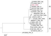
Phylogenetic tree generated with nucleotide sequences of the coat protein (CP) gene of cucumber mosaic virus (CMV) isolates. The lengths of branches are proportional to genetic distances, and branch significance is indicated at nodes for posterior probability values higher than 50 %. The nucleotide sequence of the CP gene of peanut stunt virus (PSV) (JN135292) was used as an outgroup for comparison. The nucleotide sequence of the CMV HP22 isolate is highlighted in red. Subgroups of CMV isolates: IA, IB, and II.
The mechanical transmissions were performed using the CMV HP22 isolate and plants of varying species (Table 1). The symptomatic leaves were ground in a 0.01 M potassium phosphate buffer (pH 7.0) containing 0.1 % sodium sulfite (1:10 w/v). The extract was applied by rubbing it onto the leaves of the seedlings (2-3 true leaves), which had previously been dusted with 600-mesh carborundum. A total of three to ten plants of each species or cultivar was inoculated and subsequently kept in a glasshouse for observation of symptoms within 30 days post-inoculation (dpi) for plants from the Amaranthaceae, Cucurbitaceae, and Solanaceae families, and 180 dpi for plants from the Bignoniaceae family. To confirm viral infection, RT-PCR using the 5’CP/3’CP primer pair and PTA-ELISA were performed.
Reactions of different plant species after mechanical inoculation with leaf extract of Handroanthus heptaphyllus infected with cucumber mosaic virus. The analysis was conducted based on symptoms, Reverse Transcription Polymerase Chain Reaction (RT-PCR), and Plate-Trapped Antigen Enzyme-Linked Immunosorbent Assay (PTA-ELISA).
The CMV HP22 isolate was mechanically transmitted to Handroanthus albus (66.6 % efficiency), H. heptaphyllus (33.3 % efficiency), H. impetiginosus (66.6 % efficiency), Tabebuia heptaphylla (33.3 % efficiency), and T. roseoalba (33.3 % efficiency) (Table 1). RT-PCR yielded positive results in one of three H. albus and H. impetiginosus plants. The mean absorbance values for extracts from infected plants were compared to those of the respective healthy controls (same plant species), and the results are as follows: H. albus (0.212/0.016), H. heptaphyllus (0.276/0.074), H. impetiginosus (0.297/0.047), T. heptaphylla (0.249/0.078), and T. roseoalba (0.238/0.078).
The vector transmission of the CMV HP22 isolate was investigated using Aphis gossypii Glover reared on cotton (Gossypium hirsutum L.). The aphids were initially fasted for 60 min and subsequently transferred to a symptomatic leaf of H. heptaphyllus (HP22) for a 15-minute virus acquisition access period. The aphids (25 insects per plant) were transferred individually to seedlings (2-3 true leaves) of test plants for a virus inoculation access period (IAP) of 15 min. Four plants of each species (H. heptaphyllus, H. impetiginosus, T. heptaphylla, and T. roseoalba) were individually inoculated. Following the IAP, the aphids were manually killed, and the plants were treated with an insecticide (thiamethoxan). All plants were subsequently kept in a glasshouse to evaluate symptoms. Virus infection was confirmed at 180 dpi by RT-PCR using the 5’CP/3’CP primer pair and PTA-ELISA.
The A. gossypii successfully transmitted the CMV HP22 isolate to all H. heptaphyllus and T. heptaphylla plants (Table 1). The RT-PCR results were negative for all samples. The mean absorbance values for extracts from infected plants in comparison to the respective healthy controls were as follows: 0.276/0.074 for H. heptaphyllus and 0.306/0.085 for T. heptaphylla.
A minimum of 80 species of aphids are capable of transmitting CMV, with A. gossypii and Myzus persicae Sulz. being the most significant and effective (Palukaitis and García-Arenal, 2003). It is uncommon for aphids to colonize ipê plants. However, as the relationship between CMV and aphids is nonpersistent, the virus can be acquired and transmitted from and to H. heptaphyllus and T. heptaphylla plants during aphid feeding probes to identify the host plants.
Interveinal chlorosis was documented in plants of the H. albus, H. impetiginosus, and T. roseoalba species that had been mechanically inoculated and manifested at 150 dpi. Additionally, H. impetiginosus plants exhibited leaf distortion (Figure 1C-E). No symptoms were observed in plants of H. heptaphyllus and T. heptaphylla that had been inoculated mechanically or by aphids. None of the mechanically inoculated plants from the families Cucurbitaceae (five species) and Solanaceae (four species) exhibited symptoms. They tested negative for CMV infection by RT-PCR and PTA-ELISA (Table 1). Despite the broad host range of CMV, the isolate from H. heptaphyllus did not infect all potential hosts, particularly those belonging to the Cucurbitaceae and Solanaceae families, as might occur in nature. Some common CMV isolates, which typically infect solanaceous and cucurbit crops, have also been reported to be unable to infect legume species (Jacquemond, 2012; Palukaitis et al., 1992). Notably, the CMV HP22 isolate could not infect Chenopodium amaranticolor H.J. Coste & A. Reyn. and C. quinoa Willd. plants, which are commonly used as indicator plants for CMV infection (Sastry et al., 2020). This observation leads to the hypothesis that the CMV from H. heptaphyllus may have a specific host range, infecting only tree species of the genera Handroanthus and Tabebuia.
The study of diseases caused by viruses in forest trees is hindered by the sporadic occurrence of such diseases, which has precluded the demonstration of their impacts (Nienhaus and Castello, 1989). Despite the difficulties in detecting viral pathogens in forest trees (Buttner et al., 2023), the present study successfully demonstrated the pathogenicity of the CMV HP22 isolate through mechanical and aphid transmission tests. Additionally, the virus infection was effectively detected through PTA-ELISA.
Among different tree species, CMV has only been documented to infect the karaka tree (Corynocarpus laevigatus J.R. & G. Forst.) in New Zealand in 1997 (Ashby et al., 1977) and the wisteria sinensis in Serbia in 2010 (Milojević et al., 2016). Given that H. heptaphyllus is a perennial plant with an exceptionally long lifespan, it is plausible that it may serve as a reservoir for CMV in the field. To the best of our knowledge, this represents the first report on CMV infecting H. heptaphyllus plants on a global scale.
Acknowledgments
This research received financial support from Fundação de Amparo à Pesquisa do Estado de São Paulo – FAPESP (grant number 2018/18274-3, and 2021/02179-4). The first author has a Postdoctoral scholarship from FAPESP (2020/05563-7).
Data availability statement
The data supporting this study's findings are available upon reasonable request to the corresponding author.
References
-
Amaral JB, Martins L, Forti VA, Cícero SM, Marcos Filho J. 2011. X-ray test to evaluate the physiological potental of Tabebuia heptaphylla seeds. Revista Brasileira de Sementes 33: 601-607 (in Portuguese, with abstract in English). https://doi.org/10.1590/S0101-31222011000400001
» https://doi.org/10.1590/S0101-31222011000400001 -
Ashby JW. 1977. Infection of karaka (Corynocarpus laevigatus J.R. & G. Forst.) by cucumber mosaic virus. New Zealand Journal of Agricultural Research 20: 533-534. https://doi.org/10.1080/00288233.1977.10427370
» https://doi.org/10.1080/00288233.1977.10427370 - Auer C. 2001. Diseases of Ipes: Identification and Control = Doenças em Ipês: Identificação e Controle. Embrapa Florestas, Colombo, PR, Brazil (in Portuguese).
-
Bello VH, Carpim L, Lorenzi HJ, Rezende JAM, Kitajima EW. 2023. Natural occurrence of cucumber mosaic virus infecting Physalis peruviana plants in Brazil. Journal of Phytopathology 171: 143-144. https://doi.org/10.1111/jph.13165
» https://doi.org/10.1111/jph.13165 -
Bujarski J, Gallitelli D, García-Arenal F, Pallás V, Palukaitis P, Reddy MK, et al. 2019. ICTV virus taxonomy profile: Bromoviridae Journal of General Virology 100: 1206-1207. https://doi.org/10.1099/jgv.0.001282
» https://doi.org/10.1099/jgv.0.001282 -
Buttner C, Landgraf M, Colino HLF, von Bargen S, Bandte M. 2023. Virus diseases of forest and urban trees. p. 61-97. In: Asiegbu FO, Kovalchuk A eds. Forest microbiology: tree diseases and pests. Elsevier, Amsterdam, The Netherlands. https://doi.org/10.1016/B978-0-443-18694-3.00011-0
» https://doi.org/10.1016/B978-0-443-18694-3.00011-0 -
Eiras M, Resende RO, Missiaggia AA, Ávila AC. 2001. RT-PCR and Dot Blot hybridization methods for a universal detection of tospoviruses. Fitopatologia Brasileira 26: 170-175. https://doi.org/10.1590/S0100-41582001000200009
» https://doi.org/10.1590/S0100-41582001000200009 -
Ha C, Coombs S, Revill PA, Harding RM, Vu M, Dale JL. 2008. Design and application of two novel degenerate primer pairs for the detection and complete genomic characterization of potyviruses. Archives of Virology 153: 25-36. https://doi.org/10.1007/s00705-007-1053-7
» https://doi.org/10.1007/s00705-007-1053-7 -
Jacquemond M. 2012. Cucumber mosaic virus. p. 439-504. In: Maramorosch K, Shatkin AJ, Murphy FA eds. Advances in Virus Research. Elsevier, Amsterdam, The Netherlands. https://doi.org/10.1016/B978-0-12-394314-9.00013-0
» https://doi.org/10.1016/B978-0-12-394314-9.00013-0 -
Kitajima EW. 2020. An annotated list of plant viruses and viroids described in Brazil (1926-2018). Biota Neotropica 20: 20190932. https://doi.org/10.1590/1676-0611-BN-2019-0932
» https://doi.org/10.1590/1676-0611-BN-2019-0932 -
Milojević K, Radović N, Stanković I, Vučurović A, Nikolić D, Bulajić A, et al. 2016. First report of Cucumber mosaic virus infecting Wisteria sinensis in Serbia. Plant Disease 100: 1799. https://doi.org/10.1094/PDIS-01-16-0096-PDN
» https://doi.org/10.1094/PDIS-01-16-0096-PDN -
Ng JCK, Falk BW. 2006. Virus-vector interactions mediating nonpersistent and semipersistent transmission of plant viruses. Annual Review of Phytopathology 44: 183-212. https://doi.org/10.1146/annurev.phyto.44.070505.143325
» https://doi.org/10.1146/annurev.phyto.44.070505.143325 -
Nienhaus F, Castello JD. 1989. Viruses in forest trees. Annual Review of Phytopathology 27: 165-186. https://doi.org/10.1146/annurev.py.27.090189.001121
» https://doi.org/10.1146/annurev.py.27.090189.001121 -
Rizos H, Gunn LV, Pares RD, Gillings MR. 1992. Differentiation of cucumber mosaic virus isolates using the polymerase chain reaction. Journal of General Virology 73: 2099-2103. https://doi.org/10.1099/0022-1317-73-8-2099
» https://doi.org/10.1099/0022-1317-73-8-2099 -
Palukaitis P, Roossinck MJ, Dietzgen RG, Francki RIB. 1992. Cucumber mosaic virus. Advances in Virus Research 41: 281-348. https://doi.org/10.1016/S0065-3527(08)60039-1
» https://doi.org/10.1016/S0065-3527(08)60039-1 -
Palukaitis P, García-Arenal F. 2003. Cucumoviruses. Advances in Virus Research 62: 241-323. https://doi.org/10.1016/s0065-3527(03)62005-1
» https://doi.org/10.1016/s0065-3527(03)62005-1 -
Roossinck MJ. 2002. Evolutionary history of Cucumber mosaic virus deduced by phylogenetic analyses. Journal of Virology 76: 3382-3387. https://doi.org/10.1128/jvi.76.7.3382-3387.2002
» https://doi.org/10.1128/jvi.76.7.3382-3387.2002 -
Sastry KS, Mandal B, Hammond J, Scott SW, Briddon RW. 2020. Encyclopedia of Plant Viruses and Viroids. Springer Nature, New Delhi, India. https://doi.org/10.1007/978-81-322-3912-3_1041
» https://doi.org/10.1007/978-81-322-3912-3_1041 - Scarante AG, Matos MFS, Soares MTS, Aguiar AV, Wrege MS. 2017. Distribution of Handroanthus heptaphyllus in Brazil and future projections according to global climate change. Revista Geama 3: 201-207.
Edited by
-
Edited by:
Alice Kazuko Inoue-Nagata