Integrated surveillance of arboviruses in febrile patients from the Brazilian Amazon reveals complex co-circulation dynamics and hidden viral diversity
ABSTRACT
Background: Arboviral infections continue to be a significant public health challenge in the Brazilian Amazon. Overlapping symptoms, limited laboratory access, and the circulation of multiple arboviruses hamper clinical diagnosis. This study aimed to characterize the epidemiological, clinical, laboratory and genomic profiles of arboviral infections in febrile patients in Manaus, Brazil, and explore additional viral agents using metagenomic sequencing.
Methods: A cross-sectional study was conducted between February 2021 and February 2023 at a tertiary reference center in Manaus, Brazil. Patients aged ≥ 5 years of age presenting with a rash and either a fever or a history of fever lasting <7 days and a negative thick blood smear for malaria were enrolled. Serum samples were tested for dengue virus (DENV), Zika virus (ZIKV), Chikungunya virus (CHIKV), yellow fever virus (YF), Oropouche virus (OROV), and Mayaro virus (MAYV) using ELISA and RT-qPCR. Positive samples were subjected to amplicon-based genome sequencing for phylogenetic analysis. A subset of RT-qPCR negative samples was analyzed using de novo shotgun metagenomic sequencing.
Results: Among the 708 enrolled participants, 243 (34.3%) had a laboratory-confirmed arboviral infection: 92 (37.9%) DENV, 64 (26.3%) CHIKV, and 4 (1.6%) ZIKV, while 83 (34.2%) laboratory profiles were compatible with coinfection, predominantly DENV+CHIKV (55/83; 66.3%). Circulation of DENV-1 genotype V and DENV-2 genotypes III (Asian American) and II (Cosmopolitan) was identified. Metagenomic analysis of 35 samples detected Pegivirus hominis and Erythroparvovirus primate 1.
Conclusions: These findings demonstrate complex arbovirus co-circulation in Manaus and support integrated surveillance strategies combining molecular, serological, and genomic approaches.
Keywords:
Arboviruses; Surveillance; Acute febrile illness; Amazon; Brazil
INTRODUCTION
Arboviral diseases such as dengue, Zika, and chikungunya, caused respectively by dengue virus (DENV), Zika virus (ZIKV), and chikungunya virus (CHIKV), remain a major public health challenge in tropical and subtropical regions, since ecological and social conditions favor the proliferation of Aedes aegypti and Aedes albopictus1,2. Recurrent outbreaks of arboviral infections have occurred in the Americas over the past few decades, contributing to the burden of acute febrile diseases3-5. Brazil, particularly its Amazon region, presents favorable conditions for the circulation and maintenance of arboviruses due to its climate, widespread vector distribution, intense human mobility, and limited access to timely diagnostic resources6.
DENV circulates endemically throughout Brazil, with the sustained presence of all four serotypes7-10, while the introduction of CHIKV and the emergence of ZIKV further increased the complexity of arbovirus transmission dynamics11,12. Furthermore, the Oropouche virus (OROV), an orthobunyavirus frequently reported in the Amazon region, has gained prominence as a significant cause of acute fever cases. Its increasing presence further highlights the complexity of the arbovirus transmission scenario in northern Brazil13-15. These viruses co-circulate nationwide and frequently produce overlapping clinical manifestations and, in some cases, coinfections, which complicate diagnosis and can lead to underestimation of incidence12,16,17. To address this, laboratory surveillance using molecular and serological methods is necessary to complement clinical assessment, accurately identify circulating viral pathogens, estimate outbreaks, and establish monitoring and control measures18-20. Genomic surveillance is relevant for monitoring viral diversity, detecting new introductions, and characterizing the arbovirus transmission at regional and international levels21.
Manaus, capital of the Amazonas state in the northern region of Brazil, has an estimated population of approximately 2.3 million inhabitants22. There have been marked fluctuations in the incidence of arboviruses over the last decade, with sustained transmission of DENV, CHIKV and ZIKV23-26. Integrated surveillance combining clinical, laboratory, and genomic data remains limited. To this end, non-specific metagenomic tools can complement these approaches by identifying viruses not typically screened for, contributing to a comprehensive picture of viral spread. This study aimed to analyze the epidemiological, clinical, laboratory and genomic aspects of arboviral infections in febrile patients in Manaus, Brazil, and, in an exploratory manner, identify additional viral agents using metagenomic sequencing.
METHODS
Ethics Statement
This study was approved by the Ethics Committee of the Fundação de Medicina Tropical Doutor Heitor Vieira Dourado (FMT-HVD), CAAE:67595417.0.0000.0005, approved on November 24, 2017). It is in accordance with the Declaration of Helsinki. All participants provided written informed consent or assent.
Study design and setting
The study was a cross-sectional investigation conducted at the Malaria Outpatient Clinic of FMT-HVD in Manaus, Brazil, between February 2021 and February 2023, a national and international reference center for tropical disease care and febrile syndromes27.
Study population and sample collection
Patients aged ≥5 years were eligible if they had a fever or a recent fever lasting <7 days, a negative thick blood smear for malaria at presentation, and at least one symptom compatible with arboviral infection. Participants were included in the study during a clinical consultation at the outpatient clinic. At that time, clinical and epidemiological data were recorded, and blood samples were collected for laboratory analysis. Each participant was included only once during the acute febrile episode. Re-enrollment of the same individual in the study during the same illness episode was not permitted.
Following this initial consultation, participants were followed longitudinally, with visits scheduled on days 28 and 270 after inclusion. However, the analyses presented in this study were restricted to clinical and laboratory data collected during the initial consultation, which corresponded to the acute phase of illness.
A 5-10 mL venous blood sample was collected for serology and molecular analysis. Arboviral infections were defined based on laboratory evidence obtained through molecular and/or serological testing. Possible coinfections were defined by molecular detection and/or serological evidence of more than one arbovirus; due to flavivirus cross-reactivity, DENV- and ZIKV-IgM-positive cases without molecular confirmation were classified as FLAV.
Laboratory diagnosis
Blood samples collected during the acute phase, at the time of participant inclusion, were subjected to serological analysis and molecular screening (RT-qPCR) for arboviruses. RT-qPCR assays were performed on these samples to detect DENV, ZIKV, CHIKV, OROV, and yellow fever (YFV).
Serological investigation
Serum samples were screened for specific IgM antibodies against DENV, ZIKV, and CHIKV using commercially available enzyme-linked immunosorbent assay (ELISA) kits according to the manufacturers' instructions. SERION ELISA Classic Dengue Virus IgM kits were used for DENV detection, Novagnost Zika Virus IgM µ-capture for ZIKV detection, and the Euroimmun Anti-Chikungunya Virus ELISA for CHIKV detection. For each essay, serum samples were diluted according to the protocols provided by the respective kits. All necessary controls, including positive, negative, and calibrators/cut-off controls, were included on each plate. Results were interpreted qualitatively based on the manufacturer’s cutoff values. Equivocal or borderline results were retested, and if they remained equivocal, they were classified as inconclusive.
Nucleic acid extraction and molecular investigation
A total of 100 µL of each serum sample was submitted to RNA extraction using the Extracta kit fast DNA and RNA viral (MVXA-P096 FAST; Loccus, Brazil) according to the manufacturer’s instructions, utilizing an Extracta 96 - DNA and RNA extractor and purifier (Loccus, Brazil). A negative template control was included in each extraction batch. One-step real-time polymerase chain reaction (RT-qPCR) was performed using the GoTaq 1-Step RT-qPCR System (Promega, USA) and oligos targeting the four DENV serotypes (DENV1-4), ZIKV, CHIKV, YFV, OROV and MAYV (Table S1)28-31. Reactions were run on a real-time PCR platform with appropriate positive and negative controls, and samples with Ct values ≤38 were considered positive30,31. Positive and negative (nuclease-free water) template controls were used.
Amplicon-based sequencing
DENV-positive samples by RT-qPCR sufficient viral load were selected for amplicon-based sequencing. The cDNA synthesis, DENV genome amplification, and library preparation were carried out using the Illumina COVIDSeq Test (Illumina, USA) modified by replacing primers as part of the kit for the primer pools primer panel for DENV-1 and DENV-2 designed by the CADDE (Brazil-UK Centre for Arbovirus Discovery, Diagnosis, Genomics and Epidemiology) project (the generated sequences from the positive DENV-1 and DENV-2 samples, Tables S2 and Table S3). Each sequencing run included a negative (nuclease-free water) and a negative template control during cDNA synthesis and amplicon generation. Library quantification was performed using the Qubit dsDNA HS assay kit on a Qubit 2.0 fluorometer (Thermo Fisher Scientific, USA). Library quality was verified using a High Sensitivity D1000 ScreenTape kit on the 4150 TapeStation system (Agilent Technologies, USA). PhiX Control v3 was used as quality control and validation for sequencing runs. Pooled libraries were normalized to 4 nM, denatured with 0.2 N NaOH, and sequenced on the Illumina MiSeq System (Illumina, USA), using a MiSeq reagent kit v2 (2 x 150 bp cycles) (Illumina, USA) with a paired-end strategy.
Genome assembly and consensus generation
Strict quality control of the raw demultiplexed sequencing reads was performed using Trimmomatic (v.0.39)32, which removed low-quality bases (minimum Phred score of Q30), sequencing adapters, primer sequences and short reads (<50 nucleotides). The cleaned paired-end reads were mapped to the DENV-1 or DENV-2 reference genome sequences (NCBI accession numbers NC_001477 for DENV-1 and NC_001474 for DENV-2) using BWA (v.0.7.17)33. BAM files were sorted, indexed and used for consensus sequence generation with SAMtools (v.1.10) and iVar (v.1.3.1)34,35.
Shotgun sequencing and bioinformatic analysis of metagenomic data
To enhance viral surveillance, RT-qPCR-negative samples with sufficient nucleic acid concentration were selected for shotgun metagenomic sequencing. Total nucleic acids (DNA and RNA) were extracted using the MagMAX™ CORE Nucleic Acid Purification kit (Thermo Fisher Scientific, USA), following the manufacturer’s protocol. Negative extraction controls were included throughout the process to monitor potential contamination. RNA was reverse-transcribed into complementary DNA (cDNA) using a 15-nucleotide degenerate random primer (15N), enabling broad-spectrum detection of RNA viruses without prior sequence information.
Metagenomic libraries were prepared using the Illumina® DNA Prep kit (Illumina, USA). Library concentrations were quantified using the Qubit dsDNA High Sensitivity assay kit on a Qubit 2.0 fluorometer (Thermo Fisher Scientific, USA), and library quality was assessed via the High Sensitivity D1000 ScreenTape kit on the 4150 TapeStation system (Agilent Technologies, USA). Libraries were normalized to equimolar concentrations (4 nM), pooled, and sequenced on the NextSeq 1000 platform (Illumina, USA) using a P1 flow cell with paired-end 2 × 150 bp reads.
Raw FASTQ files were screened using the Chan Zuckerberg ID (CZID). Based on the taxonomic classification results, the raw sequencing data were then imported into Geneious Prime (v.2025.0.2). Reads were trimmed to remove low-quality bases and sequencing adapters, retaining only those with an average Phred quality score ≥ Q30 and a minimum length of 50 nucleotides. The resulting high-quality reads were mapped to the reference genomes of the viral taxa identified by CZID, enabling targeted reconstruction and refinement of viral sequences (NCBI accession numbers: ON340918.1 for Pegivirus hominis and NC_000883.2 for Erythroparvovirus primate 1). Consensus sequences were generated from the mapped alignments and analyzed using Genome Detective36.
Phylogenetic analysis
Consensus nucleotide sequences were submitted to Genome Detective for DENV-1 and 2 genotype assignment36. For the phylogenetic analysis, the newly generated sequences (Table S3) were combined with DENV-1-2 complete genomes with high coverage (≤ 5 % of Ns), date, and country of collection retrieved from the EpiArbo, GISAID database (https://gisaid.org) up to July 202537. The generated sequences were submitted to genotyping, which was performed using a dengue virus typing tool (Tables S4 and Table S5). To investigate the phylodynamics of DENV-1 and DENV-2, we combined the newly generated sequences with publicly available DENV-1 and DENV-2 genome sequences (Tables S6, Table S7 and Table S8).
Phylogenetic analyses of Pegivirus hominis and Erythroparvovirus primate 1 were performed using the newly generated sequences, together with representative reference sequences retrieved from GenBank (https://www.ncbi.nlm.nih.gov/genbank)(Tables S9,S10,S11 and S12).
The nucleotide sequences were aligned using MAFFT (v.7.526)38 and manually curated in AliView (v.1.28)39. A maximum likelihood (ML) phylogenetic tree was inferred using IQ-TREE (v.1.6.12)40 under the best-fit substitution model implied by the ModelFinder41 model-selection method implemented in IQ-TREE40. The reliability of branching patterns was tested using the ultrafast bootstrap approximation (UFBoot), with 1,000 bootstrap alignments, and the SH-like Approximate Likelihood-ratio test (SH-aLRT), with 1,000 replicates42. The time-scaled ML phylogenetic trees were inferred using TreeTime43. Time-resolved trees were not generated for the Pegivirus hominis and Erythroparvovirus primate 1 datasets due to limited temporal signal and sample size. All trees were visualized and edited using R statistical software (v.4.3.1)44.
Statistical analysis
Continuous variables with a normal distribution were presented as mean and standard deviation, while categorical variables were described as n/N (percentage). For exploratory comparisons between groups, ANOVA was used for continuous variables with a normal distribution, and Fisher's exact test was used for categorical variables. Given the descriptive nature of the study, the absence of pre-established hypotheses, and the lack of a formal sample size calculation, these analyses should be understood as exploratory. For this reason, the p-values presented need to be interpreted with caution, as no formal adjustment for multiple comparisons was performed. All statistical analyses were performed using R software (version 4.4.2)44 .
RESULTS
Demographic and clinical characteristics
Between February 2021 and February 2023, 708 patients with symptoms suggestive of arboviruses were recruited, and 243 (34.3%) had laboratory-confirmed infection: 92 (37.9%) were positive for DENV, 64 (26.3%) for CHIKV, and 4 (1.6%) for ZIKV, while 83 (34.2%) had serological and/or molecular profiles compatible with possible coinfection , predominantly DENV+CHIKV (55; 66.3%) (Figure S1 and Table 1). No infections caused by OROV, YFV or MAYV were detected. DENV-1 (18/243; 7.4%) and DENV-2 (3/243; 1.2%) were detected. Most cases occurred in 2022 (458; 63.4%). The distribution of infections by year is shown in Table 2.
Most participants were men (138; 57%), with a mean age of 40 years, and were of mixed race (168; 70%) (Table 1). The most frequent comorbidity was hypertension (24; 9.9%), with a difference for hyperlipidemia, which was prevalent in DENV infection (8; 8.8%). Previous contact with individuals with recent arbovirus infections was common among patients with DENV (18; 20%) and ZIKV (1; 25%).
Facial flushing (39; 42%), rash/skin changes (52; 57%), generalized rash (31; 34%), conjunctivitis (21; 23%), pruritus (43; 47%), peripheral edema (23; 25%), mottled skin (19; 21%), and cold and clammy skin (11; 12%) were frequently observed among individuals with DENV infection. The overall analysis indicated a difference in the distribution of these clinical manifestations across all study groups (p < 0.005). Bone and/or joint pain (69; 75% in DENV; 29; 53% in DENV+CHIKV) and retro-orbital pain (59; 64% in DENV; 20; 36% in DENV+CHIKV), as well as sore throat (24; 26% in DENV; 16; 29% in DENV+CHIKV), dyspnea (20; 22% in DENV; 6; 11% in DENV+CHIKV), oliguria (16; 17% in DENV; 8; 15% in DENV+CHIKV), and choluria (17; 19% in DENV; 7; 13% in DENV+CHIKV), showed variation in frequency among the groups. Exploratory analysis indicated differences in symptom distribution across groups (p ≤ 0.017).. Differences in hydration status were observed in the global comparison, with a higher proportion of dehydration observed in individuals infected with CHIKV (16; 25%) and those with DENV+CHIKV coinfection (17; 31%) (p = 0.003).
Regarding laboratory parameters, lower erythrocyte, hemoglobin, and hematocrit levels were observed in coinfections (DENV+CHIKV; erythrocytes: 4.68-4.75 million/µL; hemoglobin: 13.29-13.73 g/dL; hematocrit: 39.4-41.7% and FLAV+CHIKV; erythrocytes: 4.68-4.75 million/µL; hemoglobin: 13.29-13.73 g/dL; hematocrit: 39.4-41.7%) compared with the other groups. The statistical analysis indicated differences across groups for erythrocytes (p = 0.03), hemoglobin (p = 0.006), and hematocrit (p = 0.003), based on global tests only. Leukocyte levels were lowest in the DENV group (5,221/µL), with difference across groups (p < 0.001). Platelet counts varied among groups, being lower in the DENV/FLAV group (163,018-177,574/µL) and higher in the FLAV group, with an overall group effect (p = 0.034). Plateletcrit was also higher in the CHIKV group, with a global difference across groups (p = 0.009). Mean corpuscular volume showed moderate variation among groups (CHIKV and DENV+CHIKV: 85.0-85.8 fL; FLAV: 88.5 fL), with an overall difference (p = 0.013). Mean corpuscular hemoglobin concentration (MCHC) showed minimal variation (32.95-33.98 g/dL), although a global difference across groups was detected (p = 0.005). Red Cell Distribution Width values were higher in the FLAV and FLAV+CHIKV groups (12.33-12.65%) compared with the DENV and CHIKV groups (11.54-11.89%), with a marginal difference across groups (p = 0.05).
Phylodynamics of DENV
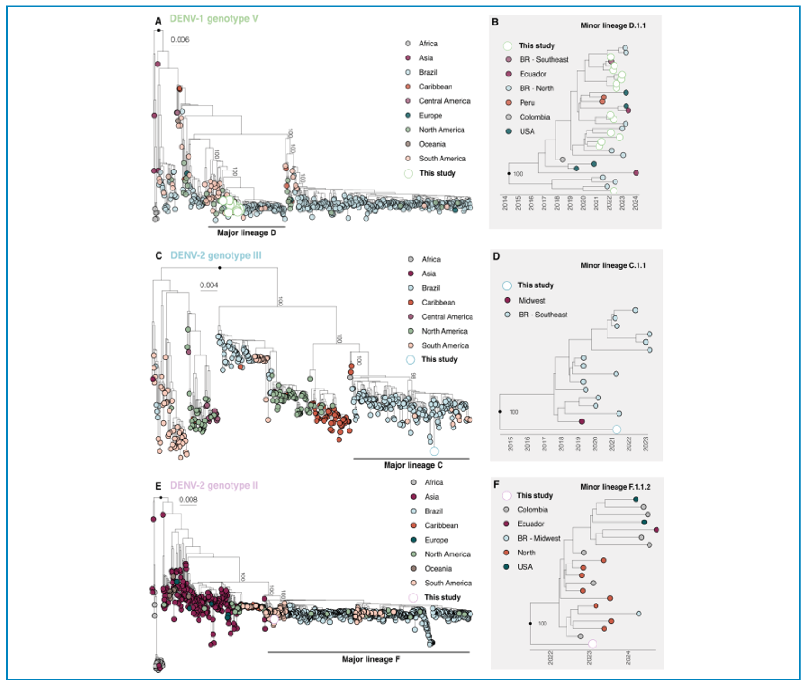
The newly generated DENV-1 sequences were classified as belonging to genotype V (DENV-1-V), while DENV-2 sequences were classified into genotypes III (Asian American) and II (Cosmopolitan) (Figures 1A, 1C and 1E). Phylogenetic analysis revealed that the generated sequences clustered within the major lineage D and the minor lineage D.1.1 (Figures 1A and 1B). The DENV-GIII sequence was found within clade C, specifically minor lineage C.1.1 (Figures 1C and 1D), Phylogenetic analyses revealed clustering within lineages closely related to strains circulating in South America and multiple regions of Brazil, suggesting regional and interregional viral dispersion (Figure 1; Tables S4 and Table S5).

Phylogenetic analysis of DENV in Manaus 2021-2023. (A) Maximum likelihood (ML) phylogenetic analysis of 14 complete genome sequences from DENV-1 genotype V (DENV-1-GV) generated in this study (2021-2023), plus 549 publicly available sequences. The sequences grouped within the major clade E. (B) Time-scaled DENV-1-GV phylogenetic subtree of major clade E, minor lineage D.1.1. (C) ML phylogenetic analysis of one (2021) complete genome sequences from DENV-2 genotype III (Asian American) (DENV-2-GIII) generated in this study, plus 482 publicly available sequences. The sequence is grouped within the major clade C. (D) Time-scaled DENV-2-GIII phylogenetic subtree of major clade C, minor lineage C.1.1. (E) ML phylogenetic analysis of one (2023) complete genome sequence from DENV-2 genotype II (Cosmopolitan) (DENV-2-GII) generated in this study, plus 1,005 publicly available sequences. The sequence is grouped within the major clade F. (F) Time-scaled DENV-2-GII phylogenetic subtree of major clade F, minor lineage F.1.1.2. The trees are mid-point rooted, and the scale bar is in units of nucleotide substitutions per site (s/s). Colors of the dots represent different sampling locations and genome sequences generated in this study.
Metagenomic analyses
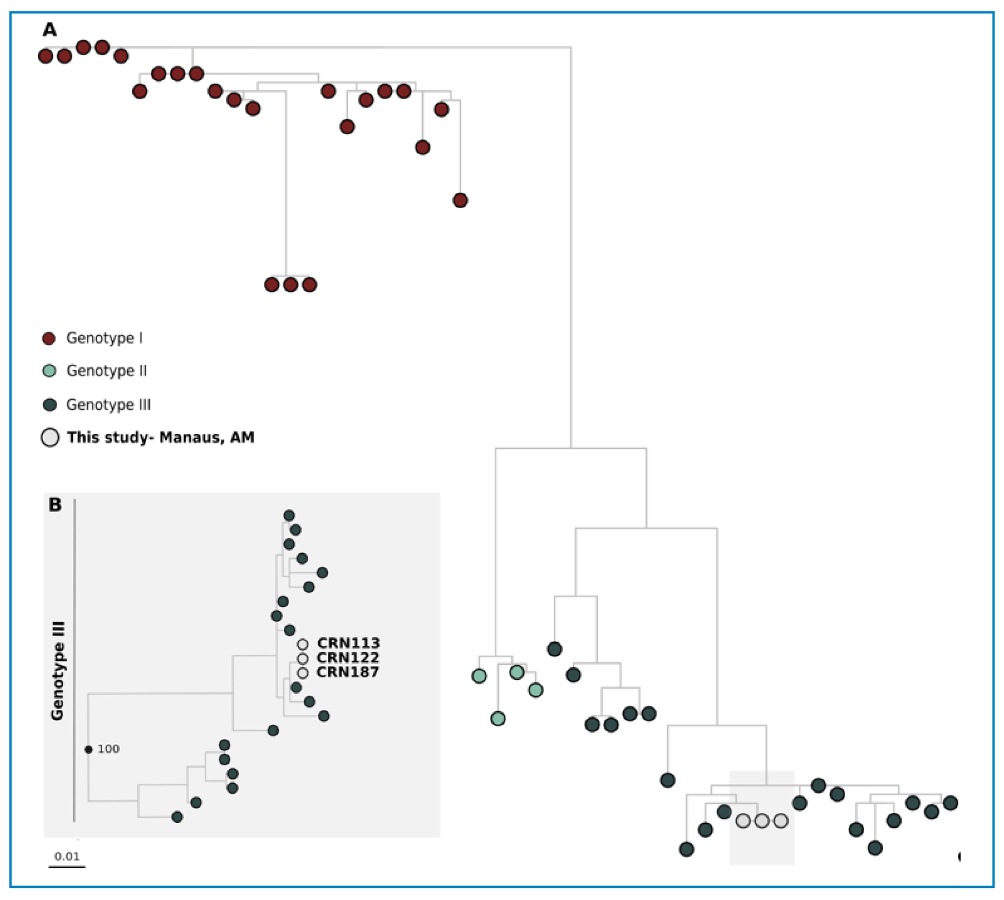
Shotgun metagenomic sequencing of 35 serum samples identified DENV-1 in two cases, both clustering within genotype V and confirmed by serotype-specific RT-PCR (Figure 1). In addition, Pegivirus hominis (genotype II in 2 samples, Figure 2; Table S10) and Erythroparvovirus primate 1 (genotype III, Figure 3; Table S11) were detected in a subset of samples and confirmed through phylogenetic analysis (Figure 2 and Figure 3).

Phylogenetic analysis of Pegivirus hominis in Manaus. (A) Maximum likelihood (ML) phylogenetic tree based on an 8,988-nucleotide coding region of the Pegivirus hominis genome. The dataset comprises 47 sequences, including one sequence generated in this study (Manaus, Brazil, 2022) and 46 reference sequences retrieved from GenBank, representing different genotypes. (B) Enlarged view of the clade containing the one sequence from this study (CRN188), which clustered within Genotype II.

Phylogenetic analysis of Erythroparvovirus primate 1 in Manaus. (A) Maximum likelihood (ML) phylogenetic tree based on a 423-nucleotide fragment of the partial VP1/VP2 coding region of Erythroparvovirus primate 1. The dataset includes three sequences obtained in this study (Manaus, Brazil, 2022) and 50 reference sequences retrieved from GenBank. Sequences are grouped by genotype, represented by distinct colors. The sequences from Manaus clustered within Genotype III. (B) Detail of the clade corresponding to Genotype III, showing the placement of the three sequences generated in this study (CRN113, CRN122 and CRN187).
DISCUSSION
This cross-sectional study examined the circulation of ZIKV, DENV, CHIKV, OROV, YFV, and MAYV among patients with suspected arboviral infection attending a tertiary healthcare unit in Manaus between February 2021 and February 2023. Infections with DENV, CHIKV, and ZIKV were confirmed using serological and molecular techniques, with DENV predominating; infections with Erythroparvovirus primate 1 and, probably incidentally, Pegivirus hominis were also identified. Furthermore, serological profiles suggest potential coinfections; these findings should be interpreted cautiously, as they do not constitute definitive evidence of simultaneous infection. A marked increase in arboviral cases was observed in 2022, coinciding with reports of increased OROV activity in the region, coinciding45. These findings highlight the dynamic nature of arbovirus circulation and the need for continuous surveillance. Although essential, laboratory surveillance of arboviruses in Brazil is primarily limited to scientific research and academic initiatives.
Manaus has historically faced recurring dengue outbreaks, characterized by the sequential emergence and co-circulation of multiple DENV serotypes23,46-48. Detection of DENV-1 and DENV-2 aligns with previous regional reports. Frequent laboratory profiles indicating possible DENV+CHIKV coinfections support findings from other studies in the Brazilian49-51, and highlight the diagnostic challenges in endemic areas. The presence of probable coinfections poses an additional challenge for both diagnosis and clinical management, which may be associated with more pronounced hematological alterations.
OROV is considered an important cause of acute febrile illnesses in the Amazon region13,52. In recent years, especially between 2023 and 2024, OROV has regained epidemiological prominence due to the expansion of outbreaks recorded not only in Brazil but also in other parts of South America53-56. Even after submitting all acute-phase samples from participants to RT-qPCR screening for OROV, no positive results were identified. The absence of detections may be associated with temporal fluctuations in viral circulation, differences inherent to local transmission dynamics, or the epidemiological context of the study period. Nevertheless, our findings reinforce the need to maintain broad molecular surveillance strategies for arboviruses in the Amazon, considering that several pathogens can circulate simultaneously and contribute in different ways to acute fever in the region13-15.
Regarding signs and symptoms, while overlap among arboviruses, DENV infections frequently display cutaneous and vasomotor signs, consistent with classic dengue57-59. Bone and joint pain, as well as retro-orbital pain, are common in both DENV infections and cases with laboratory evidence of possible DENV and CHIKV coinfection. Both viruses trigger a strong systemic inflammatory response, with CHIKV inducing a more pronounced reaction60-62. Although some signs may aid clinical suspicion, the overlap in symptoms limits the value of clinical features alone, underscoring the need for combined serological and molecular testing.
Laboratory parameters vary depending on the virus causing the infection. DENV+CHIKV and FLAV+CHIKV showed reductions in erythrocyte, hemoglobin, and hematocrit levels. Experimental studies involving arbovirus coinfections demonstrate substantial reductions in red blood cell counts and hematocrit values during the acute phase. These alterations are consistent with intense systemic inflammatory responses and possible impairment of erythropoiesis in coinfections63. These results are consistent with research that points to a possible immune dysregulation during arbovirus co-infections64. However, due to the cross-sectional design of this study, it is not possible to establish cause-and-effect relationships between the observed phenomena. However, these findings should be interpreted with caution, given the study’s cross-sectional design and the absence of data on clinical outcomes. Leukopenia and thrombocytopenia, typical of DENV infection, were confirmed, while the reduction in platelets in the DENV+FLAV group suggests a combined effect on their consumption or destruction. The increase in plateletcrit in CHIKV cases may reflect a compensatory response or platelet activation associated with the CHIKV-induced inflammatory process.
Phylogenetic analyses revealed the co-circulation of different DENV-1 and DENV-2 genotypes in Manaus, with a predominance of DENV-1 genotype V and detection of DENV-2 genotypes II (Cosmopolitan) and III (Asian American). The circulation of DENV-1-V in Manaus is consistent with patterns observed in recent years in other regions of Brazil65. The origin of DENV-1-V is traced to the Caribbean and Venezuela; however, multiple independent introductions over time have led to high genetic variability and intense viral exchange in South America, with the Brazilian Amazon potentially acting as a regional connectivity hub57-60. The clustering of samples from Manaus with sequences from the southeast and north of Brazil reinforces the notion that the country is nationally interconnected in terms of viral dissemination, raising concerns about the speed at which new genomic variants may spread in large urban centers and peripheral areas.
In Brazil, DENV-2 genotype III was first detected in 1990, followed by additional introductions through 2022, primarily from Caribbean countries and northern South America66. In the Amazon, genotype III was first reported in Acre in 202167,68. Its introduction into the state of Amazonas likely occurred between May 2020 and August 202266. In the present study, DENV-2 genotype II sequences clustered within clade F, sublineage F.1.2, and are closely related to sequences from several South American countries, consistent with regional dispersal. In contrast, the identification of a DENV-2 genotype III sequence within clade C, sublineage C.1.1, is consistent with intranational dissemination, likely originating in the northern region of Brazil and subsequently expanding to the southeastern and midwestern regions. This may indicate the Amazon region as an important area for the emergence and spread of viral lineages.
Metagenomic analysis of 35 serum samples identified Pegivirus hominis and Erythroparvovirus primate 1. The detection of Pegivirus hominis reflects an incidental finding due to the high sensitivity of metagenomic techniques, rather than a causal relationship with the clinical presentation61-63. Although its direct pathogenicity has not been established, the identification of this virus underscores the importance of non-targeted approaches to laboratory surveillance of febrile syndromes in the Amazon, which may reveal the diversity of circulating viruses and their potential interactions with endemic infections such as dengue and other arboviruses. Further studies with larger samples and improved sequencing are needed to determine the clinical significance of this virus. Analysis of the viral sequences placed the isolate within genotype 2, which is well-documented in Brazil69-77.
The detection of Erythroparvovirus primate 1 in three serum samples, all belonging to genotype III, suggests the circulation of a specific lineage in the studied population. Genotype 3 of this species appeared to be restricted to Africa but has already been detected in countries outside this continent, including Brazil78-80. Although parvovirus Erythroparvovirus primate 1 is not typically considered a febrile illness, fever may occur in some cases. The clinical presentation is variable and often nonspecific, making clinical suspicion difficult and contributing to underdiagnosis. The low frequency observed limits inferences on the magnitude of local transmission. The finding highlights the importance of molecular surveillance, particularly because viremia is typically short-lived and may be underestimated in serum samples.
This study has limitations. First, the cross-sectional design of this study, coupled with the fact that the clinical and laboratory data analyzed were collected during a single consultation, limits the ability to monitor symptom evolution and laboratory changes throughout the course of the infection. Although the protocol provided for follow-up visits, longitudinal data were not considered in the analysis presented here. Furthermore, the selection of participants from a tertiary referral center may have introduced selection biases. More restrictive clinical inclusion criteria and the likelihood of low viremia at the time of collection may have reduced case detection, while cross-reactivity observed in flavivirus serology may have affected the correct classification of infections. The absence of clinical follow-up prevented confirmation of recent infections, and metagenomic analyses limited to a subset of samples likely underestimated viral diversity. Furthermore, arbovirus transmission patterns may have been influenced by the COVID-19 pandemic. These limitations reinforce the need for further longitudinal studies with integrated approaches to better understand the dynamics of these infections.
In conclusion, this study highlights the circulation of ZIKV, CHIKV, and DENV in Manaus from 2021 to 2023 and underscores the importance of integrated surveillance across clinical, molecular, and serological dimensions. The application of metagenomic strategies revealed that febrile syndromes in the Amazon may be associated with viruses other than known arboviruses, suggesting viral diversity that may not yet be recognized. Phylogenetic analysis helped track the current and past circulation of these viruses in the region. Future studies should focus on coinfections, their clinical management and associated clinical outcomes, and the role of emerging or re-emerging viruses in the profile of febrile illnesses in the Amazon.
ACKNOWLEDGMENTS
We express our sincerest gratitude to the institutions that provided essential technical support for the development and implementation of this study. ChatGPT (OpenAI, Inc.), Manus AI, and Grammarly (Grammarly Inc.) were used solely to assist with linguistic review and improve clarity, readability, and style. All scientific content, analyses, and interpretations of results are the sole responsibility of the authors.
SUPPLEMENTARY MATERIAL
Supplementary material
Supplementary material
Supplementary material
Supplementary material
Supplementary material
Supplementary material
Supplementary material
Supplementary material
Supplementary material
Supplementary material
Supplementary material
Supplementary material
Supplementary material
REFERENCES
-
1 de Jesus Santos T, de Araújo KCGM, Góes MA de O, Bezerra-Santos M, Ribeiro CJN, dos Santos AD, et al. Spatial and Temporal Dynamics of Chikungunya Incidence in Brazil and the Impact of Social Vulnerability: A Population-Based and Ecological Study. Diseases. 2024;12:135. doi:10.3390/DISEASES12070135/S1
» https://doi.org/10.3390/DISEASES12070135/S1 -
2 Soni S, Gill VJS, . Anusheel, Singh J, Chhabra J, Gill GJS, et al. Dengue, Chikungunya, and Zika: The Causes and Threats of Emerging and Re-emerging Arboviral Diseases. Cureus. 2023;15:e41717. doi:10.7759/CUREUS.41717 PubMed PMID: 37575782.
» https://doi.org/10.7759/CUREUS.41717 PubMed PMID: 37575782 -
3 Dye-Braumuller KC, Prisco RA, Nolan MS. (Re)Emerging Arboviruses of Public Health Significance in the Brazilian Amazon. Microorganisms 2025, Vol 13, Page 650. 2025;13:650. doi:10.3390/MICROORGANISMS13030650
» https://doi.org/10.3390/MICROORGANISMS13030650 -
4 Forshey BM, Guevara C, Laguna-Torres VA, Cespedes M, Vargas J, Gianella A, et al. Arboviral Etiologies of Acute Febrile Illnesses in Western South America, 2000-2007. PLoS Negl Trop Dis. 2010;4:e787. doi:10.1371/JOURNAL.PNTD.0000787 PubMed PMID: 20706628.
» https://doi.org/10.1371/JOURNAL.PNTD.0000787 -
5 Watts DM, Russell KL, Wooster MT, Sharp TW, Morrison AC, Kochel TJ, et al. Etiologies of Acute Undifferentiated Febrile Illnesses in and near Iquitos from 1993 to 1999 in the Amazon River Basin of Peru. Am J Trop Med Hyg. 2022;107:1114-28. doi:10.4269/AJTMH.22-0259 PubMed PMID: 36162442.
» https://doi.org/10.4269/AJTMH.22-0259 -
6 Daudt-Lemos M, Ramos-Silva A, Faustino R, Noronha TG de, Vianna RA de O, Cabral-Castro MJ, et al. Rising Incidence and Spatiotemporal Dynamics of Emerging and Reemerging Arboviruses in Brazil. Viruses. 2025;17:158. doi:10.3390/V17020158/S1 PubMed PMID: 40006913.
» https://doi.org/10.3390/V17020158/S1 -
7 Daudt-Lemos M, Ramos-Silva A, Faustino R, Noronha TG de, Vianna RA de O, Cabral-Castro MJ, et al. Rising Incidence and Spatiotemporal Dynamics of Emerging and Reemerging Arboviruses in Brazil. Viruses. 2025;17:158. doi:10.3390/V17020158/S1 PubMed PMID: 40006913.
» https://doi.org/10.3390/V17020158/S1 -
8 Nunes PCG, Daumas RP, Sánchez-Arcila JC, Nogueira RMR, Horta MAP, Dos Santos FB. 30 years of fatal dengue cases in Brazil: a review. BMC Public Health. 2019;19. doi:10.1186/S12889-019-6641-4 PubMed PMID: 30898104.
» https://doi.org/10.1186/S12889-019-6641-4 -
9 De Figueiredo RMP, Naveca FG, Bastos MDS, Melo MDN, Viana SDS, Mourão MPG, et al. Dengue Virus Type 4, Manaus, Brazil. Emerg Infect Dis. 2008;14:667. doi:10.3201/EID1404.071185 PubMed PMID: 18394292.
» https://doi.org/10.3201/EID1404.071185 -
10 Gurgel-Gonçalves R, Kleber de Oliveira W, Croda J, Oswaldo Cruz F, Grande C. The greatest Dengue epidemic in Brazil: Surveillance, Prevention, and Control. Rev Soc Bras Med Trop. 2024;57:e00203-2024. doi:10.1590/0037-8682-0113-2024 PubMed PMID: 39319953.
» https://doi.org/10.1590/0037-8682-0113-2024 -
11 Nunes MRT, Faria NR, de Vasconcelos JM, Golding N, Kraemer MUG, de Oliveira LF, et al. Emergence and potential for spread of Chikungunya virus in Brazil. BMC Med. 2015;13:102. doi:10.1186/S12916-015-0348-X PubMed PMID: 25976325.
» https://doi.org/10.1186/S12916-015-0348-X -
12 Lowe R, Barcellos C, Brasil P, Cruz OG, Honório NA, Kuper H, et al. The Zika Virus Epidemic in Brazil: From Discovery to Future Implications. Int J Environ Res Public Health. 2018;15:96. doi:10.3390/IJERPH15010096 PubMed PMID: 29315224.
» https://doi.org/10.3390/IJERPH15010096 -
13 Moreira HM, Sgorlon G, Queiroz JAS, Roca TP, Ribeiro J, Teixeira KS, et al. Outbreak of Oropouche virus in frontier regions in western Amazon. Microbiol Spectr. 2024;12. doi:10.1128/spectrum.01629-23 PubMed PMID: 38323826.
» https://doi.org/10.1128/spectrum.01629-23 -
14 Mourão MPG, de Melo GC, Nascimento J, Mwangi VI, Sacchetto L, Gardinassi LG, et al. Clinical and laboratory profiles of Oropouche virus disease from the 2024 outbreak in Manaus, Brazilian Amazon. PLoS Negl Trop Dis. 2025;19. doi:10.1371/journal.pntd.0013604 PubMed PMID: 41042827.
» https://doi.org/10.1371/journal.pntd.0013604 -
15 de Lima RC, Dias HG, de Souza TMA, Familiar-Macedo D, Ribeiro ED, Corrêa VC, et al. Oropouche Virus Exposure in Febrile Patients during Chikungunya Virus Introduction in the State of Amapá, Amazon Region, Brazil. Pathogens. 2024;13. doi:10.3390/pathogens13060469 PubMed PMID: 38921767.
» https://doi.org/10.3390/pathogens13060469 -
16 Oidtman RJ, España G, Alex Perkins T. Co-circulation and misdiagnosis led to underestimation of the 2015-2017 Zika epidemic in the Americas. PLoS Negl Trop Dis. 2021;15:e0009208. doi:10.1371/JOURNAL.PNTD.0009208 PubMed PMID: 33647014.
» https://doi.org/10.1371/JOURNAL.PNTD.0009208 -
17 de França Cirilo MV, Pour SZ, de Fatima Benedetti V, Farias JP, Fogaça MMC, da Conceição Simões R, et al. Co-circulation of Chikungunya virus, Zika virus, and serotype 1 of Dengue virus in Western Bahia, Brazil. Front Microbiol. 2023;14:1240860. doi:10.3389/FMICB.2023.1240860/BIBTEX
» https://doi.org/10.3389/FMICB.2023.1240860/BIBTEX -
18 Ribeiro MO, Godoy DT, Fontana-Maurell M, Costa EM, Andrade EF, Rocha DR, et al. Analytical and clinical performance of molecular assay used by the Brazilian public laboratory network to detect and discriminate Zika, Dengue and Chikungunya viruses in blood. Braz J Infect Dis. 2021;25. doi:10.1016/J.BJID.2021.101542 PubMed PMID: 33587923.
» https://doi.org/10.1016/J.BJID.2021.101542 -
19 Polanía-Espinosa D, Castro-Trujillo S, Narváez CF. Clinical, virological, and antibody profiles of overlapping dengue and chikungunya virus infections in children from southern Colombia. PLoS Negl Trop Dis. 2025;19:e0013260. doi:10.1371/JOURNAL.PNTD.0013260 PubMed PMID: 40920827.
» https://doi.org/10.1371/JOURNAL.PNTD.0013260 -
20 Hall RA, Blitvich BJ, Johansen CA, Blacksell SD. Advances in Arbovirus Surveillance, Detection and Diagnosis. Biomed Res Int. 2012;2012:512969. doi:10.1155/2012/512969 PubMed PMID: 22665984.
» https://doi.org/10.1155/2012/512969 -
21 Casimiro-Soriguer CS, Lara M, Aguado A, Loucera C, Ortuño FM, Lorusso N, et al. A Genomic Surveillance Circuit for Emerging Viral Pathogens. Microorganisms. 2025;13:912. doi:10.3390/MICROORGANISMS13040912/S1
» https://doi.org/10.3390/MICROORGANISMS13040912/S1 -
22 Manaus (AM) | Cidades e Estados | IBGE [Internet]. [cited 2026 Jan 5]. Available from: https://www.ibge.gov.br/cidades-e-estados/am/manaus.html
» https://www.ibge.gov.br/cidades-e-estados/am/manaus.html -
23 De Figueiredo RMP, Bastos MDS, Melo MDN, Mourão MPG, Naveca FG, Costa CA, et al. Dengue virus type 4, Manaus, Brazil. Emerg Infect Dis. 2008. doi:10.3201/eid1404.071185
» https://doi.org/10.3201/eid1404.071185 -
24 Figueiredo RM, Naveca FG, Oliveira CM, Bastos Mde S, Mourão MP, Viana Sde S, et al. Co-infection of Dengue virus by serotypes 3 and 4 in patients from Amazonas, Brazil. Rev Inst Med Trop Sao Paulo. 2011;53(6):321-3. doi: 10.1590/s0036-46652011000600004. PMID: 22183455.
» https://doi.org/10.1590/s0036-46652011000600004 -
25 Redivo E de F, Menezes CB, Castilho M da C, Brock M, Magno E da S, Saraiva MDGG, et al. Zika Virus Infection in a Cohort of Pregnant Women with Exanthematic Disease in Manaus, Brazilian Amazon. Viruses. 2020;12:1362. doi:10.3390/V12121362 PubMed PMID: 33260784.
» https://doi.org/10.3390/V12121362 -
26 Naveca FG, Claro I, Giovanetti M, de Jesus JG, Xavier J, Iani FC de M, et al. Genomic, epidemiological and digital surveillance of Chikungunya virus in the Brazilian Amazon. PLoS Negl Trop Dis. 2019;13:e0007065. doi:10.1371/JOURNAL.PNTD.0007065 PubMed PMID: 30845267.
» https://doi.org/10.1371/JOURNAL.PNTD.0007065 -
27 Mourão MPG, De Bastos MS, de Figueiredo RMP, De Gimaque JBL, Alves V do CR, Saraiva M das GG, et al. Arboviral diseases in the Western Brazilian Amazon: a perspective and analysis from a tertiary health & research center in Manaus, State of Amazonas. Rev Soc Bras Med Trop. 2015;48:20-6. doi:10.1590/0037-8682-0133-2013 PubMed PMID: 26061367.
» https://doi.org/10.1590/0037-8682-0133-2013 -
28 Domingo C, Patel P, Yillah J, Weidmann M, Méndez JA, Nakouné ER, et al. Advanced yellow fever virus genome detection in point-of-care facilities and reference laboratories. J Clin Microbiol. 2012;50:4054-60. doi:10.1128/JCM.01799-12 PubMed PMID: 23052311.
» https://doi.org/10.1128/JCM.01799-12 -
29 Naveca FG, do Nascimento VA, de Souza VC, Nunes BTD, Rodrigues DSG, da Costa Vasconcelos PF. Multiplexed reverse transcription real-time polymerase chain reaction for simultaneous detection of Mayaro, Oropouche, and Oropouche-like viruses. Mem Inst Oswaldo Cruz. 2017;112:510-3. doi:10.1590/0074-02760160062 PubMed PMID: 28591313.
» https://doi.org/10.1590/0074-02760160062 - 30 Centers for Disease Control and Prevention. Trioplex Real-time RT-PCR Assay For use under an Emergency Use Authorization only Instructions for Use. 2021.
-
31 Johnson BW, Russell BJ, Lanciotti RS. Serotype-specific detection of dengue viruses in a fourplex real-time reverse transcriptase PCR assay. J Clin Microbiol. 2005;43:4977-83. doi:10.1128/JCM.43.10.4977-4983.2005 PubMed PMID: 16207951.
» https://doi.org/10.1128/JCM.43.10.4977-4983.2005 -
32 Bolger AM, Lohse M, Usadel B. Trimmomatic: a flexible trimmer for Illumina sequence data. Bioinformatics. 2014;30:2114-20. doi:10.1093/BIOINFORMATICS/BTU170 PubMed PMID: 24695404.
» https://doi.org/10.1093/BIOINFORMATICS/BTU170 -
33 Li H, Durbin R. Fast and accurate short read alignment with Burrows-Wheeler transform. Bioinformatics. 2009;25:1754-60. doi:10.1093/BIOINFORMATICS/BTP324 PubMed PMID: 19451168.
» https://doi.org/10.1093/BIOINFORMATICS/BTP324 -
34 Grubaugh ND, Gangavarapu K, Quick J, Matteson NL, De Jesus JG, Main BJ, et al. An amplicon-based sequencing framework for accurately measuring intrahost virus diversity using PrimalSeq and iVar. Genome Biol. 2019;20. doi:10.1186/S13059-018-1618-7 PubMed PMID: 30621750.
» https://doi.org/10.1186/S13059-018-1618-7 -
35 Li H, Handsaker B, Wysoker A, Fennell T, Ruan J, Homer N, et al. The Sequence Alignment/Map format and SAMtools. Bioinformatics. 2009;25:2078-9. doi:10.1093/BIOINFORMATICS/BTP352 PubMed PMID: 19505943.
» https://doi.org/10.1093/BIOINFORMATICS/BTP352 -
36 Vilsker M, Moosa Y, Nooij S, Fonseca V, Ghysens Y, Dumon K, et al. Genome Detective: an automated system for virus identification from high-throughput sequencing data. Bioinformatics. 2019;35:871-3. doi:10.1093/BIOINFORMATICS/BTY695 PubMed PMID: 30124794.
» https://doi.org/10.1093/BIOINFORMATICS/BTY695 -
37 Wallau GL, Abanda NN, Abbud A, Abdella S, Abera A, Ahuka-Mundeke S, et al. Arbovirus researchers unite: expanding genomic surveillance for an urgent global need. Lancet Glob Health. 2023;11:e1501-2. doi:10.1016/S2214-109X(23)00325-X PubMed PMID: 37541265.
» https://doi.org/10.1016/S2214-109X(23)00325-X -
38 Katoh K, Standley DM. MAFFT multiple sequence alignment software version 7: improvements in performance and usability. Mol Biol Evol. 2013;30:772-80. doi:10.1093/MOLBEV/MST010 PubMed PMID: 23329690.
» https://doi.org/10.1093/MOLBEV/MST010 -
39 Larsson A. AliView: a fast and lightweight alignment viewer and editor for large datasets. Bioinformatics. 2014;30:3276-8. doi:10.1093/BIOINFORMATICS/BTU531 PubMed PMID: 25095880.
» https://doi.org/10.1093/BIOINFORMATICS/BTU531 -
40 Nguyen LT, Schmidt HA, Von Haeseler A, Minh BQ. IQ-TREE: a fast and effective stochastic algorithm for estimating maximum-likelihood phylogenies. Mol Biol Evol. 2015;32:268-74. doi:10.1093/MOLBEV/MSU300 PubMed PMID: 25371430.
» https://doi.org/10.1093/MOLBEV/MSU300 -
41 Kalyaanamoorthy S, Minh BQ, Wong TKF, Von Haeseler A, Jermiin LS. ModelFinder: fast model selection for accurate phylogenetic estimates. Nat Methods. 2017;14:587-9. doi:10.1038/NMETH.4285 PubMed PMID: 28481363.
» https://doi.org/10.1038/NMETH.4285 -
42 Minh BQ, Nguyen MAT, Von Haeseler A. Ultrafast approximation for phylogenetic bootstrap. Mol Biol Evol. 2013;30:1188-95. doi:10.1093/MOLBEV/MST024 PubMed PMID: 23418397.
» https://doi.org/10.1093/MOLBEV/MST024 - 43 Sagulenko P, Puller V, Neher RA. TreeTime: Maximum-likelihood phylodynamic analysis. Virus Evol. 2018;4:vex042. doi:10.1093/VE/VEX042 PubMed PMID: 29340210.
-
44 R Core Team (2021). R: A language and environment for statistical computing. R Foundation for Statistical Computing, Vienna, Austria. Available from: https://www.R-project.org/
» https://www.R-project.org/ -
45 Okesanya OJ, Amisu BO, Adigun OA, Ahmed MM, Agboola AO, Kab T, et al. Addressing the emerging threat of Oropouche virus: implications and public health responses for healthcare systems. Trop Dis Travel Med Vaccines. 2025;11. doi:10.1186/S40794-024-00236-X PubMed PMID: 39748388.
» https://doi.org/10.1186/S40794-024-00236-X -
46 de Figueiredo RMP, Oliveira CM, Bastos M de S, Mourão MPG, Viana S de S, Melo M do N, et al. Co-infection of dengue virus by serotypes 3 and 4 in patients from Amazonas, Brazil. Rev Inst Med Trop Sao Paulo. 2011. doi: 10.1590/S0036-46652011000600004
» https://doi.org/10.1590/S0036-46652011000600004 -
47 do Nascimento VA, de Souza VC, Naveca FG. Complete genome of a dengue virus serotype 4 strain from amazonas, Brazil. Mem Inst Oswaldo Cruz. 2016;111:141-3. doi:10.1590/0074-02760150416 PubMed PMID: 26841048.
» https://doi.org/10.1590/0074-02760150416 - 48 Bastos M de S, Figueiredo RMP de, Ramasawmy R, Itapirema E, Gimaque JBL, Santos LO, et al. Simultaneous circulation of all four dengue serotypes in Manaus, State of Amazonas, Brazil in 2011. Rev Soc Bras Med Trop. 2012;45:393-4. PubMed PMID: 22760143.
-
49 Dos R, Marinho SS, Sanz Duro RL, Santos GL, Hunter J, Da Aparecida M, et al. Detection of coinfection with Chikungunya virus and Dengue virus serotype 2 in serum samples of patients in State of Tocantins, Brazil. J Infect Public Health. 2020;13:724-9. doi:10.1016/j.jiph.2020.02.034
» https://doi.org/10.1016/j.jiph.2020.02.034 -
50 Nodari JZ, Gatti FD, Sousa T de J, de Mendonça GC, Damasceno TCD, da Silva SMP, et al. Genomic and epidemiologic investigation of arboviral coinfections, Espírito Santo, Brazil. Am J Infect Control. 2025;53:1177-82. doi:10.1016/J.AJIC.2025.08.008 PubMed PMID: 40819672.
» https://doi.org/10.1016/J.AJIC.2025.08.008 -
51 Sousa SS da S, Cruz ACR, Aragão CF, Cereja GJGP, Silva SP da, Sousa RMM de, et al. Retrospective Study of Arbovirus Circulation in Northeast Brazil in 2019 and 2022: Insights into the Re-Emergence of DENV-3 and the Co-Infection of DENV-1 and CHIKV. Viruses. 2025;17:475. doi:10.3390/V17040475/S1 PubMed PMID: 40284918.
» https://doi.org/10.3390/V17040475/S1 -
52 Rosa JFT da, Souza WM de, Pinheiro F de P, Figueiredo ML, Cardoso JF, Acrani GO, et al. Oropouche Virus: Clinical, Epidemiological, and Molecular Aspects of a Neglected Orthobunyavirus. Am J Trop Med Hyg. 2017;96:1019-30. doi:10.4269/ajtmh.16-0672 PubMed PMID: 28167595.
» https://doi.org/10.4269/ajtmh.16-0672 -
53 Scachetti GC, Forato J, Claro IM, Hua X, Salgado BB, Vieira A, et al. Re-emergence of Oropouche virus between 2023 and 2024 in Brazil: an observational epidemiological study. Lancet Infect Dis. 2025;25:166-75. doi:10.1016/S1473-3099(24)00619-4 PubMed PMID: 39423838.
» https://doi.org/10.1016/S1473-3099(24)00619-4 -
54 Salvato RS. Re-emergence of Oropouche virus as a novel global threat. Curr Res Microb Sci. 2025;8:100406. doi:10.1016/j.crmicr.2025.100406
» https://doi.org/10.1016/j.crmicr.2025.100406 -
55 Cruz CD, Espejo V, Espinoza A, Castillo-Oré RM, Troncos G, Ampuero JS, et al. Co-Circulation of 2 Oropouche Virus Lineages during Outbreak, Amazon Region of Peru, 2023-2024. Emerg Infect Dis. 2025;31:879-81. doi:10.3201/eid3104.241748 PubMed PMID: 40064474.
» https://doi.org/10.3201/eid3104.241748 -
56 Godinho IP, Dória ÍF, Rocha V de M, Miranda BA, Salomão LFC, Stancioli B, et al. Insights into the expansion of Oropouche virus in Brazil: epidemiological and environmental aspects. Exp Biol Med. 2025;250:10647. doi:10.3389/ebm.2025.10647 PubMed PMID: 40776978.
» https://doi.org/10.3389/ebm.2025.10647 -
57 Halsey ES, Williams M, Laguna-Torres VA, Vilcarromero S, Ocanã V, Kochel TJ, et al. Occurrence and Correlates of Symptom Persistence Following Acute Dengue Fever in Peru. Am J Trop Med Hyg. 2014;90:449. doi:10.4269/AJTMH.13-0544 PubMed PMID: 24470564.
» https://doi.org/10.4269/AJTMH.13-0544 -
58 Simmons CP, Farrar JJ, van Vinh Chau N, Wills B. Dengue. N Engl J Med. 2012;366:1423-32. doi:10.1056/NEJMRA1110265 PubMed PMID: 22494122.
» https://doi.org/10.1056/NEJMRA1110265 -
59 Chen HR, Chuang YC, Lin YS, Liu HS, Liu CC, Perng GC, et al. Dengue Virus Nonstructural Protein 1 Induces Vascular Leakage through Macrophage Migration Inhibitory Factor and Autophagy. PLoS Negl Trop Dis. 2016;10:e0004828. doi:10.1371/JOURNAL.PNTD.0004828 PubMed PMID: 27409803.
» https://doi.org/10.1371/JOURNAL.PNTD.0004828 -
60 de Araújo S, de Melo Costa VR, Santos FM, de Sousa CDF, Moreira TP, Gonçalves MR, et al. Annexin A1-FPR2/ALX Signaling Axis Regulates Acute Inflammation during Chikungunya Virus Infection. Cells. 2022;11:2717. doi:10.3390/CELLS11172717/S1 PubMed PMID: 36078125.
» https://doi.org/10.3390/CELLS11172717/S1 -
61 Amaral JK, Taylor PC, Schoen RT. Bone erosions and joint damage caused by chikungunya virus: a systematic review. Rev Soc Bras Med Trop. 2024;57:e00404-2024. doi:10.1590/0037-8682-0433-2023 PubMed PMID: 38597523.
» https://doi.org/10.1590/0037-8682-0433-2023 -
62 Sun W, Shi S, Liao S, Zhai M. Chikungunya fever: pathogenesis and mechanisms underlying pain symptoms. Front Immunol. 2025;16:1679385. doi:10.3389/FIMMU.2025.1679385/BIBTEX PubMed PMID: 41306980.
» https://doi.org/10.3389/FIMMU.2025.1679385/BIBTEX - 63 Valiant WG, Mattapallil MJ, Higgs S, Huang YJS, Vanlandingham DL, Lewis MG, et al. Simultaneous Coinfection of Macaques with Zika and Dengue Viruses Does not Enhance Acute Plasma Viremia but Leads to Activation of Monocyte Subsets and Biphasic Release of Pro-inflammatory Cytokines. Sci Rep. 2019;9:7877. doi:10.1038/S41598-019-44323-Y PubMed PMID: 31133721.
-
64 Kumar N, Sharma S, Barua S, Tripathi BN, Rouse BT. Virological and Immunological Outcomes of Coinfections. Clin Microbiol Rev. 2018;31:e00111-17. doi:10.1128/CMR.00111-17 PubMed PMID: 29976554.
» https://doi.org/10.1128/CMR.00111-17 -
65 de Bruycker-Nogueira F, Souza TMA, Chouin-Carneiro T, da Costa Faria NR, Santos JB, Torres MC, et al. DENV-1 Genotype V in Brazil: Spatiotemporal dispersion pattern reveals continuous co-circulation of distinct lineages until 2016. Sci Rep. 2018;8(1):17160. doi: 10.1038/s41598-018-35622-x. PMID: 30464188; PMCID: PMC6249214.
» https://doi.org/10.1038/s41598-018-35622-x -
66 Graf T, Do Nascimento Ferreira C, de Lima GB, de Lima RE, Machado LC, de Lima Campos T, et al. Multiple introductions and country-wide spread of DENV-2 genotype II (Cosmopolitan) in Brazil. Virus Evol. 2023;9. doi:10.1093/VE/VEAD059 PubMed PMID: 38288387.
» https://doi.org/10.1093/VE/VEAD059 -
67 Amorim MT, Hernández LHA, Naveca FG, Essashika Prazeres IT, Wanzeller ALM, Silva EVP da, et al. Emergence of a New Strain of DENV-2 in South America: Introduction of the Cosmopolitan Genotype through the Brazilian-Peruvian Border. Trop Med Infect Dis. 2023;8. doi:10.3390/TROPICALMED8060325 PubMed PMID: 37368743.
» https://doi.org/10.3390/TROPICALMED8060325 -
68 Giovanetti M, Pereira LA, Santiago GA, Fonseca V, Paquita M, Mendoza G, et al. Emergence of Dengue Virus Serotype 2 Cosmopolitan Genotype, Brazil. Emerg Infect Dis. 2022;28:1725. doi:10.3201/EID2808.220550 PubMed PMID: 35876608.
» https://doi.org/10.3201/EID2808.220550 -
69 Savassi-Ribas F, Pereira JG, Horta MAP, Wagner TCS, Matuck TA, Monteiro de Carvalho DB, et al. Human pegivirus-1 infection in kidney transplant recipients: A single-center experience. J Med Virol. 2020;92:2961-8. doi:10.1002/JMV.25764 PubMed PMID: 32167183.
» https://doi.org/10.1002/JMV.25764 -
70 Slavov SN, Silveira RM, Rodrigues ES, Diefenbach CF, Zimmermann AM, Covas DT, et al. Human pegivirus-1 (HPgV-1, GBV-C) RNA prevalence and genotype diversity among volunteer blood donors from an intra-hospital hemotherapy service in Southern Brazil. Transfus Apher Sci. 2019;58:174-8. doi:10.1016/J.TRANSCI.2019.01.002 PubMed PMID: 30709590.
» https://doi.org/10.1016/J.TRANSCI.2019.01.002 -
71 de Cássia Compagnoli Carmona R, Cilli A, Charlys da Costa A, Caldeira Reis F, Leal É, Cristina Pereira dos Santos F, et al. Microorganisms Pegivirus Detection in Cerebrospinal Fluid from Patients with Central Nervous System Infections of Unknown Etiology in Brazil by Viral Metagenomics. Microorganisms. 2023;12(1):19. doi: 10.3390/microorganisms12010019.
» https://doi.org/10.3390/microorganisms12010019 -
72 Oliveira LA, Martins RMB, Carneiro MAS, Teles SA, Silva SA, Cardoso DDP, et al. Prevalence and genotypes of GB Virus C/Hepatitis G virus among blood donors in Central Brazil. Mem Inst Oswaldo Cruz. 2002;97:953-7. doi:10.1590/S0074-02762002000700005 PubMed PMID: 12471420.
» https://doi.org/10.1590/S0074-02762002000700005 -
73 Da Mota LD, Finger-Jardim F, Silva CM, Germano FN, Nader MM, Gonçalves CV, et al. High prevalence and autochtonous transmission of human pegivirus (HPgV-1) in blood donors in the extreme southern of Brazil. J Med Virol. 2019;91:31-7. doi:10.1002/JMV.25291 PubMed PMID: 30133818.
» https://doi.org/10.1002/JMV.25291 -
74 Alcalde R, Nishiya A, Casseb J, Inocêncio L, Fonseca LAM, Duarte AJS. Prevalence and distribution of the GBV-C/HGV among HIV-1-infected patients under anti-retroviral therapy. Virus Res. 2010;151:148-52. doi:10.1016/J.VIRUSRES.2010.04.008 PubMed PMID: 20420864.
» https://doi.org/10.1016/J.VIRUSRES.2010.04.008 -
75 Pereira LMMB, Spinelli V, Ximenes RA, Cavalcanti MS, Melo R, Juc N, et al. Chronic hepatitis C infection: influence of the viral load, genotypes, and GBV-C/HGV coinfection on the severity of the disease in a Brazilian population. J Med Virol. 2002;67:27-32. doi:10.1002/JMV.2188 PubMed PMID: 11920814.
» https://doi.org/10.1002/JMV.2188 -
76 Nunes PF, da Cruz Coelho E, da Silva JR, da Silva Costa CC, Sampaio RMA, Frade PCR, et al. Hepatitis C and Human Pegivirus Coinfection in Patients with Chronic Hepatitis C from the Brazilian Amazon Region: Prevalence, Genotypes and Clinical Data. Viruses. 2023;15. doi:10.3390/V15091892 PubMed PMID: 37766298.
» https://doi.org/10.3390/V15091892 -
77 dos Santos Bezerra R, Santos EV, Maraninchi Silveira R, Silva Pinto AC, Covas DT, Kashima S, et al. Molecular prevalence and genotypes of human pegivirus-1 (HPgV-1) and SENV-like viruses among multiply transfused patients with beta-thalassemia. Transfus Apher Sc. 2020;59. doi:10.1016/j.transci.2019.102697 PubMed PMID: 31859221.
» https://doi.org/10.1016/j.transci.2019.102697 -
78 Wermelinger MC, Oelemann WMR, Lima de Mendonça MC, Naveca FG, Von Hubinger MG. Detection of human parvovirus B19 infection: a study of 212 suspected cases in the state of Rio de Janeiro, Brazil. J Clin Virol. 2002;25:223-30. doi:10.1016/S1386-6532(02)00014-8 PubMed PMID: 12367658.
» https://doi.org/10.1016/S1386-6532(02)00014-8 -
79 Keller LW, Barbosa ML, Melo FL, Pereira LM, David-Neto E, Lanhez LE, et al. Phylogenetic analysis of a near-full-length sequence of an erythrovirus genotype 3 strain isolated in Brazil. Arch Virol. 2009;154(10):1685-7. doi: 10.1007/s00705-009-0482-x. PMID: 19701602.
» https://doi.org/10.1007/s00705-009-0482-x -
80 Alves ADR, Amado LA. A Retrospective Analysis of Clinical and Epidemiological Aspects of Parvovirus B19 in Brazil: A Hidden and Neglected Virus Among Immunocompetent and Immunocompromised Individuals. Viruses. 2025;17:303. doi:10.3390/V17030303 PubMed PMID: 40143234.
» https://doi.org/10.3390/V17030303
-
Data Availability Statement:
The data is available for access at https://doi.org/10.5281/zenodo.18321098. Sequences and sequencing data generated in this study have been deposited in GISAID and GenBank databases, under the accession numbers listed in Supplementary Tables 4, 10 and 11
-
Financial Support:
This work was supported by the Centers for Research on Emerging Infectious Diseases (CREID) program through the “Coordinating Research on Emerging Arboviral Threats Encompassing the Neotropics (CREATE-NEO)” grant (1U01AI151807), both funded by the National Institutes of Health (NIH, USA) and awarded to NV. This study was partly funded by the INCT Viral Genomic Surveillance and One Health (grant #405786/2022-0) and the INCT Dengue Program (grant #465425/2014-3). VSS, MVGL and MLN are recipients of Conselho Nacional de Desenvolvimento Científico e Tecnológico (CNPq) research fellowships. KJSC received a doctoral scholarship from Coordenação de Aperfeiçoamento de Pessoal de Nível Superior (CAPES). DCBS is a recipient of a research fellowship from Fundação de Amparo à Pesquisa do Estado do Amazonas (FAPEAM). MLN is supported by the Fundação de Amparo à Pesquisa do Estado de São Paulo (FAPESP; grants #2022/03645-1 and #25/00398-1). VBC is supported by a FAPESP PhD Scholarship #2024/20412-6.
Edited by
-
Section Editor:
Wuelton Marcelo Monteiro. Orcid: https://orcid.org/0000-0002-2174-5058
-
Editor-in-Chief:
Prof. Dalmo Correia Filho. Orcid: https://orcid.org/0000-0002-2174-5058
The data is available for access at https://doi.org/10.5281/zenodo.18321098. Sequences and sequencing data generated in this study have been deposited in GISAID and GenBank databases, under the accession numbers listed in Supplementary Tables 4, 10 and 11