Bioactive glass for bone reconstruction and osseointegration of dental implants
Resumo
Introdução Os vidros bioativos (BGs) são materiais altamente ativos capazes de induzir a regulação positiva da proliferação e diferenciação de osteoblastos, promovendo a formação de uma camada biologicamente ativa semelhante ao osso, denominada hidroxicarbonato apatita (HCA). A formação dessa camada está diretamente relacionada à ligação óssea e à neoformação óssea.
Objetivo Apresentar e discutir as propriedades biológicas dos vidros bioativos e suas principais aplicações na Implantodontia e na prática odontológica.
Material e método Trata-se de uma revisão da literatura baseada em estudos in vivo, modelos animais, ensaios clínicos e evidências acumuladas ao longo de mais de 35 anos de aplicação clínica dos vidros bioativos na Odontologia.
Resultado Os estudos demonstram que os BGs apresentam propriedades de bioatividade, biocompatibilidade, remodelação óssea e ação antimicrobiana. Na Implantodontia, os BGs comerciais têm sido amplamente utilizados em enxertos e aumentos ósseos, com resultados positivos na formação óssea. Além disso, o uso de BGs como revestimento de implantes dentários de titânio ou zircônia mostrou melhora na osteointegração, com respostas favoráveis de células osteoblásticas e fibroblásticas. Os BGs também demonstraram capacidade de interferir na adesão e no crescimento do biofilme bacteriano.
Conclusão Com base nas evidências disponíveis, os vidros bioativos apresentam elevado potencial para aplicações odontológicas, especialmente na Implantodontia, devido às suas propriedades osteogênicas e antimicrobianas, sendo considerados materiais promissores para a prática clínica atual e futura.
Descritores:
Materiais biocompativeis; regeneração óssea; osseointegração; implantes dentários
Abstract
Introduction Bioactive glasses (BGs) are highly active materials capable of inducing positive regulation of osteoblast proliferation and differentiation, promoting the formation of a biologically active layer similar to bone, called hydroxycarbonate apatite (HCA). The formation of this layer is directly related to bone bonding and bone neoformation.
Objective To present and discuss the biological properties of bioactive glasses and their main applications in implant dentistry and dental practice.
Material and method This is a literature review based on in vivo studies, animal models, clinical trials, and evidence accumulated over more than 35 years of clinical application of bioactive glasses in dentistry.
Result Studies show that BGs have properties of bioactivity, biocompatibility, bone remodeling, and antimicrobial action. In implant dentistry, commercial BGs have been widely used in bone grafts and augmentations, with positive results in bone formation. In addition, the use of BGs as a coating for titanium or zirconia dental implants has shown improvement in osseointegration, with favorable responses from osteoblastic and fibroblastic cells. BGs have also demonstrated the ability to interfere with the adhesion and growth of bacterial biofilm.
Conclusion Based on the available evidence, bioactive glasses show high potential for dental applications, especially in implant dentistry, due to their osteogenic and antimicrobial properties, and are considered promising materials for current and future clinical practice.
Descriptors:
Biocompatible materials; bone regeneration; osseointegration; dental implants
INTRODUCTION
Bone substitutes can be applied, such as xenografts and synthetic grafts. Nowadays xenografts are widely used, however, due to religious limitations and the manufacturers’ inability to prove that inorganic xenografts are absent of prions, synthetic biomaterials have increased in clinical use1. Besides being capable of filling bone defects, synthetic materials have been in recent years, optimized not only to be biocompatible and safe but also to present a regenerative potential and to provide structural support2. Nowadays, a wide range of manufactured synthetic grafts are available on the market, as well as the literature is abundant with numerous pre-clinical and clinical studies. This was mostly driven by the fact that xenografts generally demonstrate deficient bone quality formation, which makes research and the use of synthetic grafts increasingly attractive and relevant3,4.
Synthetic grafts can present different compositions but are normally made of calcium phosphates, glass or glass ceramics, or polymers, or even a combination of these materials. The most known substitutes are composed of hydroxyapatite (HA), beta-tricalcium phosphate (β-TCP), biphasic calcium phosphate (BCP), polymers, calcium sulfate, glass-ceramics, and bioactive glass (BG)3,4.
In terms of regenerative properties, the bone induction potential of synthetic grafts can be determined by cell differentiation and ectopic bone formation, when compared to the autogenous graft which has the advantages of cell recruitment, proliferation, and differentiation, in addition to the ectopic bone formation5.
For bone regeneration, the literature indicates that HA is the ideal scaffold for bone repair, but not for all applications of alveolar bone augmentation6. Bioceramics was introduced to be used in determined implant applications. The concept of bioactivity was first defined as a bioactive material eliciting a specific biological response at the interface of the material, forming a bond between the tissues and the material7,8.
Bone graft resorption is one of the important issues to be considered, it is reported that HA takes a while to be replaced by native bone, while β-TCP is known to resorb quickly9, and when reabsorbed, with the osteoclastic activity of natural bone, constituent ions are released that stimulate bone formation. Likewise, bioactive glasses belong to the third generation of biomaterials and are highly active biomaterials that can induce the upregulation of genes for the adhesion, proliferation, and differentiation of cells, and also release several ions to the medium that results in the formation of a biological active bone-like apatite layer on their surface10.
BG are absorbable and their dissolution leaches to the medium ions, such as silicon and calcium, that have been described to up-regulate seven families of genes in osteoblasts11.
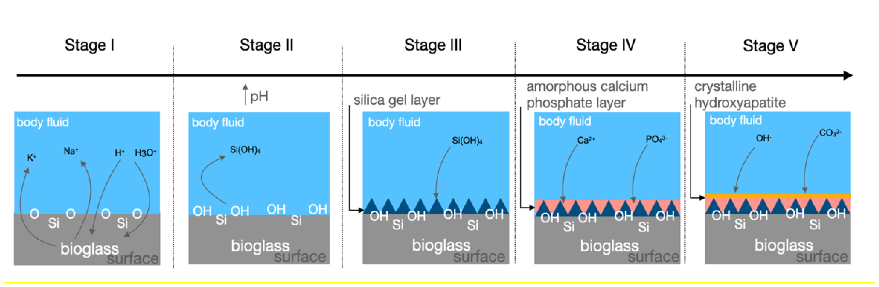
When BG is in contact with body fluids, the process of hydrolysis begins, and a cascade of reactions takes place. For didactical reasons, these reactions are divided into five stages. In stage I, alkali and alkali earth ions are released into the medium and are replaced in the glass structure by H+ or H3O+ ions from the fluid, this reaction will cause an increase in the local pH resulting in a rupture of Si-O-Si bonds. In stage II the silicon is released into the fluid in the form of silanol (Si(OH)4). Later in stage III, the, Si(OH)4 groups condense, forming a polymerized silica gel layer on the surface of the glass. The open structure of silica gel allows the continuity of ionic exchange between the glass and the body fluid. Calcium and phosphate ions diffuse from the glass and in conjunction with the calcium and phosphate ions from the fluid, form an amorphous calcium phosphate layer over the silica gel (stage IV). After the period, the thickness of these layers increases, and carbonate species are incorporated in the latter and begin to crystallize into the hydroxycarbonate apatite layer (HCA). The HCA is chemically and structurally like the mineral apatite phase found in our bone tissue (Figure 1)12,13.
Mechanism of formation of hydroxyapatite layer into bioglass surface when in contact with body fluid.
After the formation of HCA, a chemical bond between the material and bone is established12,13. Then, the stages linked to material-cell interaction begin, firstly with the biochemical adsorption of growth factors on the HCA layer, later with macrophage action, attachment and differentiation of stem cells, matrix generation, and crystallization, and finally growth and bone proliferation (Figure 2)12,13 . Bioglass 45S5 is still considered the gold standard of bioactive materials12, due to its high bioactivity. However, 45S5 presented shortcomings related to its high alkali content, fast dissolution, and rates of resorption14, the glass transition temperature (Tg ~550°C), the onset of crystallization (Tc ~ 610°C)1, and high pH value created by the high doses of sodium leached to the culture medium may demonstrate cytotoxic effects in vitro15.

Therefore, modifications to the 45S5 formulation have been long studied so different properties and improvements are achieved, such as processability and antimicrobial activity. For this reason, BG has a wide range of applications, for example for toothpastes, demineralizing agents, hypersensitivity treatment, bone grafting, scaffolds, drug delivery, soft tissue engineering, and biomaterial coating.
Given the importance of providing safe, predictable, and successful dental implant treatment for patients, the use of bioactive glass surface treatment can be employed. Some studies in animals have shown that titanium implants coated with bioactive glass have demonstrated more favorable osseointegration results compared to implants without bioactive glass treatment.
Despite the good qualities of the BG, the need to overcome autogenous graft in bone regeneration is highly expected among researchers, BG can chemically bond to hard and soft tissue and is a promising alternative to bone regeneration and coating of implants. In this context, the present study reviewed and contextualized some applications of BG focused on the oral surgeries field for bone augmentation and for coating dental implants to improve osseointegration.
METHOD
The articles were selected through a comprehensive search utilizing the PubMed database. The search encompassed a period from articles 1990 to 2023. The key terms, medical subject headings (MeSH) terms, and boolean operators ("AND" and "OR") were used across each database to refine the search. Search terms included "bioactive glass", "bioglass", "bone regeneration", "bone augmentation", "osseointegration" and "dental implants". The search strategy was collectively reviewed by members (IFB and MTS) to execute using the Peer Review of Electronic Search Strategies (PRESS) checklist. Inclusion criteria for this review encompassed full-text, peer-reviewed research articles that reported primary data on the application of bioactive glass for bone augmentation or coating into dental implants. Studies were included if they involved in vivo, animal, or clinical studies. Exclusion criteria were non-peer-reviewed articles and studies not reporting specific outcomes related to the application of bioglass for oral surgeries and implant dentistry, for example, for the use of medical applications for the orthopedics field. Commentaries, editorials, and reviews without original data were also excluded.
RESULT
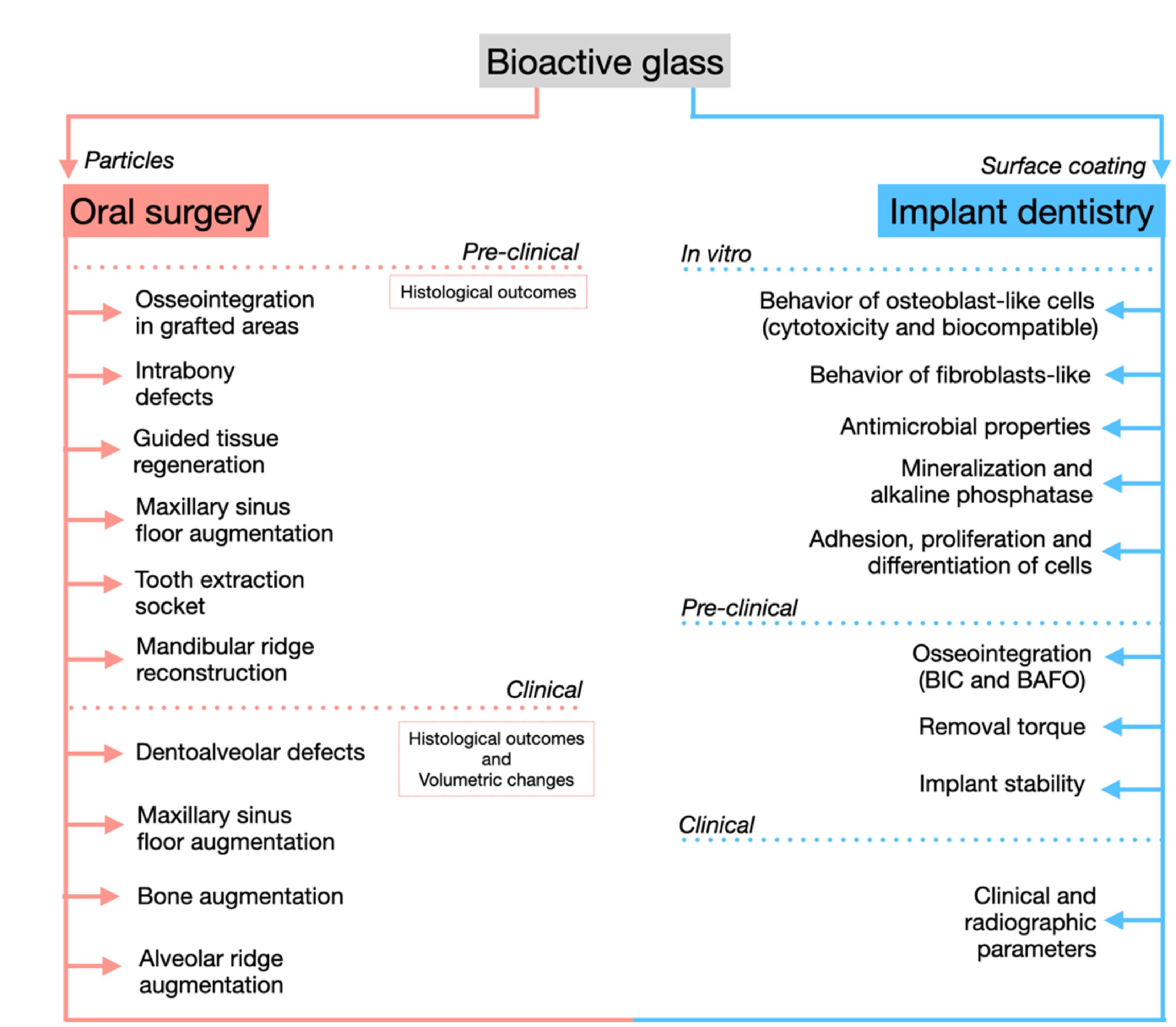
A total of 18 articles were selected for this narrative review. The division was composed of a total of 12 articles that were included for the bone regeneration part of this review. A total of 6 articles were included for osseointegration of dental implants, 3 articles were pre-clinical studies, and only 3 were clinical studies. Figure 3 illustrates the BG findings according to the application in particle shape for oral surgeries approach for bone augmentation and coating BG into dental implants to improve cell response and osseointegration.

Applications of bioactive glass as regenerative surgical treatment in oral surgery and coating of dental implants for improved osseointegration.
Bone Augmentation Applications Focused on Oral Surgery Proposals
Clinical Studies
Table 1 summarizes all relevant clinical studies on bioactive glass application for bone augmentation in oral surgeries.
Summary of relevant clinical studies on bioactive glass application for bone augmentation in oral surgeries
In 1993, a clinical trial enrolled several patients for different applications using Biogran®. Biogran® ranged from 300 to 360 µm narrow size. Several indications were selected for treatment, such as apical resection areas, cystic defects, extraction sites, and defects of the alveolar ridge caused by surgery or resorption. A total of 87 patients with 106 application sites were enrolled for this study. For apical resections, the results were very satisfactory, as the glass particles integrated into the repaired bone defects, on the other hand, in some cases infection reoccurred and the glass particles became fibrously encapsulated. For extraction sites, the follow-up ranged from 6 to 35 months and demonstrated that healing was uneventful in most cases and prevented the collapse of the alveolar ridge. The correction of the alveolar ridge using Biogran® demonstrated after 3 months of the application that sites appeared to have stabilized, and for cystic defects, 16 defects were filled with Biogran®, and only in two cases was the cystic area reinfected16.
Three cases of maxillary sinus floor augmentation using the bone-added osseous sinus floor elevation by osteotomy technique procedure with Biogran® biomaterial, and then dental implant placement. For case two, an implant was removed, and immediate grafting with Biogran®. These surgical procedures in these three cases were demonstrated to provide good outcomes after bone augmentation and dental implant placement18.
A randomized clinical study evaluated patients who submitted to extraction sites for the repair of dentoalveolar defects and ridge maintenance at the site of extraction sockets with PerioGlas® or Biogran® before dental implant placement (6 or 7 months). At 6 months bone biopsies were collected to evaluate tissue response by histological analysis and grafts demonstrated the glass to be intimately related to an adherent connective tissue, there was no evidence of an inflammatory infiltrate in specimens harvested with BG, indicating a biocompatible material. However, at 7 months, some biopsies demonstrated that bone appeared to lie close to the surface of glass particles, suggesting growth by apposition. The characteristic fissuring of the glass particles was routinely seen, in close apposition with the amorphous silica gel layer. Implants survived up to 3 years of follow-up in sites grafted with BG, even when such grafts appeared to only slowly conduct new bone growth19.
In case reports enrolled with 7 patients, teeth were extracted, and the alveolar socket was filled with BioGran®, after 6 months biopsies were collected. The histological findings showed fibro-osseous fragments prevailing, there was a dense conjunctive pattern in the cervical half, and trabeculated bone in the apical portion, demonstrating multiple growth sites, rapidly filling the bone defect. In general, the central areas demonstrated an invagination of the adjacent conjunctive surrounded by a halo of homogeneous mineralized conjunctive tissue with regions in the process of ossification and retention of osteocytes and osteoblasts on the surface23.
Bone augmentation for alveolar preservation before dental implants was performed in three case reports using Perioglas® particles or Perioglas® mixed with AB or blood clot. After 6 months dental implants were placed, and biopsies collected. The two case reports that use Perioglas®+AB, demonstrated minimal inflammatory reaction, that the cellular reaction around the particles was characterized by normal connective tissue with little infiltrate, demonstrating the compatibility of bioactive glass particles in contact with the soft tissues and intimately connected to the newly formed bone. The third case that used Perioglas®+blood clot, demonstrated less bone formation, although the particle reactivity appeared to be comparable to the other cases, thus, the early evidence of lower bone formation in the biopsy did not seem to have an adverse effect on the clinical result21.
Biogran® was used compared with autogenous bone (AB) from iliac crest bone for sinus floor elevation in 3 female patients with severe maxillary atrophy indicated for maxillary sinus floor augmentation. An 80% Biogran® particle was mixed with 20% AB compared to 100% AB on the other control side. The histological assessment demonstrated a lesser percentage of total bone volume at 4 months for Biogran® when compared to AB (26.9%±1.7% and 39.3%±0.5%, respectively), the Biogran® particles at this period were surrounded by bone as well as by a cell-rich stroma of loose connective tissue. At 6 months, the bone had increased in quantitated inside the Biogran® particles as well as around them, bone was found all over the transformed glass particles, however, Biogran® continued with the lower percentage when compared to AB (35.6%±1.5% and 40.8%±0.6%, respectively). At 15 months of healing, mature lamellar bone is indicated by the absence of empty lacunae and normally appearing mature bone marrow. Bone formation continued as evidenced by osteoid layers lining the excavated center as well as the periphery of the former particles while osteoblasts were bordering the osteoid layers. At 15 months Biogran® showed 38.8%±2.1% when compared to AB (41.7%±1.2%)20.
In cases of maxillary sinus floor augmentation, Perioglas® was used to compare bone formation after 6 months with other synthetic biomaterials and AB. The histomorphometry of the percentage of newly formed bone demonstrated higher values for AB (40.1%±3.2%) when compared to synthetic biomaterials, such as Perioglas® (31%±1.9%), and HA (32%±2.5%), however, the analyze of residual graft material demonstrate similar values for AB and Perioglas® (18%±2.3% and 18%±2.4%, respectively) when compared to HA (34%±1.6%). The descriptive histological assessment showed that Perioglas® has the potential to promote bone regeneration because the particles were surrounded by newly formed bone, no gaps or fibrous tissues were present at the bone-biomaterial interface, and graft particles were connected by newly formed bone22.
The clinical approach of trans alveolar sinus augmentation filling with BioGran® mixed with AB was assessed in 31 patients. After 4 months, biopsies were collected representing the new tissues formed inside the sinus, and the transalveolar osteotomy and the dental implants were placed. Sinus augmentation grafted with BioGran® mixed with AB, was demonstrated to be biocompatible with the bone formation that, is a short distance from the sinus floor. Inside the transalveolar osteotomy, new bone fraction was almost twice as much as in the sinus cavity, and the residual biomaterial occupied a much smaller fraction than that inside the sinus24.
A prospective clinical study approached using Biogran®+AB to analyze in comparison with AB alone in maxillary sinus elevation surgery. After 6 months of healing biopsies were collected for histomorphometry and immunohistochemical analysis. The results demonstrated that bone formation was greater in the amount of bone percentage at the apical region for both groups, Biogran®+AB (45.8%±13.8) and AB alone (42.0%±16.6), in the intermediate region Biogran® +AB (33.2%±13.3) and AB alone (35.0%±13.9), and pristine bone greater percentage for Biogran®+AB (36.6%±12.9) and AB alone (34.4%±14.4). The bone resorption was smaller for Biogran®+AB when compared to AB alone. According to immunohistochemistry to detect TRAP gene response, a low level of osteoclast-specific staining presented in both groups, indicating that both groups were in a remodeling phase25. In sequence, the same authors published the volumetric changes and trabecular microarchitecture of the same study. The cone beam computed tomography (CBCT) demonstrated a greater percentage of bone graft resorption for AB (45.7%±18.5), and Biogran® (44.0%±16.0) the association of Biogran®+AB resulted in a lesser percentage (37.9%±18.9) with no statistically significant difference. %BV/TV resulted in similar values for all groups Biogran® (52.06%), Biogran®+AB (57.04%), and AB (57.19%). These results of volumetric changes indicated that BG+AB and AB alone have similar resorption26.
Maxillary sinus floor augmentation using BG (S53P4®) alone or with the association of autogenous cortical cancellous bone in maxillary sinus floor augmentation, after 21-34 weeks and 49-62 weeks biopsies were collected, and the results demonstrated a few small chronic inflammatory cells in some specimens in the BG-AB group. For all groups, mature trabecular bone with Harversian systems, osteoid, and mature bone marrow with hematopoietic cells were observed at both time points. These grafted areas demonstrated resorption as well as new bone formation on the old trabeculae as a sign of bone remodeling. The bone area evaluated through histomorphometry analysis demonstrated that the association of the BG-AB group demonstrated at 25.7%±7.4 for 21-34 weeks and 28.8%±17.1 for 49-62 weeks, however, for the AB group alone the values were 25.1%±7.2 and 25.1%±6.3, respectively17.
Biogran® was associated with AB through maxillary sinus floor augmentation in 40 patients, and they were divided into five groups, 1) AB; 2) Biogran®; 3) Biogran®+AB; 4) BioOss® and 5) BioOss®+AB. After 6 months of bone repair, bone biopsies were collected and demonstrated the new bone formed for an intermediate area with greater values for the autogenous group (38.1%±21.4) and the xenograft group demonstrated a lesser value (32.5%±10.8) however no statistical difference. The association of Biogran®+AB demonstrated a value of 34.8%±14.5, however, group AB demonstrated a high level of osteocalcin protein presence, which points to a mature bone that can receive dental implants for groups Biogran®+AB; BioOss® alone or BioOss®+AB27.
Dental Implant Surface Treatment Applications
Pre-Clinical Studies
The tibia and femur of rabbits were analyzed for osseointegration of implants with Titanium (cpTi), HA, and BG surfaces. Screw-shaped with a diameter of 4.1 mm was prepared with cpTi surface and the preparation with HA and BG. After 12 weeks, removal torque was performed, and biopsies were collected for histomorphometry. The results demonstrated surface roughness (Sa) with higher values for HA (0.76±0.17), BG (0.70±0.16), and Titanium (0.64±0.41). Peak removal torque (N/cm) resulted in the femur’s highest value for HA when compared to BG (81±17 and 53±7, respectively). Furthermore, for the tibia, HA showed (73±22) and BG (61±29). In the histomorphometry (BIC) analysis for tibia bone, Ti showed the lowest percentage (17%±7) when compared to HA (29%±7) and BG (24%±13) at 12 weeks28.
The analysis of bioactive fiber-reinforced composite implant (FRC) with BG was applied in the tibias of pigs to analyze histomorphometry of the values of BIC after 4 and 12 weeks. The samples were divided into groups, such as threaded FRC implant, threaded BG containing FRC implant, and threaded custom-made titanium implant (Ti). At 4 weeks, BIC values demonstrated a higher value for FRC/BG (33.1%±11.1) when compared to FRC and Ti (27.5%±5.1 and 19.3%±6.9, respectively). At 12 weeks, all values increased demonstrating the greatest value for FRC/BG (46.9%±9.0) and similar values for FRC and Ti implants (40.2%±15.3 and 41.8%±12.3, respectively). Newly formed bone tissue was visible for all groups, but woven bone appeared to be in direct contact with both FRC and FRC/BG surfaces, additionally, adding BG particles improves the bone response29.
In maxillary areas, dental implants were installed with BG coating by air air-abrasion or in zinc oxide (ZnO) surfaces in comparison with implants with inert glass surfaces. After 8 weeks of healing, biopsies were collected. ZnO demonstrated greater values of BIC (70.49%±12.74) when compared to BG (57.91%±24.10) and inert implants (16.20%±25.53), the same for a percentage of the bone volume within the defect (73.81%±15.06 and 62.50%±20.51, respectively), inert demonstrated the lesser values (5.03%±0.92). these findings demonstrated that the presence of BG or ZnO by air abrasion results in better osseointegration and bone regeneration compared to inert glass air-abrasion treatment surfaces30.
Clinical Studies
Table 2 summarizes all relevant clinical studies on bioactive glass (BG) application for coating into dental implants
Summary of relevant clinical studies on bioactive glass (BG) application for coating into dental implants
A study investigated an indigenous HA-coated and BG-coated titanium dental implant system. Implants with the composition of Ti-6Al-4V were coated through the air plasma spray technique to the HA surface and the vitreous enameling technique to the BG surface. After in vitro analysis of different surfaces to identify their biocompatible, and also demonstrating no cytotoxicity34 the prospective clinical study which involved 14 patients was submitted to install the implants in the maxilla and mandible with 6 months of prosthetic loading. Marginal bone loss, HA-coated implants demonstrated in maxilla greater bone loss at 6 months when compared to baseline (1.85±0.08 and 1.13±0.17, respectively), the same for mandible (1.69±0.08 and 1.09±0.12, respectively). For BG-coated implants installed in the maxilla at 6 months when compared to baseline (1.78±0.11 and 1.12±0.17, respectively), the same for mandible (1.55±0.09 and 1.06±0.12, respectively), all values showed no differences between groups. Suppuration, resorption of entire areas of HA coating, and abnormal mobility with vertical pressure were observed with only one HA implant in the lower jaw31.
In addition, the same group of researchers investigated the same surfaces (HA and BG) in another short-term, prospective clinical study that enrolled 31 patients with 12 months of prosthetic loading. A total of 28 implants were placed in the anterior maxilla and 34 implants were placed in the anterior mandible. No inflammation was found after 6 and 12 months of follow-up. For marginal bone loss, both surfaces within both periods increased values when compared to baseline.
For marginal bone loss, the HA group for maxilla at 12 months demonstrated higher value when compared to baseline (1.92 ±0.10 and 1.13, respectively), the same for mandible (1.73±0.09 and 1.09±0.12, respectively). The same for the BG group when installed in maxilla at 12 months follow-up demonstrated higher value when compared to baseline (1.81±0.15 and 1.12±0.17, respectively), for mandible the same increased values for 12 months (1.60±0.09 and 1.06±0.12, respectively). The number of implant failures was one for the HA group when installed in the maxilla and two when installed in the mandible, for the BG group only one in the maxilla and one for the mandible32.
In the third study by the same research group33, a total of 62 patients were enrolled in 126 dental implant placements, comparing three groups, (45 for HA, 41 for BG, and 40 for titanium) in anterior maxillary. The outcomes of 12 months after prosthetic rehabilitation through clinical and radiological parameters, demonstrated that both surfaces were nontoxic and biocompatible, marginal bone loss in the radiograph, and less interfacial gaps were observed in computed tomography with bioactive glass at the anterior maxilla. All groups showed marginal bone loss after 12 months in the maxilla and mandible when compared to baseline33.
DISCUSSION
The clinical studies included in the topic “Bone Augmentation Applications focused on Oral surgery proposals” show considerable heterogeneity in terms of design, sample size, clinical application, and evaluation methods. The designs ranged from case reports and case series to randomized controlled clinical trials, reflecting different levels of scientific evidence.
Clinical trials, such as those conducted by Norton, Wilson19 (2002), Turunen et al.17 (1997), Scarano et al.22 (2006), Stavropoulos et al.24 (2012), and Pereira et al.26,27 (2018, 2020), included a larger number of patients and provided more robust data on the performance of bioactive glass. In contrast, case reports and case series, for example, Leonetti et al.18 (2000); Gatti et al.21 (2006); Margonar et al.23 (2012), presented more limited evidence due to the smaller number of patients included in the study, focusing on descriptive clinical outcomes.
Regarding the composition of biomaterials, most studies evaluated Biogran® or similar formulations. Some variations were observed, such as S53P4 and PerioGlas®, which differ slightly in composition and particle size. Despite these differences, the reported biological behavior was relatively consistent among the studies. Clinical applications were predominantly related to maxillary sinus elevation17,18,20,22,24-27 and alveolar ridge augmentation16,19,21,23.
Overall, bioactive glass demonstrated consistent osteoconductive properties and the ability to promote bone formation18,20,26,27. Several studies reported sufficient bone regeneration for dental implant placement, especially in maxillary sinus elevation procedures26,27.
However, differences were observed in the use of bioactive glass alone or in combination with autogenous bone. Some studies20 indicated that isolated bioactive glass can promote bone formation, even in severe atrophy conditions. On the other hand, other studies such as Gatti et al.21 (2006); Menezes et al.25 (2018); Pereira et al.26 (2018) reported better results when bioactive glass was combined with autogenous bone, including greater bone volume and better repair characteristics. Long-term outcomes were also addressed in some studies. Norton, Wilson19 (2002) reported implant survival for up to 3 years.
Despite promising results, direct comparison between studies is limited by various factors: Heterogeneity of study designs; variability in sample sizes; variations in biomaterial composition and particle size; different evaluation methods (clinical, radiographic, and histological); varied follow-up periods.
The preclinical experimental studies included in the topic "Dental Implant Surface Treatment Applications" show variations in terms of animal models, composition of bioactive glass, surface modification techniques, and experimental design.
Regarding animal models, rabbits28, pigs29, and rats30 were used, which implies important biological differences in bone response and repair processes. Evaluation periods also varied between 4 and 12 weeks, which can directly impact the interpretation of osseointegration results.
In terms of bioactive glass composition, there are variations among studies. Ramires et al.28 (2003) used a particle-based coating containing 65% SiO2, while Ballo et al.29 (2009) employed bioactive glass fibers with different oxide proportions. Abushahba et al.30 (2023) compared different compositions, including 45S5 bioactive glass and formulations containing zinc oxide (ZnO).
Ballo et al.29 (2009) demonstrated that implants reinforced with bioactive glass fibers showed adequate biocompatibility and promoted increased peri-implant osteogenesis and bone maturation compared to conventional titanium. Abushahba et al.30 (2023) found that surfaces treated with abrasive blasting, especially those containing ZnO, resulted in improved osseointegration and bone regeneration. These results suggest that modifications in bioactive glass composition can enhance its biological effects.
As these are preclinical studies, caution should be exercised when extrapolating the results to clinical practice in humans.
The clinical studies included in the topic "Dental Implant Surface Treatment Applications" analyzed titanium implants with surfaces modified by hydroxyapatite and bioactive glass coatings, allowing direct comparisons between these biomaterials. The implants used had standardized characteristics, being titanium with a cylindrical shape, internal hexagonal connection, and surface previously treated by acid etching and blasting.
The clinical outcomes evaluated mainly focused on marginal bone loss around the implants over time. Mistry et al.31 (2011) observed lower marginal bone loss in hydroxyapatite-coated implants (0 mm) compared to bioactive glass-coated implants (0.7 mm) after 6 months, suggesting superior initial performance of hydroxyapatite. However, in a subsequent study (Mistry et al.32, 2011), both hydroxyapatite-coated and bioactive glass-coated implants showed an increase in marginal bone loss after 12 months, indicating that both materials may undergo changes over time.
In the study by Mistry et al.33 (2016), the results after 12 months demonstrated that, despite marginal bone loss, bioactive glass showed satisfactory clinical performance, being considered a promising and biocompatible alternative for implant coating.
Overall, the studies indicate that both hydroxyapatite and bioactive glass can promote osseointegration, although hydroxyapatite may have initial advantages in terms of marginal bone preservation. On the other hand, bioactive glass shows consistent performance and relevant clinical potential.
Comparing the studies has some important limitations such as relatively short follow-up periods (6 to 12 months) and variability in sample sizes.
CONCLUSION
This review provides a literature overview of different applications of some of the most common bioactive glass compositions (in particular, 45S5 commercially as BioGran® or PerioGlas®), and their clinical outcomes in vivo approaches for bone augmentation demonstrating to be safe, well-tolerated for oral surgeries.
Equivalent findings for coating titanium and zirconia dental implants, due to chemical bonding and application in pre-clinical studies demonstrated the cytocompatibility and formation of bone around BG surface.
Future perspectives including analyzing the novel compositions and protocols of BG coating in different applications and procedures are more intensively explored through clinical trials and are still necessary.
ACKNOWLEDGMENTS
The authors are thankful for Brazilian funding supported by the Sao Paulo Research Foundation (FAPESP) grants numbers 2021/00632-3, and 2013/07793-6–CeRTEV- Center for Research, Technology, and Education in Vitreous Materials.
How to cite:
DATA AVAILABILITY
REFERENCES
-
1 Kim Y, Rodriguez AE, Nowzari H. The risk of prion infection through bovine grafting materials. Clin Implant Dent Relat Res. 2016 Dec;18(6):1095-102. https://doi.org/10.1111/cid.12391 PMid:26856530.
» https://doi.org/10.1111/cid.12391 -
2 Langer R, Tirrell DA. Designing materials for biology and medicine. Nature. 2004 Apr;428(6982):487-92. https://doi.org/10.1038/nature02388 PMid:15057821.
» https://doi.org/10.1038/nature02388 -
3 Park CH, Rios HF, Jin Q, Sugai JV, Padial-Molina M, Taut AD, et al. Tissue engineering bone-ligament complexes using fiber-guiding scaffolds. Biomaterials. 2012 Jan;33(1):137-45. https://doi.org/10.1016/j.biomaterials.2011.09.057 PMid:21993234.
» https://doi.org/10.1016/j.biomaterials.2011.09.057 -
4 Jensen SS, Bornstein MM, Dard M, Bosshardt DD, Buser D. Comparative study of biphasic calcium phosphates with different HA/TCP ratios in mandibular bone defects. A long-term histomorphometric study in minipigs. J Biomed Mater Res B Appl Biomater. 2009;90B(1):171-81. https://doi.org/10.1002/jbm.b.31271 PMid:19085941.
» https://doi.org/10.1002/jbm.b.31271 -
5 Miron RJ, Sculean A, Shuang Y, Bosshardt DD, Gruber R, Buser D, et al. Osteoinductive potential of a novel biphasic calcium phosphate bone graft in comparison with autographs, xenografts, and DFDBA. Clin Oral Implants Res. 2016 Jun;27(6):668-75. https://doi.org/10.1111/clr.12647 PMid:26227281.
» https://doi.org/10.1111/clr.12647 -
6 Dorozhkin SV. Calcium orthophosphate-based bioceramics. Materials. 2013 Sep;6(9):3840-942. https://doi.org/10.3390/ma6093840 PMid:28788309.
» https://doi.org/10.3390/ma6093840 -
7 Cao W, Hench LL. Bioactive materials. Ceram Int. 1996;22(6):493-507. https://doi.org/10.1016/0272-8842(95)00126-3
» https://doi.org/10.1016/0272-8842(95)00126-3 -
8 Hench LL, Splinter RJ, Allen WC, Greenlee TK. Bonding mechanisms at the interface of ceramic prosthetic materials. J Biomed Mater Res. 1971;5(6):117-41. https://doi.org/10.1002/jbm.820050611
» https://doi.org/10.1002/jbm.820050611 -
9 Cordaro L, Bosshardt DD, Palattella P, Rao W, Serino G, Chiapasco M. Maxillary sinus grafting with Bio-Oss or Straumann Bone Ceramic: histomorphometric results from a randomized controlled multicenter clinical trial. Clin Oral Implants Res. 2008 Aug;19(8):796-803. https://doi.org/10.1111/j.1600-0501.2008.01565.x PMid:18705811.
» https://doi.org/10.1111/j.1600-0501.2008.01565.x -
10 Xynos ID, Edgar AJ, Buttery LD, Hench LL, Polak JM. Gene-expression profiling of human osteoblasts following treatment with the ionic products of Bioglass 45S5 dissolution. J Biomed Mater Res. 2001 May;55(2):151-7. https://doi.org/10.1002/1097-4636(200105)55:2<151::AID-JBM1001>3.0.CO;2-D PMid:11255166.
» https://doi.org/10.1002/1097-4636(200105)55:2<151::AID-JBM1001>3.0.CO;2-D -
11 Xynos ID, Edgar AJ, Buttery LD, Hench LL, Polak JM. Ionic products of bioactive glass dissolution increase proliferation of human osteoblasts and induce insulin-like growth factor II mRNA expression and protein synthesis. Biochem Biophys Res Commun. 2000 Sep;276(2):461-5. https://doi.org/10.1006/bbrc.2000.3503 PMid:11027497.
» https://doi.org/10.1006/bbrc.2000.3503 -
12 Hench LL. The story of Bioglass. J Mater Sci Mater Med. 2006 Nov;17(11):967-78. https://doi.org/10.1007/s10856-006-0432-z PMid:17122907.
» https://doi.org/10.1007/s10856-006-0432-z -
13 Hench LL. Bioceramics: from concept to clinic. J Am Ceram Soc. 1991 Jul;74(7):1487-510. https://doi.org/10.1111/j.1151-2916.1991.tb07132.x
» https://doi.org/10.1111/j.1151-2916.1991.tb07132.x -
14 Sepulveda P, Jones JR, Hench LL. In vitro dissolution of melt-derived 45S5 and sol-gel derived 58S bioactive glasses. J Biomed Mater Res. 2002 Aug;61(2):301-11. https://doi.org/10.1002/jbm.10207 PMid:12007211.
» https://doi.org/10.1002/jbm.10207 -
15 Kansal I, Reddy A, Muñoz F, Choi S-J, Kim H-W, Tulyaganov DU, et al. Structure, biodegradation behavior and cytotoxicity of alkali-containing alkaline-earth phosphosilicate glasses. Mater Sci Eng C. 2014;44:159-65. https://doi.org/10.1016/j.msec.2014.08.016 PMid:25280692.
» https://doi.org/10.1016/j.msec.2014.08.016 -
16 Schepers EJ, Ducheyne P, Barbier L, Schepers S. Bioactive glass particles of narrow size range: a new material for the repair of bone defects. Implant Dent. 1993;2(3):151-6. https://doi.org/10.1097/00008505-199309000-00002 PMid:8142934.
» https://doi.org/10.1097/00008505-199309000-00002 -
17 Turunen T, Peltola J, Helenius H, Yli-Urpo A, Happonen RP. Bioactive glass and calcium carbonate granules as filler material around titanium and bioactive glass implants in the medullar space of the rabbit tibia. Clin Oral Implants Res. 1997 Apr;8(2):96-102. https://doi.org/10.1034/j.1600-0501.1997.080204.x PMid:9758960.
» https://doi.org/10.1034/j.1600-0501.1997.080204.x -
18 Leonetti JA, Rambo HM, Throndson RR. Osteotome sinus elevation and implant placement with narrow size bioactive glass. Implant Dent. 2000;9(2):177-82. https://doi.org/10.1097/00008505-200009020-00012 PMid:11307397.
» https://doi.org/10.1097/00008505-200009020-00012 - 19 Norton MR, Wilson J. Dental implants placed in extraction sites implanted with bioactive glass: human histology and clinical outcome. Int J Oral Maxillofac Implants. 2002 Mar-Apr;17(2):249-57. PMid:11958408.
-
20 Tadjoedin ES, de Lange GL, Lyaruu DM, Kuiper L, Burger EH. High concentrations of bioactive glass material (BioGran) vs. autogenous bone for sinus floor elevation. Clin Oral Implants Res. 2002 Aug;13(4):428-36. https://doi.org/10.1034/j.1600-0501.2002.130412.x PMid:12175381.
» https://doi.org/10.1034/j.1600-0501.2002.130412.x -
21 Gatti AM, Simonetti LA, Monari E, Guidi S, Greenspan D. Bone augmentation with bioactive glass in three cases of dental implant placement. J Biomater Appl. 2006 Apr;20(4):325-39. https://doi.org/10.1177/0885328206054534 PMid:16443622.
» https://doi.org/10.1177/0885328206054534 -
22 Scarano A, Degidi M, Iezzi G, Pecora G, Piattelli M, Orsini G, et al. Maxillary sinus augmentation with different biomaterials: a comparative histologic and histomorphometric study in man. Implant Dent. 2006 Jun;15(2):197-207. https://doi.org/10.1097/01.id.0000220120.54308.f3 PMid:16766904.
» https://doi.org/10.1097/01.id.0000220120.54308.f3 -
23 Margonar R, Queiroz TP, Luvizuto ER, Marcantonio E, Lia RC, Holzhausen M, et al. Bioactive glass for alveolar ridge augmentation. J Craniofac Surg. 2012 May;23(3):e220-2. https://doi.org/10.1097/SCS.0b013e31824de5a4 PMid:22627439.
» https://doi.org/10.1097/SCS.0b013e31824de5a4 -
24 Stavropoulos A, Sima C, Sima A, Nyengaard J, Karring T, Sculean A. Histological evaluation of healing after transalveolar maxillary sinus augmentation with bioglass and autogenous bone. Clin Oral Implants Res. 2012 Jan;23(1):125-31. https://doi.org/10.1111/j.1600-0501.2011.02161.x PMid:21504477.
» https://doi.org/10.1111/j.1600-0501.2011.02161.x -
25 Menezes JD, Pereira RDS, Bonardi JP, Griza GL, Okamoto R, Hochuli-Vieira E. Bioactive glass added to autogenous bone graft in maxillary sinus augmentation: a prospective histomorphometric, immunohistochemical, and bone graft resorption assessment. J Appl Oral Sci. 2018 Jun;26(0):e20170296. https://doi.org/10.1590/1678-7757-2017-0296 PMid:29898173.
» https://doi.org/10.1590/1678-7757-2017-0296 -
26 Pereira RS, Menezes JD, Bonardi JP, Griza GL, Okamoto R, Hochuli-Vieira E. Comparative study of volumetric changes and trabecular microarchitecture in human maxillary sinus bone augmentation with bioactive glass and autogenous bone graft: a prospective and randomized assessment. Int J Oral Maxillofac Implants. 2018 May;47(5):665-71. https://doi.org/10.1016/j.ijom.2017.11.016 PMid:29246424.
» https://doi.org/10.1016/j.ijom.2017.11.016 -
27 Pereira RDS, Bonardi JP, Ouverney FRF, Campos AB, Griza GL, Okamoto R, et al. The new bone formation in human maxillary sinuses using two bone substitutes with different resorption types associated or not with autogenous bone graft: a comparative histomorphometric, immunohistochemical and randomized clinical study. J Appl Oral Sci. 2020 Dec;29:e20200568. https://doi.org/10.1590/1678-7757-2020-0568 PMid:33331393.
» https://doi.org/10.1590/1678-7757-2020-0568 -
28 Ramires PA, Wennerberg A, Johansson CB, Cosentino F, Tundo S, Milella E. Biological behavior of sol-gel coated dental implants. J Mater Sci Mater Med. 2003 Jun;14(6):539-45. https://doi.org/10.1023/A:1023412131314 PMid:15348438.
» https://doi.org/10.1023/A:1023412131314 -
29 Ballo AM, Akca EA, Ozen T, Lassila L, Vallittu PK, Närhi TO. Bone tissue responses to glass fiber-reinforced composite implants--a histomorphometric study. Clin Oral Implants Res. 2009 Jun;20(6):608-15. https://doi.org/10.1111/j.1600-0501.2008.01700.x PMid:19515036.
» https://doi.org/10.1111/j.1600-0501.2008.01700.x -
30 Abushahba F, Areid N, Gürsoy M, Willberg J, Laine V, Yatkin E, et al. Bioactive glass air-abrasion promotes healing around contaminated implant surfaces surrounded by circumferential bone defects: An experimental study in the rat. Clin Implant Dent Relat Res. 2023 Apr;25(2):409-18. https://doi.org/10.1111/cid.13172 PMid:36602418.
» https://doi.org/10.1111/cid.13172 -
31 Mistry S, Kundu D, Datta S, Basu D, Soundrapandian C. Indigenous hydroxyapatite coated and bioactive glass coated titanium dental implant system - fabrication and application in humans. J Indian Soc Periodontol. 2011 Jul;15(3):215-20. https://doi.org/10.4103/0972-124X.85663 PMid:22028507.
» https://doi.org/10.4103/0972-124X.85663 -
32 Mistry S, Kundu D, Datta S, Basu D. Comparison of bioactive glass coated and hydroxyapatite coated titanium dental implants in the human jaw bone. Aust Dent J. 2011 Mar;56(1):68-75. https://doi.org/10.1111/j.1834-7819.2010.01305.x PMid:21332743.
» https://doi.org/10.1111/j.1834-7819.2010.01305.x -
33 Mistry S, Roy R, Kundu B, Datta S, Kumar M, Chanda A, et al. Clinical outcome of hydroxyapatite coated, bioactive glass coated, and machined Ti6Al4V threaded dental implant in human jaws: a short-term comparative study. Implant Dent. 2016 Apr;25(2):252-60. https://doi.org/10.1097/ID.0000000000000376 PMid:26741743.
» https://doi.org/10.1097/ID.0000000000000376 -
34 Ghosh SK, Nandi SK, Kundu B, Datta S, De DK, Roy SK, et al. In vivo response of porous hydroxyapatite and beta-tricalcium phosphate prepared by aqueous solution combustion method and comparison with bioglass scaffolds. J Biomed Mater Res B Appl Biomater. 2008 Jul;86(1):217-27. https://doi.org/10.1002/jbm.b.31009 PMid:18161811.
» https://doi.org/10.1002/jbm.b.31009
CONFLICTS OF INTERESTS
*
CORRESPONDING AUTHOR Elcio Marcantonio Junior, UNESP – Universidade Estadual Paulista “Júlio de Mesquita Filho”, Faculdade de Odontologia de Araraquara, Departamento de Diagnostico e Cirurgia, R. Humaitá, 1680, 14801-385 Araraquara - SP, Brasil, e-mail: elcio.marcantonio@unesp.brEdited by