
 Hydroxyapatite/zinc oxide functionalized with folic acid as a curcumin carrier for cancer treatment
ABSTRACT
The primary treatment for cancer is chemotherapy; however, it causes side effects, which motivates research focused on the development of delivery systems for antineoplastic agents. One example is the hydroxyapatite (HAp) carrier, as it is a biocompatible and non-toxic biomaterial that can acquire antibacterial properties with the addition of zinc oxide (ZnO) nanoparticles, preventing possible infections during surgical procedures. Furthermore, functionalization with folic acid (FA) enables active targeting of tumor cells, while curcumin (Cur) exhibits antitumor activity. The objective of this study was to develop a carrier with antibacterial properties using FA-functionalized HAp@ZnO for curcumin delivery. HAp synthesized by the hydrothermal method was evaluated through in vitro bioactivity assays. Subsequently, ZnO nanoparticles were incorporated into HAp, and antibacterial activity was assessed against Staphylococcus aureus and Escherichia coli. The material was then functionalized with FA, curcumin was incorporated, and hemolysis and cytotoxicity assays were performed. Characterization techniques included Fourier Transform Infrared Spectroscopy (FTIR), X-ray Diffraction (XRD), Scanning Electron Microscopy (SEM), Brunauer–Emmett–Teller (BET) surface area analysis, and Ultraviolet–Visible (UV–Vis) absorption spectroscopy. The synthesized HAp exhibited a crystalline, bioactive material with high surface area. HAp@ZnO showed well-defined inhibition halos for both bacteria, except for the HAp@20%ZnO sample against E. coli. The amount of curcumin adsorbed was 54.79 ± 3.52% for HAp@10%ZnO.AF.Cur and 45.49 ± 3.79% for HAp@20%ZnO.AF.Cur. HAp effectively modulated the toxicity of the active components, significantly reducing the hemolytic effects of ZnO and curcumin. The results indicate biocompatibility in non-tumor cells and suggested differential cytotoxic response in tumor cells. Therefore, a promising carrier for targeted antitumor therapy was developed, combining antibacterial activity, active targeting, controlled release, and biocompatibility.
Keywords:
Hydroxyapatite; Zinc oxide nanoparticles; Drug delivery system; Curcumin; Folic acid
1. INTRODUCTION
Cancer comprises a group of diseases characterized by uncontrolled cell proliferation and the ability to invade surrounding tissues and metastasize [1]. Among the main cancer treatments are surgery, radiotherapy, and chemotherapy, with the latter being widely used in cases where other approaches are not indicated [2, 3]. However, chemotherapy presents several limitations, including the low aqueous solubility of many drugs, the lack of targeted delivery to tumor cells, and the reduced fraction of the drug that reaches the tumor site, resulting in severe side effects and a significant impairment of patients’ quality of life [3,4,5,6].
These challenges have driven the development of drug delivery systems capable of promoting the selective targeting of antineoplastic agents, thereby enhancing therapeutic efficacy and reducing adverse effects [2]. In this context, nanocarriers based on proteins, polymers, and inorganic materials have been extensively investigated. However, such systems present certain limitations, including difficulties in drug conjugation, issues related to nanoparticle (NP) morphology, and potential toxicity risks associated with metallic NPs [5, 7]. Consequently, there is a growing need to develop biocompatible biomaterials with structural stability and an appropriate safety profile for clinical applications.
Hydroxyapatite [Ca10(PO4)6(OH)2] (HAp) stands out as a promising candidate due to its chemical and crystallographic similarity to bone tissue, high biocompatibility, and ability to adsorb therapeutic molecules through its surface ionic sites [8, 9]. In addition, its crystalline structure allows for ionic substitutions, enabling the incorporation of specific functionalities into the material [8].
In the orthopedic–oncological context, the presence of antibacterial activity constitutes a strategic clinical requirement. Procedures such as tumor resections, implant placement, and bone reconstruction are associated with a high risk of postoperative infections, often related to biofilm formation and, in severe cases, implant removal [10]. Since pure HAp does not exhibit antimicrobial activity, the incorporation of zinc oxide (ZnO) nanoparticles has been widely investigated as a strategy to impart antibacterial properties to the composite. ZnO exhibits low toxicity, high biocompatibility, and is not classified as a heavy metal, in contrast to silver [11, 12]. Previous studies have demonstrated that the addition of ZnO to HAp significantly reduces the proliferation of bacteria such as Staphylococcus aureus, Staphylococcus epidermidis, and Escherichia coli, with efficacy proportional to the oxide content used [13, 14]. Therefore, the HAp/ZnO combination can simultaneously integrate bone support, drug delivery, and infection prevention within a single functional system.
The therapeutic efficiency of these systems can be further enhanced through active targeting strategies. Folic acid (FA) stands out due to its high affinity for folate receptors that are overexpressed in various tumor cells, promoting receptor-mediated internalization and increased intracellular accumulation [15,16,17,18,19]. HAp/ZnO systems functionalized with FA have been primarily reported for the delivery of conventional chemotherapeutic agents, such as doxorubicin [20, 21].
In parallel, natural compounds have been investigated as alternative or adjuvant therapeutic agents. Curcumin exhibits well-established antitumor activity by modulating pathways associated with cell proliferation and angiogenesis [22, 23]. However, its clinical application is limited by low solubility and bioavailability, making its incorporation into nanostructured systems a rational strategy for molecular protection and controlled release [24,25,26,27,28]. HAp-based systems functionalized with FA and combined with curcumin have already demonstrated high cellular internalization and significant cytotoxicity against osteosarcoma cells [29].
Although previous studies have explored HAp/ZnO-based systems functionalized with folic acid for antitumor drug delivery, the literature has predominantly focused on conventional chemotherapeutic agents. To date, there are no reports integrating, in a single platform, an HAp matrix incorporated with ZnO nanoparticles to provide antibacterial activity, functionalized with folic acid for active tumor targeting, and employed as a carrier for curcumin, while simultaneously addressing controlled drug release, cellular selectivity, and the prevention of infections associated with orthopedic oncological procedures.
In view of this gap, the present study proposes the development of a multifunctional system based on HAp/ZnO functionalized with folic acid for the loading and controlled release of curcumin. This strategy aims to integrate, within a single platform, antibacterial properties, tumor-targeting potential, and drug delivery performance, with potential applications in bone oncology and reconstructive orthopedics.
2. MATERIALS AND METHODS
2.1. HAp synthesis
In this synthesis, the hydrothermal method was employed, which was adapted from the methodology of DARYAN et al. [30]. For the synthesis, solutions of ammonium hydrogen phosphate ((NH4)2HPO4) at 28.57 mmol.L–1 and calcium nitrate tetrahydrate (Ca(NO3)2.4H2O- 99%, Dinâmica) at 47.71 mmol.L–1 were prepared. Subsequently, the hydrogen phosphate solution was introduced into the nitrate solution under continuous stirring (500 rpm) for 1 h. Then, the regulatory agent sodium citrate (Na3C6H5O7, P.A., Synth) was added at a citrate/calcium mass ratio of 0.3, and the pH was adjusted to 6.0 with hydrochloric acid (HCl) at 1.0 mol.L–1. The prepared sus-pension was transferred to a reactor at 180°C for twenty-four hours, where it remained under hydrothermal treatment. After the sample cooled, it was centrifuged at 3000 rpm for 10 min. Immediately after, the super-natant was discarded and the precipitate was washed with deionized water and alcohol. The material was placed in an oven at 70°C for 48 hours for drying.
2.2. In vitro bioactivity assay
Initially, the Simulated Body Fluid (SBF) solution was prepared according to the methodology of KOKUBO and TAKADAMA [31], in order to evaluate the bioactive potential of the material through the formation of an apatite layer on its surface after immersion in a simulated physiological medium. For this purpose, the reagents sodium chloride (NaCl), sodium bicarbonate (NaHCO3), potassium chloride (KCl), dipotassium hydrogen phosphate trihydrate (K2HPO4.3H2O), magnesium chloride hexahydrate (MgCl2.6H2O), calcium chloride (CaCl2), sodium sulfate (Na2SO4), and tris (hydroxymethyl) aminomethane [(HOCH2)3CNH2] were completely dissolved, and the pH was adjusted to 7.4 with hydrochloric acid (HCl).
The pellets (as described in section 2.2) were immersed in the SBF solution (15 mL) for determined time intervals: 0, 3, 7, 14, and 21 days and kept in a Shaker incubator at 37°C and 100 rpm. The solutions were renewed every 3 days throughout the test, and the samples were washed with distilled water, dried in an oven at 40°C, and stored for subsequent characterization.
2.3. Synthesis of HAp NPs coated with ZnO
The synthesis of ZnO NPs was a precipitation reaction involving potassium hydroxide (KOH, Êxodo Científica) and the precursor zinc nitrate hexahydrate Zn(NO3)2.6H2O- 98%, Dinâmica). The resulting sus-pension was centrifuged, and the precipitate was washed with deionized water. Subsequently, the NPs were calcined at 500°C for 3.0 h following reference [32].
The incorporation of ZnO NPs into hydroxyapatite (HAp@ZnO) was based on the methodology of MARTÍNEZ et al. [33], which employs a viscous medium to keep the particles uniformly dispersed and suspended. A solution containing 5.0 mL of Glycerin (95%, Proquímios) and 10.0 mL of distilled water was used under stirring at 90°C. Then, the ZnO NP content was added; in this study, the amounts were 5%, 10%, and 20% relative to the mass of the HAp. Afterward, the HAp (non-calcined) was added to the dispersion. After 10 minutes of stirring, the material was dried and calcined at 900°C for 3 h to promote the interaction between HAp and ZnO.
2.4. Antibacterial assay of HAp@ZnO
The antibacterial activity was performed using HAp incorporated with 0%, 5%, 10%, and 20% ZnO NPs against Staphylococcus aureus (ATCC 25923, ATCC 33591) and Escherichia coli (ATCC 25922) strains. The assay was conducted using the agar diffusion method, in which bacterial colonies of S. aureus and E. coli were suspended in sterile saline solution (0.85% NaCl) until a turbidity compatible with 0.5 on the McFarland scale (1 × 10⁶ UFC/mL) was obtained. A sterile swab was soaked in the bacterial suspension, pressed against the tube walls to remove excess suspension, and then gently streaked in five directions across the plate to ensure full surface coverage. Subsequently, the HAp@ZnO disks were placed on the surface of the inoculated medium and pressed gently to ensure complete contact with the agar surface. The plates containing the disks were incubated in an oven at 35°C for 24 hours, after which the presence or absence of halos around the samples was observed. The antibacterial assay was performed in independent triplicates (n = 3) for each concentration and bacterial strain.
2.5. Functionalization of HAp@ZnO with folic acid
Initially, 0.05 g of folic acid was added to 100 mL of PBS solution, followed by 0.1 g of HAp@ZnO, which was kept under stirring, in the dark, and at room temperature for 48 hours. The mixture was centrifuged at 5000 rpm for 20 minutes and then dried at 70°C for 48 h. The functionalization of HAp@ZnO with folic acid followed adaptations of the methodology described in the literature by SANTOS et al. [34].
The quantification of FA present in the HAp@ZnO.AF samples was calculated from a standard curve (concentration × absorbance) previously obtained by dissolving the material in an HCl solution. For this, the HAp@ZnO.AF biomaterial under study (0.01 g) was dissolved in 0.1 mol.L–1 HCl (3 mL), and the resulting solutions were read in a UV-vis spectrophotometer using the maximum wavelength (\lambda_{max}) for folic acid of 310 nm. It is noteworthy that the FA quantification was performed in independent triplicates (n = 3), and the values are presented as mean ± standard deviation.
2.6. Incorporation of the therapeutic agent curcumin
A curcumin (Cur) solution was prepared with water/ethanol (4:1) at a concentration of 100 μg.mL–1. For every 100 mL of these solutions, 1.0 g of the biomaterial under study (HAp@ZnO.AF) was added, keeping it under continuous stirring at 20 °C for 72 hours—the maximum stability period for absorption readings of the supernatant. The suspension was then centrifuged at 4000 rpm for 20 min, and an aliquot of the respective supernatants was removed for absorption reading using the maximum wavelength (λmáx.) for curcumin (440 nm).
The amount of therapeutic agent incorporated into the material was determined based on a concen-tration versus absorbance standard curve previously established for curcumin. The percentage of loaded curcumin was calculated based on the initial curcumin concentration (100 μg.mL–1). All experiments were performed in independent triplicates (n = 3), and the values are presented as mean ± standard deviation.
2.7. In vitro hemolysis assay
The hemolytic activity of HAp, ZnO, AF, Cur, HAp@ZnO, and HAp@ZnO.AF.Cur related to their ability to cause damage to the plasma membrane of erythrocytes—was evaluated according to the method described by PITA et al. [35] and YEDGAR et al. [36] with some modifications. Red blood cells (RBCs) from two Swiss mice were used (Approved by the Animal Ethics Committee, protocol No 003/260924). The mice were anesthetized with ketamine (150 mg/kg) and xylazine (15 mg/kg) and subjected to cardiac puncture to collect approximately 2 mL of blood, which was immediately stored in tubes containing ethylenediaminetetraacetic acid (EDTA) as an anticoagulant. The collected blood was diluted in 10 mL of PBS and centrifuged at 3000 rpm for 4 minutes to separate the plasma. The supernatant was carefully removed, and the process was repeated two more times. Subsequently, the erythrocytes were resuspended in PBS to obtain a 1% (v/v) suspension.
The samples were tested at concentrations of 200, 100, 50, and 15 µg.mL–1. For each assay, 1 mL of the test solution was mixed with 1 mL of the 1% erythrocyte solution in triplicate. For the positive control (100% hemolysis), a 0.1% Triton X-100 solution was used, and for the negative control (0% hemolysis), PBS was used, both also incubated with the erythrocyte solution. The tubes were kept under gentle agitation for 60 minutes and subsequently centrifuged at 3000 rpm for 4 minutes. The supernatant was collected, and hemoglobin release was quantified by spectrophotometry at 540 nm. To calculate the CH50 (50% hemolytic concentration), the following equation was used, where Abs corresponds to absorbance:
The percentage of hemolysis was calculated according to the described equation, and the results were expressed as mean ± standard deviation. The classification of hemocompatibility followed the ASTM F756 standard [37]: materials intended for biomedical applications must present a maximum hemolysis rate of less than 5% to be considered safe, with values below 2% classified as highly hemocompatible, while rates higher than 5% indicate hemolytic behavior [38, 39].
2.8. In vitro cytotoxicity evaluation
Cytotoxicity was evaluated using the MTT method [3-(4,5-dimethyl-2-thiazolyl)-2,5-diphenyl-2H-tetrazolium bromide]. L929 and Sarcoma-180 cells were seeded in 96-well culture plates at densities of 5 × 10³ e 2 × 10⁴ cells per well, respectively, in Dulbecco’s Modified Eagle Medium (DMEM) supplemented with 10% fetal bovine serum (FBS), 1% antibiotics, and 0.1% Amphotericin-B. The plates were maintained in an incubator with 90% humidity at 37°C and 5% CO2. Different concentrations of the isolated substances and formulations were added to the cell cultures, corresponding to 200, 100, 50, and 25 µg.mL– 1. After 24 and 72 hours of treatment, the MTT reagent was added. Following 3 hours of incubation, the absorbance was measured at 540 nm using a Loccus® LMR-96i-4 microplate reader. Cell viability was calculated using the following formula:
2.9. Statistical data analysis
The experimental data were expressed as mean ± standard deviation. Statistical analysis was performed using Student’s t-test for independent samples and Analysis of Variance (ANOVA), followed by Tukey’s multiple comparison test. Differences were considered statistically significant when p < 0.05.
2.10. Characterization
The crystalline structure was identified by X-ray diffraction (XRD) using a Siemens Diffract ACT Series 1000, employing copper K\alpha radiation (λ = 1,4518 Å) at 40 kV and 40 mA. The 2θ range spanned from 5° to 70°. Phases were identified using data from the ICSD reference files within the X’Pert HighScore software. The identification of functional groups in the HAp was performed via Fourier-transform infrared spectroscopy (FTIR) (model IRTracer-100 – SHIMADZU®) across a wavenumber range of 4000–400 cm–1. Samples were prepared as KBr pellets using mechanical pressing. Morphology was observed using Scanning Electron Microscopy (SEM) (Hitachi TM1000 and Vega 3 Tescan). The samples were coated with a thin layer of gold (sputtering process) to increase surface conductivity. The surface area was determined using the BET technique through N2 adsorption/desorption analysis at an analysis bath temperature of –195.8°C. To measure absorbance in the UV-visible region, a UV-Vis spectrophotometer (model IL-592) was used with quartz cuvettes for liquid samples.
3. RESULTS AND DISCUSSION
The synthesized HAp was initially characterized by FTIR, and the corresponding spectrum is shown in Figure 1. The presence of characteristic phosphate (PO43–) bands is observed, confirming the formation of the apatite phase. The band located at 1034 cm–1 corresponds to the asymmetric stretching vibration (ν3) of PO43–, while the bands at 602 and 561 cm–1 are assigned to the bending vibrations (ν4), which are typical of the crystalline structure of HAp. The band at 3436 cm–1 is associated with the stretching vibrations of hydroxyl ions (OH–), mainly related to water adsorbed on the material surface. When comparing the obtained spectral profile with that reported for stoichiometric HAp, variations in the relative intensities between the ν3 and ν4 phosphate modes are observed. According to classical studies, changes in intensity ratios and band broadening of PO43– vibrations may be associated with the presence of structural defects, reduced crystallinity, non-stoichiometry, or anionic substitutions, particularly by carbonate [40, 41]. The bands at 1630 and 1384 cm–1 are attributed to carbonate ions (CO32–), possibly resulting from partial citrate decomposition during the hydrothermal treatment and subsequent CO2 incorporation into the apatite crystal lattice [42]. Carbonate substitution at PO43– or OH- sites is widely reported in the literature and may induce changes in the relative intensities and shapes of vibrational bands [43]. Therefore, although the presence of phosphate bands confirms the formation of the apatite phase, the observed spectral variations indicate possible structural and/or surface modifications.

The crystalline structure of the synthesized HAp was evaluated by XRD (Figure 2), showing well-defined and intense peaks characteristic of apatite, which indicates high crystallinity of the synthesized material. Comparison of the experimental pattern with the crystallographic database confirms the correspondence with the Ca10(PO4)6(OH)2 phase, space group P63/m, according to file ICSD 26205 [9, 44]. The absence of secondary peaks related to undesired phases, such as β-tricalcium phosphate (β-TCP) or calcium oxide (CaO), suggests high phase purity of the obtained material. Furthermore, a relative increase in the intensity of the (300) and (002) crystallographic planes was observed when compared to other peaks, suggesting preferential growth along the c-axis. In the literature, this behavior has been associated with heteroepitaxial growth induced by the presence of citrate during synthesis [30, 42]. Thus, the results indicate that sodium citrate did not promote alterations in the crystalline structure of HAp but influenced the crystal growth mechanism. Consequently, the diffractogram confirms the successful synthesis of HAp, with high phase purity and a structure compatible with reference crystallographic standards. These results are in line with the FTIR data, as both techniques confirm the presence of characteristic phosphate and hydroxyl groups, as well as the preservation of the crystalline structure.

The morphology of HAp was investigated by SEM (Figure 3), revealing micrometric agglomerates with irregular morphology and a high degree of particle coalescence. The absence of clearly individualized primary particles prevents the accurate determination of the average particle size and does not allow the material to be conclusively classified as nanometric based solely on the presented images. Complementary analyses, such as transmission electron microscopy (TEM) or quantitative image-based particle size distribution, would be required to confirm the size of the primary particles. Although the literature reports that citrate may act as a modulator of nucleation and crystal growth through the adsorption of carboxylate groups (–COO−) on the surface of forming crystals [26, 31, 42], the SEM data obtained in this study do not allow a quantitative confirmation of this effect.

The nitrogen adsorption/desorption isotherm of HAp (Figure 4) corresponds to type IV according to the IUPAC classification, being characterized by the presence of hysteresis and two inflection points, which indicate the occurrence of capillary condensation in mesoporous structures (2–50 nm) [45]. The synthesized HAp exhibited a specific surface area (SBET) of 96.46 m2·g−1, which is higher than that reported for HAp obtained in the absence of citrate (40.32 m2·g−1) and comparable to that of HAp synthesized with a citrate/calcium ratio of 0.3 (81.86 m2·g−1) [30]. Although the SBET value is relatively high when compared with highly crystalline HAp reported in the literature [46, 47], this parameter alone does not allow the inference of particle size at the nanometric scale, since high surface areas may also arise from textural porosity, interparticle voids, or particle agglomeration. The high surface area observed represents a relevant characteristic for applications in antineoplastic drug delivery systems, as it favors interaction with biological fluids and the incorporation of therapeutic molecules.

The in vitro bioactivity assay of HAp demonstrated the formation of apatite on the surface over time, as observed in the micrographs (Figure 5). After 3 days of immersion, the initial formation of discrete nuclei is observed, while at 21 days, a more continuous layer covers a significantly larger fraction of the sample surface, indicating bioactive behavior. However, to confirm the formation of a new apatite layer, complementary analyses using techniques such as atomic force microscopy (AFM), X-ray diffraction (XRD), and FTIR are required, allowing the correlation of morphological, structural, and chemical evidence and providing greater reliability to the results. The mechanism of apatite formation begins with the hydroxyl (OH–) and phosphate (PO43-) groups on the HAp surface interacting with Ca2+ ions available in the SBF (Simulated Body Fluid) solution. This promotes the nucleation of calcium-rich amorphous calcium phosphate (ACP), which possesses a positive surface charge. Subsequently, this Ca2+-rich ACP interacts with phosphate ions from the solution, resulting in the formation of a thermodynamically more stable, calcium-poor ACP. This phase progressively transforms into crystalline apatite through a process of structural reorganization and crystallization [48].

Surface micrographs of HAp after immersion in solution of simulated body fluids at different periods: (a) 0 days, (b) 3 days, and (c) 21 days.
The antibacterial assay of HAp@ZnO was evaluated against S. aureus (ATCC 33591, ATCC 25923) and E. coli (ATCC 25922), as illustrated in the photographic images obtained after the incubation period and the corresponding graph in Figure 6. The formation of an inhibition halo—a translucent region around the discs—indicates antiproliferative activity against the tested bacteria [49]. Pure HAp did not exhibit an inhibition zone for any of the strains evaluated, confirming the absence of intrinsic antimicrobial activity. In contrast, samples containing ZnO exhibited well-defined inhibition halos for both bacteria, with the exception of the HAp@20%ZnO sample against E. coli, which showed no halo formation. These results demonstrate that incorporating ZnO into the HAp matrix is a promising strategy to confer antibacterial properties to the carrier, expanding its applicability in biomedical systems. In general, the inhibition halos were smaller for E. coli compared to S. aureus, indicating that Gram-positive bacteria are more sensitive to lower concentrations of ZnO. This behavior can be attributed to structural differences in the bacterial cell wall; the outer membrane present in Gram-negative bacteria acts as an additional barrier against antimicrobial agents [50].

Assay in a bacterial media with S. aureus (ATCC 33591, ATCC 25923) and E. coli (ATCC 25922): (T) ZnO, (T2) HAp pure; (T5) HAp@5%ZnO; (T3) HAp@10%ZnO, and (T4) HAp@20%ZnO.
One-way ANOVA showed no significant difference in inhibition halos for S. aureus ATCC 33591, regardless of concentration (F = 0.33; p = 0.729). For S. aureus ATCC 25923, the results suggest that increasing ZnO content may influence the inhibition halo, particularly for HAp@10%ZnO, but without confirmed statistical difference (F = 4.20; p = 0.072). In contrast, for E. coli ATCC 25922, a statistically significant difference was observed between the evaluated concentrations (p < 0.001), highlighting the influence of ZnO content on antibacterial activity, with a loss of efficacy at higher concentrations. Tukey’s post-hoc test indicated no statistically significant differences between ZnO concentrations for both S. aureus strains (p > 0.05), demonstrating similar antibacterial behavior among the HAp@ZnO composites. Conversely, for E. coli, HAp@20%ZnO presented significantly smaller inhibition halos compared to the HAp@5%ZnO and HAp@10%ZnO formulations (p < 0.05), indicating a decrease in antibacterial effectiveness at higher concentrations
Based on the previous discussions and within the studied concentration range, it is evident that the HAp@10%ZnO formulation is sufficient to promote effective antibacterial activity. The HAp@20%ZnO was included to investigate the upper limits of ZnO incorporation into the HAp matrix and to evaluate the effects on the loading of Folic Acid (AF) and Curcumin. Furthermore, recent studies indicate that higher ZnO contents are associated with a marked reduction in the number of bacterial colonies, justifying the choice of this concentration for comparative analyses [13, 51].
The quantification of AF incorporated into the HAp@ZnO systems was performed using UV–Vis spectroscopy based on a calibration curve. The method showed excellent linearity within the concentration range of 3.2 to 19.2 µg·mL–1 with a high coefficient of determination (R2), confirming the analytical method’s suitability for AF quantification (Figure 7). The results from the destructive assay in acidic medium, performed with an initial AF concentration of 218 µg·mL–1, indicated that the HAp@10%ZnO.AF sample achieved a loading of (10.70 ± 0.12)%, while the HAp@20%ZnO.AF sample exhibited (11.27 ± 0.15)% of AF. Statistical comparison between the groups using Student’s t-test demonstrated that increasing the ZnO content from 10% to 20% did not result in a statistically significant difference in the amount of incorporated AF (p > 0.05). These results indicate that the ZnO content does not significantly influence AF loading efficiency under these conditions, suggesting that AF interaction occurs predominantly with the HAp matrix or with available surface sites similarly across both compositions. Furthermore, it has been reported that ZnO@HAp systems functionalized with AF and loaded with doxorubicin demonstrated targeted drug delivery capabilities, evidenced by cellular uptake tracking via fluorescence microscopy [20]. Such findings highlight the role of folic acid as an active targeting agent, contributing to increased therapeutic efficacy and reduced side effects associated with conventional therapies.

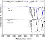
The potential of the HAp@ZnO.AF carrier as a drug delivery support was investigated using curcumin as the therapeutic agent. The biomaterials HAp@10%ZnO.AF.Cur and HAp@20%ZnO.AF.Cur were analyzed by FTIR (Figure 8) to confirm the incorporation of the organic components. The characteristic phosphate bands of HAp (~1030, ~960, ~602, and ~565 cm–1) were preserved, indicating maintenance of the crystalline structure after the incorporation of ZnO, folic acid, and curcumin. The presence of ZnO is evidenced by the band at ~500–430 cm–1, attributed to Zn–O stretching vibrations, which is more intense in the sample containing 20% ZnO, in agreement with the higher oxide content. The incorporation of folic acid and curcumin is confirmed by absorption bands in the 1700–1500 cm–1 region, assigned to C=O stretching, aromatic C=C vibrations, and conjugated systems. Notably, contributions at ~1650–1600 cm–1 (conjugated carbonyl groups of curcumin and amide/carboxyl groups of folic acid) and at ~1510–1500 cm–1 (aromatic ring vibrations) are observed. The broad band at ~3400 cm–1, associated with –OH groups, exhibits increased broadening after drug loading, suggesting the occurrence of hydrogen-bonding interactions.
Complementarily, the crystalline structure of the biomaterials was investigated by X-ray diffraction (XRD) (Figure 9). The diffractograms of the HAp@10%ZnO.AF.Cur and HAp@20%ZnO.AF.Cur samples exhibit well-defined peaks attributed to hexagonal HAp, in agreement with COD standards 010740566 and 969001234, respectively, as well as peaks assigned to hexagonal ZnO (ICSD 029272). These results indicate the preservation of the crystalline phases of the carrier after functionalization and loading of the phytotherapeutic agent. The XRD results corroborate the FTIR data, confirming that the crystalline phases of the carrier were maintained following functionalization and drug loading. Several studies have reported that HAp may undergo decomposition into secondary calcium phosphate phases, such as β-tricalcium phosphate (β-TCP), or even calcium oxide (CaO), at temperatures close to 900 °C. However, in the present study, even after calcination at 900 °C, HAp remains the predominant phase identified in the diffractograms [52]. No characteristic peaks attributable to β-TCP are observed; if present, this phase is likely in trace amounts, below the visual detection limit of the XRD technique.

a) X-ray powder diffraction for HAp@10%ZnO.AFCur, Cucumin and b) X-ray powder diffraction for HAp@20%ZnO.AFCur, Cucumin.
Scanning electron microscopy analysis of (a) HAp@10%ZnO.AF.Cur and (b) HAp@20%ZnO.AF.Cur, presented in Figure 10, reveals the formation of micrometric agglomerates with irregular morphology and a high degree of particle coalescence. No clearly individualized primary particles were observed, which prevents accurate determination of particle size by this technique. The morphological differences between the samples are subtle, suggesting that increasing the ZnO concentration does not promote significant changes in the overall morphology of the material after curcumin loading.

a) Micrographs obtained by scanning electron microscopy for HAp@10%ZnO.AFCur and b) Micrographs obtained by scanning electron microscopy for HAp@20%ZnO.AFCur.
In the curcumin loading assay, the HAp@10%ZnO.AF.Cur system showed a significantly higher in-corporation of 54.79 ± 3.52% compared to the HAp@20%ZnO.AF.Cur system, which adsorbed 45.49 ± 3.79% of the curcumin, according to Student’s t-test for independent samples (p < 0.05). Therefore, a decrease in loading efficiency is observed with increasing ZnO content, indicating changes in the surface properties of the material.
The hemocompatibility of the developed biomaterials was investigated through hemolysis assays in order to evaluate their potential safety for biomedical applications involving possible blood contact (Figure 11). According to the ASTM F756 standard, hemolysis values below 5% classify materials as non-hemolytic, while values between 5% and 25% indicate slight to moderate hemolytic behavior, and values above 25% indicate significant hemolytic potential. Pure hydroxyapatite exhibited hemolysis values below 5% at all evaluated concentrations, confirming its well-established biocompatibility reported in previous studies. This behavior is commonly attributed to its chemical similarity to the inorganic phase of bone and its relatively low reactivity toward biological membranes, which tends to minimize erythrocyte disruption [53, 54].

Hemolysis percentage of erythrocytes after exposure to different biomaterials (15–200 µg·mL⁻¹). Results expressed as mean ± standard deviation. Hemocompatibility classification based on ASTM F756 standard.
Similarly, folic acid presented negligible hemolytic activity, supporting its use as a functionalization molecule in nanostructured drug delivery systems. These findings are consistent with previous reports describing the biological safety of folate-functionalized nanomaterials [20, 55]. In contrast, ZnO nanoparticles exhibited the highest hemolysis levels, particularly at higher concentrations, reaching values above 40% at 200 µg·mL⁻¹. This behavior has been previously associated with oxidative stress mechanisms, including ROS generation and membrane lipid peroxidation, as well as direct nanoparticle interaction with erythrocyte membranes, which may compromise membrane integrity [55, 56].
Interestingly, when ZnO was incorporated into the hydroxyapatite matrix, a significant reduction in hemolytic activity was observed. This behavior may suggest that the HAp structure acts as a stabilizing matrix, possibly reducing nanoparticle aggregation and limiting direct contact between ZnO and erythrocyte membranes[55, 56]. Additionally, the composite structure may contribute to a more controlled exposure of ZnO, thereby attenuating its hemolytic effects, as suggested in previous studies involving ceramic nanocomposites.
Free curcumin showed moderate hemolysis at higher concentrations, while lower concentrations remained within acceptable limits. This effect may be related to its hydrophobic nature and its ability to interact with lipid bilayers when not associated with a carrier system [57, 58]. Notably, when curcumin was incorporated into the composite systems, a reduction in hemolysis was observed compared to the free drug, which may indicate a protective effect provided by the carrier matrix, possibly by reducing direct drug–membrane interactions.
For the multifunctional HAp@ZnO.AF.Cur systems, hemolysis remained below 10% at concentrations up to 50 µg·mL⁻¹, while slightly higher values (approximately 10–15%) were observed at 200 µg·mL⁻¹, particularly for the HAp@10%ZnO.AF.Cur formulation. These results indicate a concentration-dependent hemolytic profile, suggesting that the biological response is influenced by the composite composition and exposure level.
Taken together, these findings suggest that the developed multifunctional systems present acceptable hemocompatibility at lower concentrations, while higher concentrations may require careful dose consideration. Therefore, although the results indicate promising hemocompatibility behavior, further biological evaluation would be necessary to fully establish the safety profile of these systems.
The cytotoxicity of the developed biomaterials was initially evaluated in L929 fibroblasts, a standard non-tumoral cell line recommended by ISO 10993-5 for in vitro cytocompatibility testing (Figure 12). All tested samples showed cell viability values above 70%, indicating absence of cytotoxic effects under the evaluated conditions [59]. Interestingly, cell viability values frequently exceeded 100%, suggesting a possible increase in metabolic activity. This behavior has been previously reported for calcium phosphate-based biomaterials and may be associated with their bioactive surface properties, which can favor cell adhesion and proliferation. In addition, the release of calcium ions from hydroxyapatite structures may contribute to cellular signaling processes involved in proliferation and metabolic regulation.

Metabolic activity of L929 (non-tumoral) and Sarcoma 180 (tumoral) cells after exposure to biomaterial extracts (25–200 µg·mL⁻¹) for 24 and 72 h, evaluated by MTT assay. Results are expressed as mean ± standard deviation. Cell viability above 70% is considered non-cytotoxic according to ISO 10993-5.
Importantly, the incorporation of ZnO, folic acid and curcumin into the HAp matrix did not compromise cytocompatibility toward L929 cells. This observation may suggest that the composite structure was able to modulate the biological effects of these components, particularly ZnO and curcumin, which may exhibit higher cytotoxicity when tested individually depending on dose and exposure conditions.
These findings therefore suggest that the developed system maintains adequate cytocompatibility toward non-tumoral cells, which is an important requirement for drug delivery platforms. Nevertheless, additional studies addressing long-term exposure and mechanistic cellular responses would be necessary to further clarify these biological interactions.
The biological response of Sarcoma 180 tumor cells differed from that observed in L929 fibroblasts. After 24 hours of exposure, pure hydroxyapatite did not significantly affect tumor cell viability, while the multifunctional systems containing ZnO and curcumin showed a reduction in metabolic activity, particularly at higher concentrations. After 72 hours, a more pronounced reduction in tumor cell viability was observed for all multifunctional systems. This behavior may suggest a time-dependent biological response, possibly associated with progressive cellular interaction with the biomaterials or cumulative effects resulting from exposure to the composite components.
Interestingly, even pure hydroxyapatite showed some reduction in tumor cell viability after prolonged exposure. Although the mechanism was not investigated in this study, this finding may suggest that extended exposure to biomaterials can influence tumor cell metabolism through indirect mechanisms such as ionic exchange or surface-mediated cellular stress. However, this observation should be interpreted cautiously and would require further investigation.
The stronger reduction in viability observed for the HAp@ZnO.AF.Cur systems may be associated with the combined biological activity of ZnO nanoparticles and curcumin, both of which have been described as presenting antitumor properties, including oxidative stress induction and modulation of cell survival pathways. However, since mechanistic assays such as apoptosis evaluation, ROS quantification or cell cycle analysis were not performed, these interpretations remain speculative.
The presence of folic acid may also contribute to enhanced interaction with tumor cells, since folate receptors are frequently overexpressed in various cancer cell types. In this context, the results obtained here may suggest a possible contribution of folate functionalization to tumor cell interaction. However, specific receptor expression analysis or cellular uptake studies would be necessary to confirm receptor-mediated effects.
Overall, the differential behavior observed between L929 and Sarcoma 180 cells may suggest a preferential reduction of tumor cell viability under the tested conditions. However, rather than demonstrating definitive selectivity, these results should be interpreted as indicating a tendency toward differential cytotoxic responses between tumoral and non-tumoral cells.
Thus, the present findings suggest that the developed systems may represent promising candidates for further investigation as antitumor drug delivery platforms, although additional biological studies would be necessary to confirm selectivity mechanisms and therapeutic efficacy. Although the present study provides important initial biological evidence, further mechanistic studies will be necessary to fully elucidate the cellular pathways involved in the observed cytotoxic effects.
4. CONCLUSION
In this work, a novel multifunctional drug delivery system based on HAp with ZnO NPs, functionalized with folic acid (AF), and evaluated as a carrier for the phytotherapeutic agent curcumin, was successfully developed. The synthesis of HAp in the presence of citrate resulted in a crystalline, bioactive, and material with a high specific surface area. The incorporation of ZnO provided effective antibacterial activity against S. aureus and E. coli, with the best performance observed for the HAp@10%ZnO formulation. Functionalization with AF was efficient and independent of the ZnO content, demonstrating the system’s viability for active target-ing of tumor cells that overexpress folate receptors. In the study of loading the therapeutic agent curcumin onto the carriers, showed adsorption rates of (54.79 ± 3.52) % for HAp@10%ZnO.AF.Cur and (45.49 ± 3.79) % for HAp@20%ZnO.AF.Cur. Hemolysis assays demonstrated that the HAp matrix effectively modulates the toxicity of the active components, significantly reducing the hemolytic effects of both ZnO and curcumin. Finally, the results show that the biomaterials are biocompatible with L929 cells and exhibit suggested differential cytotoxic response against Sarcoma 180 cells, highlighting their potential for anti-tumor applications. In an integrated manner, the findings evidence that the HAp@ZnO.AF.Cur carrier It exhibited antibacterial activity, as well as promising initial results regarding biocompatibility and a differential response in in vitro cellular models. Although these findings indicate promising potential for antitumor applications, further studies are required to confirm targeting mechanisms and therapeutic selectivity.
6. BIBLIOGRAPHY
-
[1] INSTITUTO NACIONAL DE CÂNCER, O que é câncer, https://www.gov.br/inca/pt-br/assuntos/cancer/o-que-e-cancer, accessed in September, 2025.
» https://www.gov.br/inca/pt-br/assuntos/cancer/o-que-e-cancer -
[2] KHIZAR, S., ALRUSHAID, N., ALAM KHAN, F., et al, “Nanocarriers based novel and effective drug delivery system”, International Journal of Pharmaceutics, v. 632, pp. 122570, Feb. 2023. doi: http://doi.org/10.1016/j.ijpharm.2022.122570. PubMed PMID: 36587775.
» https://doi.org/10.1016/j.ijpharm.2022.122570 -
[3] CHEN, W., SHI, K., YU, Y., et al, “Drug delivery systems for colorectal cancer chemotherapy”, Chinese Chemical Letters, v. 35, n. 2, pp. 109159, Sep. 2024. doi: http://doi.org/10.1016/j.cclet.2023.109159.
» https://doi.org/10.1016/j.cclet.2023.109159 -
[4] SUN, T., ZHANG, Y.S., PANG, B., et al, “Engineered nanoparticles for drug delivery in cancer therapy”, Angewandte Chemie International Edition in English, v. 53, n. 46, pp. 12320–12364, Nov. 2014. doi: http://doi.org/10.1002/anie.201403036. PubMed PMID: 25294565.
» https://doi.org/10.1002/anie.201403036 -
[5] TRAN, S., DEGIOVANNI, P.J., PIEL, B., et al, “Cancer nanomedicine: a review of recent success in drug delivery”, Clinical and Translational Medicine, v. 6, n. 1, e44, Dec. 2017. doi: http://doi.org/10.1186/s40169-017-0175-0. PubMed PMID: 29230567.
» https://doi.org/10.1186/s40169-017-0175-0 - [6] NAVES, J.F., ARAUJO, T.C.C.F., “Qualidade de vida e bem-estar subjetivo de sobreviventes ao câncer ósseo: percepção de sobreviventes e familiares”, Interação em Psicologia, v. 19, n. 3, pp. 351–363, Sep/Dec. 2015.
-
[7] SAFITRI, N., RAUF, N., TAHIR, D., “Enhancing drug loading and release with hydroxyapatite nanoparticles for efficient drug delivery: a review synthesis methods, surface ion effects, and clinical prospects”, Journal of Drug Delivery Science and Technology, v. 90, pp. 105092, Dec. 2023. doi: http://doi.org/10.1016/j.jddst.2023.105092.
» https://doi.org/10.1016/j.jddst.2023.105092 -
[8] ELIAZ, N., METOKI, N., “Calcium phosphate bioceramics: a review of their history, structure, proper-ties, coating technologies and biomedical applications”, Materials, v. 10, n. 4, pp. 1–104, Mar. 2017. doi: http://doi.org/10.3390/ma10040334. PubMed PMID: 28772697.
» https://doi.org/10.3390/ma10040334 -
[9] LIU, Z., SHI, J., CHEN, L., et al, “3D printing of fish-scale derived hydroxyapatite/chitosan/PCL scaffold for bone tissue engineering”, International Journal of Biological Macromolecules, v. 274, n. Pt 2, pp. 133172, Aug. 2024. doi: http://doi.org/10.1016/j.ijbiomac.2024.133172. PubMed PMID: 38880458.
» https://doi.org/10.1016/j.ijbiomac.2024.133172 -
[10] SHI, C., GAO, J., WANG, M., et al, “Ultra-trace silver-doped hydroxyapatite with non-cytotoxicity and effective antibacterial activity”, Materials Science and Engineering C, v. 55, pp. 497–505, Oct. 2015. doi: http://doi.org/10.1016/j.msec.2015.05.078. PubMed PMID: 26117782.
» https://doi.org/10.1016/j.msec.2015.05.078 - [11] PORTO, R. C. T., “Nanopartículas de óxido de zinco sintetizadas pelo método poliol: caracteriza-ção e avaliação da atividade antibacteriana”, Materials Science and Engineering C, v. 22, pp. 1–8, Jan. 2018.
-
[12] CHEN, M.M., TANG, X., LI, J.J., et al, “Active targeting tumor therapy using host-guest drug delivery system based on biotin functionalized azocalix[4]arene”, Journal of Controlled Release : Official Journal of the Controlled Release Society, v. 368, pp. 691–702, Apr. 2024. doi: http://doi.org/10.1016/j.jconrel.2024.03.017. PubMed PMID: 38492860.
» https://doi.org/10.1016/j.jconrel.2024.03.017 -
[13] SAHA, N., KESKINBORA, K., SUVACI, E., et al, “Sintering, microstructure, mechanical, and antimicrobial properties of HAp-ZnO bio-composites”, Journal of Biomedical Materials Research. Part B, Applied Biomaterials, v. 95, n. 2, pp. 430–440, Nov. 2010. doi: http://doi.org/10.1002/jbm.b.31734. PubMed PMID: 20878929.
» https://doi.org/10.1002/jbm.b.31734 -
[14] BEYENE, Z., GHOSH, R., “Effect of zinc oxide addition on antimicrobial and antibiofilm activity of hydroxyapatite: a potential nanocomposite for biomedical applications”, Materials Today. Communications, v. 21, pp. 100612, Dec. 2019. doi: http://doi.org/10.1016/j.mtcomm.2019.100612.
» https://doi.org/10.1016/j.mtcomm.2019.100612 -
[15] LIU, Y., HINNANT, B., CHEN, S., et al, “Hyaluronic acid-modified extracellular vesicles for targeted doxorubicin delivery in hepatocellular carcinoma”, Experimental Cell Research, v. 443, n. 2, pp. 114332, Dec. 2024. doi: http://doi.org/10.1016/j.yexcr.2024.114332. PubMed PMID: 39551341.
» https://doi.org/10.1016/j.yexcr.2024.114332 -
[16] KABILOVA, T.O., SHMENDEL, E.V., GLADKIKH, D.V., et al, “Targeted delivery of nucleic acids into xenograft tumors mediated by novel folate-equipped liposomes”, European Journal of Pharmaceutics and Biopharmaceutics : Official Journal of Arbeitsgemeinschaft für Pharmazeutische Verfahrenstechnik e.V, v. 123, pp. 59–70, Feb. 2018. doi: http://doi.org/10.1016/j.ejpb.2017.11.010. PubMed PMID: 29162508.
» https://doi.org/10.1016/j.ejpb.2017.11.010 -
[17] LIU, Y., LI, K., PAN, J., et al, “Folic acid conjugated nanoparticles of mixed lipid monolayer shell and biodegradable polymer core for targeted delivery of Docetaxel”, Biomaterials, v. 31, n. 2, pp. 330–338, Jan. 2010. doi: http://doi.org/10.1016/j.biomaterials.2009.09.036. PubMed PMID: 19783040.
» https://doi.org/10.1016/j.biomaterials.2009.09.036 -
[18] WU, J., LIU, Q., LEE, R.J., “A folate receptor-targeted liposomal formulation for paclitaxel”, International Journal of Pharmaceutics, v. 316, n. 1–2, pp. 148–153, Jun. 2006. doi: http://doi.org/10.1016/j.ijpharm.2006.02.027. PubMed PMID: 16564654.
» https://doi.org/10.1016/j.ijpharm.2006.02.027 -
[19] ISLAM, M.R., RAUF, A., AKASH, S., et al, “Targeted therapies of curcumin focus on its therapeutic benefits in cancers and human health: Molecular signaling pathway-based approaches and future perspectives”, Biomedicine & Pharmacotherapy = Biomédecine & Pharmacothérapie, v. 170, pp. 116034, Jan. 2024. doi: http://doi.org/10.1016/j.biopha.2023.116034. PubMed PMID: 38141282.
» https://doi.org/10.1016/j.biopha.2023.116034 -
[20] BEHNAMSANI, A., MESHKINI, A., “Synthesis and engineering of mesoporous ZnO@HAP heterostruc-ture as a pH-sensitive nano-photosensitizer for chemo-photodynamic therapy of malignant tumor cells”, Journal of Drug Delivery Science and Technology, v. 53, pp. 101200, Oct. 2019. doi: http://doi.org/10.1016/j.jddst.2019.101200.
» https://doi.org/10.1016/j.jddst.2019.101200 -
[21] GU, Y., YANG, R., ZHANG, Y., et al, “Molecular mechanisms and therapeutic strategies in overcoming chemotherapy resistance in cancer”, Molecular Biomedicine, v. 6, n. 1, pp. 2, Jan. 2025. doi: http://doi.org/10.1186/s43556-024-00239-2. PubMed PMID: 39757310.
» https://doi.org/10.1186/s43556-024-00239-2 -
[22] RUBAGOTTI, S., CROCI, S., FERRARI, E., et al, “Uptake of Ga-curcumin derivatives in different cancer cell lines: toward the development of new potential 68 Ga-labelled curcuminoids-based radiotracers for tumour imaging”, Journal of Inorganic Biochemistry, v. 173, pp. 113–119, Aug. 2017. doi: http://doi.org/10.1016/j.jinorgbio.2017.05.002. PubMed PMID: 28511061.
» https://doi.org/10.1016/j.jinorgbio.2017.05.002 -
[23] SUETH-SANTIAGO, V., MENDES-SILVA, G.P., DECOTÉ-RICARDO, D., et al, “Curcumin, the golden powder from turmeric: insights into chemical and biological activities”, Química Nova, v. 38, n. 4, pp. 538–552, May. 2015. doi: http://doi.org/10.5935/0100-4042.20150035.
» https://doi.org/10.5935/0100-4042.20150035 -
[24] AYUB, H., ISLAM, M., SAEED, M., et al, “On the health effects of curcumin and its derivatives”, Food Science & Nutrition, v. 12, n. 11, pp. 8623–8650, Nov. 2024. doi: http://doi.org/10.1002/fsn3.4469. PubMed PMID: 39620006.
» https://doi.org/10.1002/fsn3.4469 -
[25] PRIYADARSINI, K., “The chemistry of curcumin: from extraction to therapeutic agent”, Molecules (Basel, Switzerland), v. 19, n. 12, pp. 20091–20112, Dec. 2014. doi: http://doi.org/10.3390/molecules191220091. PubMed PMID: 25470276.
» https://doi.org/10.3390/molecules191220091 -
[26] MA, H., ZHOU, Q., CHANG, J., et al, “Grape seed-inspired smart hydrogel scaffolds for melanoma therapy and wound healing”, ACS Nano, v. 13, n. 4, pp. 4302–4311, Apr. 2019. doi: http://doi.org/10.1021/acsnano.8b09496. PubMed PMID: 30925040.
» https://doi.org/10.1021/acsnano.8b09496 - [27] DEI CAS, M., GHIDONI, R., “Dietary Curcumin: correlation between bioavailability and health potential”, Nutrients, v. 11, n. 9, pp. 2147, Sep. 2019. PubMed PMID: 31500361.
-
[28] PRASAD, S., TYAGI, A.K., AGGARWAL, B.B., “Recent developments in delivery, bioavailability, absorption and metabolism of curcumin: the golden pigment from golden spice”, Cancer Research and Treatment, v. 46, n. 1, pp. 2–18, Jan. 2014. doi: http://doi.org/10.4143/crt.2014.46.1.2. PubMed PMID: 24520218.
» https://doi.org/10.4143/crt.2014.46.1.2 - [29] RODRIGUES, A.S., “Folic acid-functionalized hydroxyapatite nanoparticles loaded with curcumin for osteosarcoma therapy”, Materials Today. Communications, v. 38, pp. 107497, 2024.
-
[30] DARYAN, S.H., KHAVANDI, A., JAVADPOUR, J., “Surface engineered hollow hydroxyapatite micro-spheres: Hydrothermal synthesis and growth mechanisms”, Solid State Sciences, v. 106, pp. 106301, Aug. 2020. doi: http://doi.org/10.1016/j.solidstatesciences.2020.106301.
» https://doi.org/10.1016/j.solidstatesciences.2020.106301 -
[31] KOKUBO, T., TAKADAMA, H., “How useful is SBF in predicting in vivo bone bioactivity?”, Biomaterials, v. 27, n. 15, pp. 2907–2915, Jan. 2006. doi: http://doi.org/10.1016/j.biomaterials.2006.01.017. PubMed PMID: 16448693.
» https://doi.org/10.1016/j.biomaterials.2006.01.017 -
[32] GHORBANI, H., MEHR, F., PAZOKI, H., et al, “Synthesis of ZnO nanoparticles by precipitation method”, Oriental Journal of Chemistry, v. 31, n. 2, pp. 1219–1221, Jun. 2015. doi: http://doi.org/10.13005/ojc/310281.
» https://doi.org/10.13005/ojc/310281 -
[33] MARTÍNEZ, C.A., GILABERT, U., GARRIDO, L., et al, “Functionalization of hydroxyapatite scaffolds with ZnO”, Procedia Materials Science, v. 9, pp. 484–490, 2015. doi: http://doi.org/10.1016/j.mspro.2015.05.020.
» https://doi.org/10.1016/j.mspro.2015.05.020 -
[34] SANTOS, C., GOMES, P., DUARTE, J.A., et al, “Development of hydroxyapatite nanoparticles loaded with folic acid to induce oste-oblastic differentiation”, International Journal of Pharmaceutics, v. 516, n. 1–2, pp. 185–195, Jan. 2017. doi: http://doi.org/10.1016/j.ijpharm.2016.11.035. PubMed PMID: 27851979.
» https://doi.org/10.1016/j.ijpharm.2016.11.035 -
[35] PITA, J.C.L.R., XAVIER, A.L., SOUSA, T.K., et al, “In vitro and in vivo antitumor effect of trachylobane-360, a diterpene from Xylopia langsdorffiana”, Molecules, v. 17, n. 8, pp. 9573–9589, 2012. doi: http://doi.org/10.3390/molecules17089573. PubMed PMID: 22885357.
» https://doi.org/10.3390/molecules17089573 -
[36] YEDGAR, S., BARSHTEIN, G., GURAL, A., “Hemolytic activity of nanoparticles as a marker of their hemocompatibility”, Micromachines, v. 13, n. 12, pp. 1249134, 2022. doi: http://doi.org/10.3390/mi13122091. PubMed PMID: 36557391.
» https://doi.org/10.3390/mi13122091 - [37] AMERICAN SOCIETY FOR TESTING AND MATERIALS, ASTM F756-17 Standard Practice for Assessment of Hemolytic Properties of Materials, West Conshohocken, ASTM, 2017.
- [38] SHITOLE, A.A., GIRAM, P.S., RAUT, P.W., et al, “Hemocompatibility assessment of biomaterials: a critical review”, Journal of Biomaterials Applications, v. 33, n. 10, pp. 1354–1373, 2019. PubMed PMID: 30880549.
- [39] ALSAKHAWY, A.M., “Hemocompatibility and cytotoxicity evaluation of bioactive nanocompo-site materials for biomedical applications”, Materials Science and Engineering C, v. 133, pp. 112622, 2022.
-
[40] SAHADAT HOSSAIN, M., AHMED, S., “FTIR spectrum analysis to predict the crystalline and amorphous phases of hydroxyapatite: a comparison of vibrational motion to reflection”, RSC Advances, v. 13, n. 21, pp. 14625–14630, 2023. doi: http://doi.org/10.1039/D3RA02580B. PubMed PMID: 37197675.
» https://doi.org/10.1039/D3RA02580B -
[41] ZARIF, M.E., BITA, B., YEHIA-ALEXE, S.A., et al, “Biological and physicochemical analysis of sr-doped hydroxyapatite/chitosan composite layers”, Polymers, v. 16, n. 13, pp. 1922, Jul. 2024. doi: http://doi.org/10.3390/polym16131922. PubMed PMID: 39000777.
» https://doi.org/10.3390/polym16131922 -
[42] YANG, H., HAO, L., DU, C., et al, “A systematic examination of the morphology of hydroxyapatite in the presence of citrate”, RSC Advances, v. 3, n. 45, pp. 23184–23189, Dec. 2013. doi: http://doi.org/10.1039/c3ra44839h.
» https://doi.org/10.1039/c3ra44839h -
[43] DUMITRESCU, C.R., MATEI, M., DEÁK, G., et al, “Sustainable synthesis and dual-function sorption of carbonated hydroxyapatite for cadmium and nitrate removal”, International Journal of Molecular Sciences, v. 26, n. 16, pp. 7766, Aug. 2025. doi: http://doi.org/10.3390/ijms26167766. PubMed PMID: 40869086.
» https://doi.org/10.3390/ijms26167766 -
[44] MA, X., ELLIS, D.E., “Initial stages of hydration and Zn substitution/occupation on hydroxyapatite (0 0 0 1) surfaces”, Biomaterials, v. 29, n. 3, pp. 257–265, Jan. 2008. doi: http://doi.org/10.1016/j.biomaterials.2007.10.001. PubMed PMID: 17961647.
» https://doi.org/10.1016/j.biomaterials.2007.10.001 -
[45] AL-GHOUTI, M.A., Da’Ana, D.A., “Guidelines for the use and interpretation of adsorption isotherm models: a review”, Journal of Hazardous Materials, v. 393, pp. 122383, Jul. 2020. doi: http://doi.org/10.1016/j.jhazmat.2020.122383. PubMed PMID: 32369889.
» https://doi.org/10.1016/j.jhazmat.2020.122383 -
[46] LIU, Q., GUO, W., YANG, M., et al, “Release behavior of folic acid grafted hollow hydroxyapatite as drug carrier”, Advances in Polymer Technology, v. 2019, pp. 1–9, Mar. 2019. doi: http://doi.org/10.1155/2019/9562437.
» https://doi.org/10.1155/2019/9562437 -
[47] BARBOSA, A.A., ALVES JUNIOR, S., OLIVEIRA, R.A.P., et al, “Structural and photophysical properties of hydroxyapatite doped with lantha-nide ions”, Matéria, v. 25, n. 4, e12862, Jun. 2020. doi: http://doi.org/10.1590/s1517-707620200004.1162.
» https://doi.org/10.1590/s1517-707620200004.1162 -
[48] JAITA, P., RANDORN, C., WATCHARAPASORN, A., et al, “In vitro bioactivity, mechanical, and cell interaction of sodium chloride-added calcium sulfate-hydroxyapatite composite bone cements”, RSC Advances, v. 14, n. 48, pp. 35460–35474, Nov. 2024. doi: http://doi.org/10.1039/D4RA06034B. PubMed PMID: 39507684.
» https://doi.org/10.1039/D4RA06034B - [49] NATIONAL COMMITTEE FOR CLINICAL LABORATORY STANDARDS, Performance Standards for Antimicrobial Disk Susceptibility Tests, Pennsylvania, NCCLS, 2003.
-
[50] GUDKOV, S.V., BURMISTROV, D.E., SEROV, D.A., et al, “A mini review of antibacterial properties of ZnO nanoparticles”, Frontiers in Physics, v. 9, pp. 611481, Mar. 2021. doi: http://doi.org/10.3389/fphy.2021.641481.
» https://doi.org/10.3389/fphy.2021.641481 -
[51] BARBOSA, A.A., JÚNIOR, S.A., FERRAZ, A.V., “A study of the bioactivity, hemocompatibility and antimicrobial properties of a zinc oxide and calcium phosphate composite for bone regeneration”, Cerâmica, v. 69, n. 390, pp. 93–98, 2023. doi: http://doi.org/10.1590/0366-69132023693903396.
» https://doi.org/10.1590/0366-69132023693903396 -
[52] JABER, H., MOUSA, M.H., QADIR, A., et al, “Comprehensive investigation into thermal stability of AB-type biocarbonate hydroxyapatite synthesized via heat-treated bovine bone”, Open Ceramics, v. 20, pp. 100701, Dec. 2024. doi: http://doi.org/10.1016/j.oceram.2024.100701.
» https://doi.org/10.1016/j.oceram.2024.100701 -
[53] OOI, C.H., LING, Y.P., ABDULLAH, W.Z., et al, “Physicochemical evaluation and in vitro hemocompatibility study on nanoporous hydroxyapatite”, Journal of Materials Science. Materials in Medicine, v. 30, n. 4, pp. 44, Apr. 2019. doi: http://doi.org/10.1007/s10856-019-6247-5. PubMed PMID: 30929088.
» https://doi.org/10.1007/s10856-019-6247-5 - [54] BENALI, Y., “Application of nanohydroxyapatite in medicine: recent advances and challenges”, Materials Chemistry and Physics, v. 315, pp. 127081, 2023.
-
[55] ZHANG, Y., “Zinc oxide-based nanocomposites for biomedical applications: biocompatibility and toxicity aspects”, Journal of Biomedical Materials Research. Part B, Applied Biomaterials, v. 110, n. 9, pp. 2150–2163, 2022. doi: http://doi.org/10.1002/jbm.b.35071.
» https://doi.org/10.1002/jbm.b.35071 - [56] SINGH, A., “Hemocompatibility and oxidative stress induced by ZnO nanoparticles: concentra-tion-dependent effects”, Colloids and Surfaces. B, Biointerfaces, v. 204, pp. 111819, 2021.
- [57] NGUYEN, M., “Curcumin delivery systems for cancer therapy: recent advances and challenges”, Biomaterials Advances, v. 133, pp. 112598, 2021.
- [58] RODRIGUES, A.S., “Folic acid-functionalized hydroxyapatite nanoparticles loaded with curcumin for osteosarcoma therapy”, Materials Today. Communications, v. 38, pp. 107497, 2024.
- [59] INTERNATIONAL ORGANIZATION FOR STANDARDIZATION, ISO 10993-5 Biological Evaluation of Medical Devices — Part 5: Tests for In vitro Cytotoxicity, Geneva, ISO, 2009.


