 Aesthetic rehabilitation in mesiodents until the eruption of permanent successors: importance of monitoring in a case report
ABSTRACT
Mesiodens are supernumerary teeth located in the anterior maxilla region, often with a conical crown and short root. Their presence may compromise occlusion, esthetics, and the eruption path of permanent incisors, representing both functional and psychosocial challenges in early childhood. The aim was to report a pediatric dentistry case of conservative management of mesiodens through esthetic rehabilitation and systematic clinical monitoring until the optimal timing for extraction. A 4-year-old boy presented with two conoid mesiodens in the anterior maxilla region. Instead of immediate extraction, a conservative approach was adopted, consisting of esthetic rehabilitation using prefabricated acetate crowns and continuous clinical and radiographic follow-up. This strategy ensured satisfactory esthetics, preserved function, and promoted the child’s psychosocial well-being during a critical stage of development. The case highlights the importance of individualized treatment planning in pediatric dentistry. Early diagnosis and conservative rehabilitation can provide temporary but significant esthetic and emotional benefits, while delaying surgical intervention until the ideal moment facilitates proper eruption of permanent successors. Conservative rehabilitation of mesiodens, associated with systematic monitoring, represents a valuable therapeutic approach. Beyond preserving function and esthetics, it supports psychosocial development and improves quality of life, highlighting the relevance of treatment timing in pediatric patients.
Indexing terms
Oral rehabilitation; Pediatric dentistry; Psychological well-being; Tooth, deciduous; Tooth, supernumerary
RESUMO
Mesiodentes são dentes supranumerários localizados na região anterior da maxila, geralmente com coroa cônica e raiz curta. Sua presença pode comprometer a oclusão, a estética e o trajeto de erupção dos incisivos permanentes, representando desafios funcionais e psicossociais na primeira infância. Objetivou-se relatar um caso clínico em Odontopediatria envolvendo o manejo conservador de mesiodentes por meio de reabilitação estética e monitoramento clínico sistemático até o momento ideal para a extração. Um menino de 4 anos apresentou dois mesiodentes cônicos na região anterior da maxila. Em vez da extração imediata, adotou-se uma abordagem conservadora, consistindo em reabilitação estética com coroas pré-fabricadas de acetato e acompanhamento clínico e radiográfico contínuo. Essa estratégia garantiu estética satisfatória, preservação da função e bem-estar psicossocial durante uma fase crítica do desenvolvimento. O caso destaca a importância do planejamento de tratamento individualizado em Odontopediatria. O diagnóstico precoce e a reabilitação conservadora podem oferecer benefícios estéticos e emocionais temporários, porém significativos, enquanto o adiamento da intervenção cirúrgica até o momento ideal favorece a erupção adequada dos sucessores permanentes. A reabilitação conservadora de mesiodentes, associada ao monitoramento sistemático, representa uma abordagem terapêutica valiosa. Além de preservar função e estética, ela apoia o desenvolvimento psicossocial e melhora a qualidade de vida, ressaltando a relevância do momento da intervenção em pacientes pediátricos.
Termos de indexação
Reabilitação oral; Odontopediatria; Bem-estar psicológico; Dente decíduo; Dente supranumerário
INTRODUCTION
The initial stages of dental formation may generate developmental disorders that can result in anomalies. They may occur singly or multiply, unilaterally or bilaterally, and in any part of the upper or lower arch. Specifically regarding mesiodens, which are supernumerary teeth located in the region between the maxillary central incisors and are mostly conical in shape with a short root [1-3]. The prevalence of mesiodens ranges from 0.15% to 3.8% in the general population and more frequently affects the male population [4].
The complications related include delayed eruption and altered trajectory of the succeeding teeth, impaction, displacement or rotation of permanent incisors, abnormal root formation, midline diastema, cystic lesions, intraoral infection, root resorption of adjacent teeth, or even eruption into the nasal cavity [4,5]. Early diagnosis and proper treatment are essential for the preservation of occlusion, esthetics, phonetics, and the patient’s emotional well-being [6].
The aim of this study was to report a clinical case involving the esthetic and functional rehabilitation of two conical-shaped mesiodens in the anterior maxillary region, in a preschooler. A carefully planned intervention that promoted spontaneous eruption, minimized psychosocial impact, and preserved oral health.
CASE REPORT
This is a descriptive observational study. A male infant patient D.G.F., 4 years old, was brought to the Supervised Pediatric Integrated Clinic at Federal University of Juiz de Fora, Governador Valadares campus (UFJF-GV) by his guardian, with the complaint that the “baby tooth was displaced from its position.” During the anamnesis, it was reported that the day before, the child was playing during school recess and bumped into another child, hitting his mouth against the other’s head.
Ethical aspects
The ethical principles of research involving human subjects were followed, with the volunteer’s identity preserved and all clarifications regarding case disclosure provided. The present study was submitted to and approved (number: 5.390.296) by the Human Research Ethics Committee of the Federal University of Juiz de Fora. Informed consent and consent for publication were obtained from the legal guardians.
Examination and first procedure
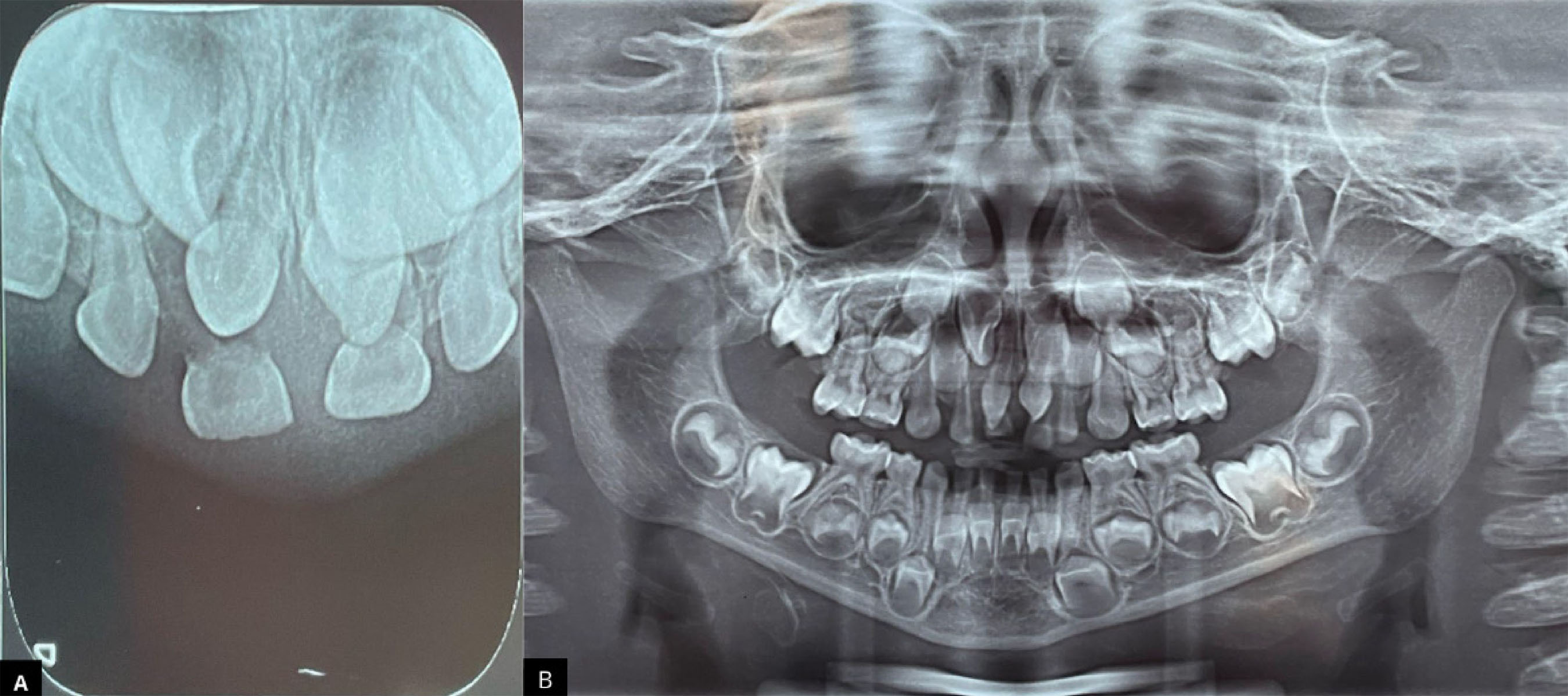
On clinical examination, it was noted that the upper right central incisor deciduous tooth presented buccal luxation, adhered to the marginal buccal gingiva. On radiographic examination (figure 1), it was clarified that there were two supernumerary teeth with conical-shaped crowns and short roots, located below the permanent maxillary central incisors. The root of upper right central incisor deciduous tooth had already undergone rhizolysis, while the left one still retained a small root portion. The tooth was extracted, and the guardian was instructed to return every six months for follow-up.
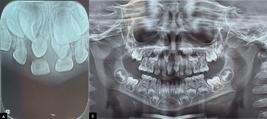
A) Periapical radiographic of the upper central region, showing the deciduous teeth, the mesiodens, and the germs of the permanent teeth. B). Panoramic radiograph.
Fist follow-up and second procedure
During the follow-up period, the upper left central incisor deciduous tooth exfoliated spontaneously, and the supernumerary teeth erupted in the site designated for the upper right and left central incisors (figure 2), which were still intraosseous. After the complete eruption of the supernumeraries, the guardians reported that the child was dissatisfied with their shape and had been subjected to bullying at school. Given this, re-anatomization of the teeth was suggested, since the permanent successors presented root development at Nolla stage 5.
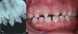
A) Periapical radiographic of the upper central region, showing the mesiodens and the permanent teeth. B) Mesiodens erupted in the position of the permanent upper central incisors.
The supernumerary teeth required no prior preparation with burs. A protocol for restoration with light-cured resin was followed, starting with prophylaxis using a Robinson brush (American Burrs − Palhoça, Brazil) in low rotation with pumice (SS White − Juiz de Fora, Brazil) and water. This was followed by conditioning with 37% phosphoric acid (FGM −Joinville, Brazil) for 30 seconds, application of the Single Bond Universal adhesive system (3M ESPE − Two Harbors, USA), and light curing for 20 seconds (Saevo −Ribeirão Preto, Brazil). Prefabricated acetate crowns shaped like upper right central incisor deciduous and upper left central incisor deciduous teeth (TDV Dental − Pomerode, Brazil) were previously selected and adapted to the cervico-occlusal height, with cervical margin trimming performed using scissors. The acetate crowns were perforated with an explorer probe (Golgran - São Caetano do Sul, Brazil) in the palatal region to allow resin overflow. Resin shade B2 (Z 350 − 3M ESPE − Two Harbors, USA) was inserted into the acetate crown until complete filling, taking care to avoid air bubble formation within the resin. The filled crown was fitted onto the supernumerary tooth, and excess resin overflowed at the cervical and perforation areas, which was removed with an explorer probe. The resin was polymerized on all surfaces for 20s. With the aid of a scalpel blade, the acetate crown was removed. Occlusal adjustment was performed using a finishing bur (2135F Fava − Franco da Rocha, Brazil), articulating paper (Accufilm Parkel − Brentwood, NY, USA), and Muller tweezers (Quinelato − Rio Claro, Brazil). The finishing and polishing were scheduled for the third session to achieve better esthetic harmonization. Figure 3 show the steps described above.

A) Conditioning with 37% phosphoric acid for 30 seconds. B) Application of the SingleBond Universal 3M ESPE adhesive system using a microbrush. C) Polymerization with a light-curing device for 20 seconds. D) Prefabricated acetate crown in the shape of the deciduous tooth, which was filled with B2 shade resin (Z 350 - 3M ESPE). E) Light curing of the resin on all surfaces. F) Removal of the acetate crown with the aid of a scalpel blade.
Subsequent follow-ups
At follow-up visits, the patient reported being very satisfied with the outcome, and the guardian noted that the child began smiling again, showing his teeth.
The patient remained under six-month follow-up (figure 4).

A) Radiograph to monitor the development of the permanent successor teeth, showing the acetate crowns still in position and well adapted. B). Mesiodens with the acetate crowns.
Surgical removal of the supernumerary teeth was indicated when the permanent successors reached Nolla stage 8, the stage at which the eruptive process begins. The follow-up period extended until the complete eruption of the permanent maxillary central incisors.
DISCUSSION
This study presents an analysis of a clinical case involving the diagnosis, management, and esthetic rehabilitation of conical-shaped mesiodens in the anterior maxillary region of a young patient, highlighting the implications for oral health, psychosocial well-being, and treatment timing considerations.
Mesiodens vary in size and shape, which influences their clinical presentation and management; in approximately two-thirds of cases, they are usually conical. This facilitates eruption into the oral cavity and contributes to surgical removal. In the reported case, the mesiodens had a conical shape, short root, and erupted into the oral cavity after exfoliation of the primary teeth [4].
In terms of eruption patterns, the literature indicates that only 25% of all mesiodens erupt spontaneously in the oral cavity [7,8], as occurred in the clinical case described. Conversely, regarding eruption of permanent teeth, in 75% of cases, the permanent incisor erupts spontaneously after removal of the mesiodens, supporting the report [9].
In the reported case, the diagnosis was made early due to an accident at school, and with the aid of radiographic examination, it was possible to identify the presence of supernumeraries in the anterior maxillary region. Considering all the complications that a supernumerary tooth can cause, diagnosis should be made early so that the professional can perform more conservative treatment [10].
Given the anterior location’s fundamental role in esthetics and function, early loss of primary anterior teeth in the maxilla or the presence of dental anomalies in this region can lead to psychological problems and impair children’s self-esteem and socialization, precisely at a very important stage of development [6,11]. It was reported that the child was suffering bullying at school due to the conical anatomical shape of the mesiodens in the anterior maxillary region.
Bullying constitutes a major social problem that harms the quality of life of affected children and adolescents; generating emotional damage and individuals with oral-facial alterations suffer from this problem [12]. When physical appearance is highly relevant for social inclusion, dental alterations can cause serious emotional impacts that interfere with self-esteem, directly influencing emotional and social well-being [13]. After esthetic rehabilitation, the guardian reported an improvement in the child’s quality of life and self-esteem.
In this context, the patient’s age and quality of life considerations guided the choice of a conservative restorative approach that would bring esthetic, functional, and emotional benefits, aiming to fully restore health. The coronal reconstruction technique used proved simple, relatively quick, low cost due to no laboratory phase needed, and efficient in achieving functionality and esthetics. Furthermore, the anatomical results obtained through its use are similar to natural teeth [14].
Another clinical observation during the case was reduced chair time [15]; it did not affect the quality of care, representing a practical benefit for both the patient and the clinician, highlighting the importance of selecting techniques that balance clinical efficiency and effectiveness.
While supernumerary teeth can cause complications across dentitions, careful consideration must be given to timing and indication for intervention. In cases of mesiodens in primary dentition, the risk of injury to developing teeth and the lack of cooperation in this phase must be considered [4,7,16]. For this reason, due to the patient’s age and the Nolla stage of the successor germs at treatment onset, the surgical procedure was postponed, keeping the supernumerary teeth in the mouth.
When supernumerary teeth do not interfere with mastication, dental or arch development, or cause pathological manifestations, surgical extraction is not necessary. However, when the presence of the extra tooth may delay, block eruption, cause deviation, rotation, or displacement of permanent teeth in the dental arch, removal is preferable, eliminating the causal factor [6]. In the reported case, surgical extraction of the mesiodens was performed because they were hindering eruption of the permanent successors.
The correct timing of surgical extraction of mesiodens is extremely important; attention should be paid to the stage of permanent tooth formation to allow normal root development, reducing the chances of root dilaceration and ankyloses [7,17]. Ideally, extraction should be before the closure of the permanent tooth apex, as this can prevent disadvantages such as loss of eruptive force, need for surgical exposure, and orthodontic traction [4].
During follow-up, normal root development was observed, and the timing for surgical removal of the mesiodens was established based on the permanent teeth’s root development stage. Extraction at the correct time can promote spontaneous eruption of the permanent successor, which may result in tooth alignment, and minimize the need for orthodontic treatment [9,18].
CONCLUSION
This case report highlights the importance of early diagnosis, conservative treatment, and regular follow-up to balance functional, esthetic, and psychosocial needs in children. Properly timed management of supernumerary teeth allowed spontaneous eruption, reduced complications, and preserved oral health. The conservative use of acetate crowns provided satisfactory esthetic and functional results, improving the patient’s quality of life and self-esteem by reducing the psychosocial effects of dental anomalies.
Ultimately, by presenting a practical, cost-effective, and patient-centered protocol, this case contributes valuable insights to clinical decision-making and highlights the need to consider not only dental health but also the emotional well-being of pediatric patients during critical stages of development.
-
How to cite this article
Neves JPF, Reis RM, Carlo FGC, Gomes JC. Aesthetic rehabilitation in mesiodents until the eruption of permanent successors: importance of monitoring in a case report. RGO, Rev Gaúch Odontol. 2026;74:e20260010. http://dx.doi.org/10.1590/1981-86372026001020250121
-
Article aligned with the Good Health and well-being goal of the Sustainable Development Goals (SDGs).
Data Availability
The research data are available in the body of the document.
REFERENCES
-
1 Verma L, Singh D. Esthetic rehabilitation in a patient with mesiodens. J Dent Allied Sci. 2012;1:88-90. doi: https://doi.org/10.4103/2277-4696.159168
» https://doi.org/10.4103/2277-4696.159168 -
2 Juuri E, Balic A. The biology underlying abnormalities of tooth number in humans. J Dent Res. 2017;96(11):1248-56. doi: https://doi.org/10.1177/0022034517720158
» https://doi.org/10.1177/0022034517720158 -
3 Fuentes R, Álvarez G, Garay I, Arias A, Dias FJ. Dientes supernumerarios suplementarios en un paciente adulto parcialmente dentado: reporte de caso. Int J Morphol. 2018;36(2):478-82. doi: https://doi.org/10.4067/S0717-95022018000200478
» https://doi.org/10.4067/S0717-95022018000200478 -
4 Dias GF, Hagedorn H, Maffezzolli MDL, Silva FF, Alves FBT. Diagnosis and treatment of supernumerary teeth in the pediatric clinic: case report. Rev CEFAC. 2019;21(6):e16318. doi: https://doi.org/10.1590/1982-0216/201921616318
» https://doi.org/10.1590/1982-0216/201921616318 -
5 Bahadure RN, Thosar N, Jain ES, Kharabe V, Gaikwad R. Supernumerary teeth in primary dentition and early intervention: a series of case reports. Case Rep Dent. 2012;2012:614652. doi: https://doi.org/10.1155/2012/614652
» https://doi.org/10.1155/2012/614652 -
6 Kouri DL, Bertaia CA, Santos R, Costa-Nobre PX, Garcia-Leite ES, Araújo FB, et al. Aesthetic and functional rehabilitation of a supernumerary peg-shaped tooth in the primary dentition. RGO, Rev Gaúch Odontol. 2014;62(2):191-5. doi: https://doi.org/10.1590/1981-86372014000200000151592
» https://doi.org/10.1590/1981-86372014000200000151592 - 7 Stringhini Junior E, Stang B, Oliveira LB. Dentes supranumerários impactados: relato de caso clínico. Rev Assoc Paul Cir Dent. 2015;69(1):8994.
-
8 Shetty V, Shetty RY, Mehrotra D, Usman A, Prabhu N. Management of impacted and inverted mesiodens: a report of two cases. J Pharm Bioallied Sci. 2024;16(Suppl 4):S4120S4122. doi: https://doi.org/10.4103/jpbs.jpbs_691_24
» https://doi.org/10.4103/jpbs.jpbs_691_24 -
9 Agarwal N, Jabin Z, Kalita S. An impacted inverted mesiodens and its surgical intervention. Int J Pedod Rehabil. 2020;5(1):22. doi: https://doi.org/10.4103/ijpr.ijpr_20_19
» https://doi.org/10.4103/ijpr.ijpr_20_19 -
10 Ferraresso L, Padovese M, De Souza K, Sakuma RH, Nagata ME, Seixas GF, et al. Diagnosis and treatment of supernumerary teeth in non-syndromic children: case series. Rev Odontol Latinoam. 2025;15:e-246671. doi: https://doi.org/10.47990/c0ax6e50
» https://doi.org/10.47990/c0ax6e50 -
11 Acharya S, Biswas R. Body image in preschool children following premature loss of primary teeth: a cross-sectional study. Int J Clin Pediatr Dent. 2022;15(3):293-8. doi: https://doi.org/10.5005/jp-journals-10005-2390
» https://doi.org/10.5005/jp-journals-10005-2390 -
12 Spezzia S. Implicações odontológicas do bullying na adolescência. Rev Flum Odontol. 2018;14(50):60-7. doi: https://doi.org/10.22409/ijosd.v2i50.36403
» https://doi.org/10.22409/ijosd.v2i50.36403 -
13 Chaudhary F, Ahmad B. The relationship between psychosocial distress and oral health status in patients with facial burns and mediation by oral health behaviour. BMC Oral Health. 2021;21(1):172. doi: https://doi.org/10.1186/s12903-021-01532-0
» https://doi.org/10.1186/s12903-021-01532-0 - 14 Miyata LB, Bonini GC, Calvo AFB, Politano GT. Reabilitação estética e funcional em paciente com cárie severa da infância: relato de caso. Rev Assoc Paul Cir Dent. 2014;68(1):22-9.
- 15 Lavor LQ, Matos KF, Paulino MR, Mendes TA, Fontes NM. Utilização da matriz de acetato na reabilitação de dentes anteriores decíduos: relato de caso clínico. Braz J Surg Clin Res. 2020;31(1):44-7.
-
16 Adisornkanj P, Chanprasit R, Eliason S, Fons JM, Intachai W, Tongsima S, et al. Genetic variants in protein tyrosine phosphatase non-receptor type 23 are responsible for mesiodens formation. Biology (Basel). 2023;12(3):393. doi: https://doi.org/10.3390/biology12030393
» https://doi.org/10.3390/biology12030393 -
17 Rahadian B, Julia V, Sulistyani L. Surgical management of mesiodens based on characteristics and complications of the condition: a systematic review. J Stomatol. 2020;73(5):261-9. doi: https://doi.org/10.5114/jos.2020.100583
» https://doi.org/10.5114/jos.2020.100583 - 18 Russell KA, Folwarczna MA. Mesiodens: diagnosis and management of a common supernumerary tooth. J Can Dent Assoc. 2003;69(6):362-6.
Edited by
-
Assistant editor
Luciana Butini Oliveira