
 Necrotizing gingivitis in patients with and without HIV: a systematic literature review and meta-analysis
ABSTRACT
Necrotizing periodontal diseases are historically established conditions that gained prominence during the First World War. Their etiology was linked to poor oral hygiene, opportunistic infections, and stress. Characterized by severity and debilitation, the disease progresses rapidly with necrotic ulcers in periodontal tissues, resulting in pain, tissue bleeding, and a foul odor. Due to the distinctive features and accelerated evolution of periodontal conditions in immunocompromised patients, individuals with Human Immunodeficiency Virus leading to Acquired Immunodeficiency Syndrome are classified as at-risk patients for the development of Necrotizing periodontal diseases. Thus, this review aims to compare the prevalence of gingivitis and necrotizing periodontitis in AIDS patients in comparison to the immunocompetent population. Additionally, it seeks to evaluate how the state of immunosuppression alters the oral cavity and provides clarity regarding the impact of AIDS on periodontal diseases. This study adhered to the methodological protocols of a systematic review as outlined in the Cochrane Handbook. The search strategy was implemented across the following databases: Pubmed, Cochrane Library, Scopus, Web of Science, and Embase. The association of necrotizing periodontal diseases in HIV+ individuals was investigated through a systematic analysis of the literature.
Indexing terms
Gingivitis, necrotizing ulcerative; HIV; Periodontal diseases
RESUMO
As doenças periodontais necrosantes são condições historicamente estabelecidas e ganharam destaque no período da primeira guerra mundial, sua etiologia estava ligada a falta de higiene oral, infecções oportunistas e quadros de estresse. De caráter grave e debilitante, a doença progride rapidamente com úlceras necróticas em tecido periodontal, gerando dor, sangramento dos tecidos e odor fétido. Em virtude de suas características e da evolução acelerada dos quadros periodontais em pacientes imunocomprometidos, pacientes portadores do vírus da imunodeficiência humana que desenvolvem a síndrome da imunodeficiência adquirida são classificados como pacientes de risco para desenvolvimento de doenças periodontais necrosantes. Dessa forma, a presente revisão busca comparar a prevalência de gengivite e periodontite necrosante em pacientes com AIDS em relação a população imunocompetente, bem com avaliar como o estado de imunossupressão altera a cavidade bucal; esclarecer a respeito do impacto da AIDS nas doenças periodontais. O presente estudo foi conduzido seguindo os protocolos metodológicos da revisão sistemática descritos no Cochrane Handbook. A estratégia de busca foi aplicada nas bases de dados: Pubmed, Cochrane Library, Scopus, Web of Science e Embase. Verificou-se a associação de doenças periodontais necrosantes em portadores de HIV+ por meio de uma análise sistemática da literatura.
Termos de indexação
Gengivite ulcerativa necrosante; HIV; Doenças periodontais
INTRODUCTION
Necrotizing Periodontal Diseases (NPDs) are historically established conditions that garnered attention during the First World War when more severe cases of the disease came to light. Referred to as ‘trench mouth’ during that period, Necrotizing Gingivitis (NG) was etiologically linked to poor oral hygiene, opportunistic infections, and the intense stress experienced by soldiers. With a severe and debilitating nature, the disease progresses rapidly, manifesting as necrotic ulcers in periodontal tissue, causing significant pain, tissue bleeding, and foul odor [1,2].
Due to its prominence in the era preceding the development of antibiotics, NPDs were typically addressed with hydrogen peroxide and improved oral hygiene. However, such measures proved insufficient for precise control of the conditions, and effective reversal began with the use of penicillins [3,4].
It is important to highlight that, following the new classification of periodontal diseases, the term ‘ulcerative’ is no longer employed in categorizing necrotizing periodontal diseases, as ulceration is considered secondary to necrosis. Consequently, the terms Necrotizing Ulcerative Gingivitis (NUG) and Necrotizing Ulcerative Periodontitis (NUP) have been replaced by Necrotizing Gingivitis (NG) and Necrotizing Periodontitis (NP). Furthermore, NG and NP can progress to more severe forms, such as Necrotizing Stomatitis (NS), when the necrotic tissue extends beyond the mucogingival junction, and ‘Noma’ in its advanced stage, both of which are now part of the NPDs group [5,6].
Effective therapy, coupled with enhanced knowledge of oral hygiene, has rendered NG and NP rare diseases with a prevalence of less than 1% in the population. However, individuals with nutritional deficits, stress conditions, and compromised immune systems become susceptible [7].
Due to their characteristics and the rapid progression of periodontal conditions in immunocompromised patients, individuals carrying the Human Immunodeficiency Virus (HIV) and developing Acquired Immunodeficiency Syndrome (AIDS) are classified as at-risk patients for the development of Necrotizing Gingivitis and Necrotizing Periodontitis (NP). Besides immunological status, additional factors such as psychological elements, including a loss of motivation for oral care, or nutritional deficiencies, further elevate the risks of NG and NP [1,8].
Despite this historical correlation, the literature presents conflicting findings regarding the prevalence of necrotizing diseases in the HIV+ population. Moreover, the constant updates in knowledge about AIDS patients often render this causal relationship outdated, necessitating more objective parameters for the classification of the at-risk population [7,9].
Thus, this review aims to compare the prevalence of NG and NP in patients with AIDS in comparison to the immunocompetent population. Seeking to characterize a deeper understanding of the etiology of these pathologies, the review also aims to provide an epidemiological perspective on the relationship between HIV and NDs.
METHODS
The scientific manuscript diligently traversed each stage of the 2020 PRISMA update, rigorously adhering to the methodological protocols for systematic reviews outlined in the Cochrane Handbook [10,11]. The chosen methodological approach was duly registered on the PROSPERO platform under registration number CRD42022321243. The search strategy, rooted in Medical Subject Headings (MeSH), was crafted as “(Patients, HIV and ‘Gingivitis, Necrotizing Ulcerative’)”, systematically expanded with the suggested entry terms in the MeSH interface. This comprehensive search strategy was then meticulously applied across various databases, including Pubmed, Cochrane Library, Scopus, Web of Science, and Embase. Notably, in Embase, MeSH terms were replaced with Emtree terms. Furthermore, no language or publication date restrictions were imposed, and the search was concluded in February 2024.
Regarding inclusion criteria, selected articles exclusively involved human samples and reported on adult patients aged over 18 years and under 60 years with HIV, explicitly mentioning the incidence of NG. Additionally, these studies incorporated patients without clinical manifestations of HIV as a defined control group. The exclusion criteria thoughtfully ruled out articles evaluating diabetic patients, those lacking patients with symptoms of pain and discomfort, and those incorporating patients with other types of immunosuppression. The systematic selection process adhered meticulously to the PRISMA protocol, systematically progressing through the scrutiny of titles, abstracts, and subsequently, full texts.
In terms of the bias risk analysis, this evaluation was conducted using the Newcastle-Ottawa scale (total score) for observational studies and the Methodological Quality. The risk of bias and the qualitative synthesis were organized into a table using Excel (Table 1). The quantitative datas were exploited also using Excel and with the following variables: author and year; group (with or whitout HIV); the number of patients in the group; the number of patients who presented with necrotizing gingivitis.
The data were dichotomized into comparable groups based on the similarity of the sample profile and outcome presentation. When feasible, a meta-analysis was conducted using RevMan 5.4 software, employing the Odds Ratio measure and a random-effects model.
RESULTS
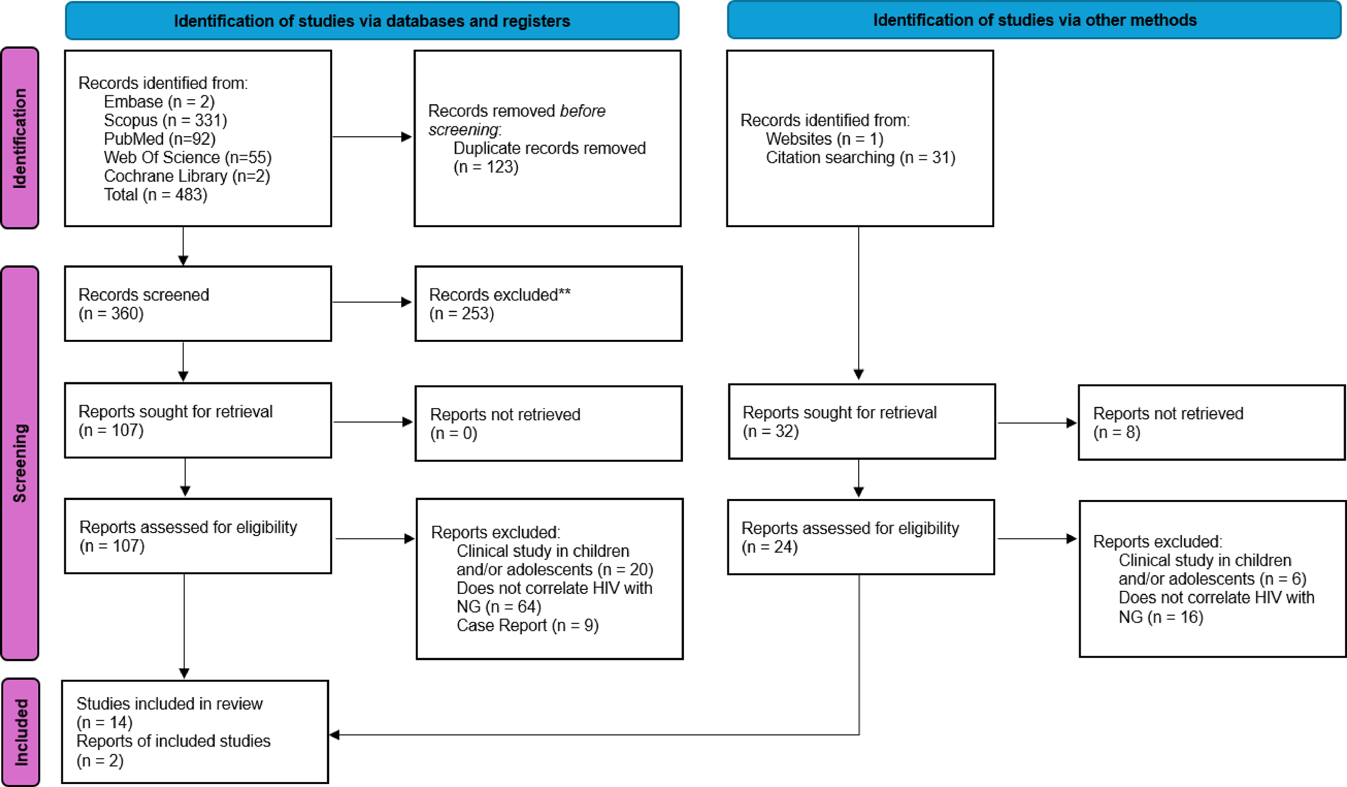
The present review initially identified 483 results in the search, of which only 14 met all the proposed inclusion criteria. After the initial screening, the selected findings had their references evaluated to identify any additional relevant literature, resulting in the inclusion of two more studies. The article selection process is outlined in figure 1.

The 16 retrieved articles were observational in nature and assessed using the Newcastle-Ottawa Scale tool. Most articles had a high risk of bias; however, those used for the meta-analysis had a moderate level of bias. The major shortcomings in the studies were related to the correct division of assessed groups and the lack of standardization in HIV and NG diagnosis. Murray’s article received a zero score due to its classification, which currently does not suit HIV studies.
The articles varied in publication year from 1988 to 2011. Clinical findings and diagnostic and prognostic observations reported in the selected articles were summarized along with the bias risk scores in table 1.
Only 11 articles provided quantitative data that could be extracted regarding the correlation between HIV and NG. These data are presented in table 2.
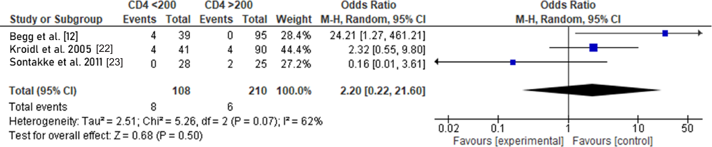
A meta-analysis was conducted comparing CD4 cell concentrations with the diagnosis of GN. The present analysis shows an Odds Ratio of 2.20 with a confidence interval from 0.22 to 21.6. Heterogeneity among the studies, as indicated by the I² test, was 62%. The comprehensive analysis can be reviewed in figure 2.
DISCUSSION
Oral lesions alone do not constitute sufficient evidence to predict a clinical manifestation of HIV infection, with their diagnosis better established through the correlation with other syndromic signs [12]. However, George Laskaris et al. [13] report that AIDS manifestations may not exhibit noticeable symptoms in the early stages of infection, and oral lesions could be the initial and sole oral presentations of the disease. Several oral findings may be associated with infection, and among the most common are oral candidiasis, necrotizing ulcerative gingivitis, hairy leukoplakia, and linear gingival erythema [12,14]. Schulten et al. [16] found Kaposi’s Sarcoma as the only oral manifestation of HIV in their report.
Murray [17] employed HIV+ homosexual men and HIV- heterosexual men as a control group for comparative analysis in his studies. Research conducted prior to gaining a deeper understanding of HIV held prejudiced information. Thus, it is crucial to highlight that perceptions of oral lesions and HIV have evolved over the years. Recent studies show that the profile of HIV/AIDS has changed, with a rise in cases among women, heterosexuals, individuals aged 15 to 24, adults over 50, and those with lower educational levels [28].
Among oral lesions strongly associated with AIDS, Necrotizing Ulcerative Gingivitis (NUG) and Necrotizing Ulcerative Periodontitis (NUP) have gained prominence due to their rapid progression and association with debilitating factors [4,29]. Our findings demonstrate that this correlation is linked to the immune system deficiency caused by HIV infection in the body, leading to a higher propensity for lesion development [18,19,30]. However, according to Laskaris et al. [13], NUG is a possible early manifestation of AIDS even in the initial stages of infection, when immunosuppression is not significant [31].
It is essential to emphasize that compromised immune cells do not necessarily signify the progression of periodontal disease but rather serve as a potential etiological source [23 21]. Nevertheless, the origin of periodontitis in HIV patients remains largely uncertain; some studies suggest the possibility of an HIV-associated periodontitis having a microbiota distinct from the traditional one, indicating a need for further research [17].
From this perspective, a concentration of viable TCD4 cells below 200 already indicates compromised immune system function [9,32], and this threshold was utilized in studies by Kroidi [22], Begg et al. [12], and Sontakke et al. [23]. At this level of immunological compromise, the characteristic signs and symptoms of HIV may manifest more prominently [33]. However, no statistical correlation with NUG/PUN was identified in the screened studies. The study by Sontakke et al. [23] did not find any indication that the low concentration of CD4 cells acts as a predictor of Necrotizing Periodontal Diseases (NPDs). It is essential to highlight that the present analysis exhibits high sample heterogeneity and compares few publications, underscoring the need for primary studies to standardize the level of immunocompromise, as well as the assessment of NUG/PUN to validate these findings.
It is known that periodontal inflammation linked to immunosuppression can trigger ulcerative periodontal diseases, such as NUG/PUN and Linear Gingival Erythema (LGE) [22]. Among the treatments for NPDs, the removal of necrotic tissue (debridement) and the crucial control of biofilm through regular oral hygiene, coupled with chlorhexidine and iodopovidone rinses, are highlighted [15,19,23]. Additionally, immediate follow-up and regular long-term maintenance are of great value for the treatment and prevention of lesion recurrence. The implementation of appropriate treatment allows for the disappearance of clinical signs and symptoms within the first few days [24].
Before the era of highly active antiretroviral therapy (HAART), lesions such as oral hairy leukoplakia (OHL) and oral candidiasis were easily discernible in HIV patients [22]. Following the implementation of HAART, a notable surge in CD4+ cell count was observed, coupled with a reduction in HIV RNA levels. This extension of the period during which patients remained free from clinical manifestations of AIDS characterized HIV infection as a chronic condition, marked by the persistent presence of the virus in the infected host [25].
Studies indicate that HIV is also detectable in the saliva of patients, albeit at very low levels—lower than those found in the blood. HIV infection is known to contribute to a reduction in salivary flow, either as a side effect of anti-HIV medication or due to its association with salivary gland diseases [23,25]. It is crucial to highlight that the use of various medications such as antibiotics, antifungal agents, and antivirals can significantly impact periodontal tissue due to alterations in the microbiota [19].
The study by Hartnett and Shiloah [14] suggests the presence of microorganisms associated with HIV-related gingivitis and periodontitis, stemming from immunodeficiency in HIV+ patients. This imbalance between host immune response and aggression potentially exacerbates the progression of periodontal disease. Therefore, a correlation can be asserted between periodontal attachment loss and a reduction in TCD4+ cell count, as the attenuation of the immune response in HIV+ patients, combined with intrinsic host factors, poses an additional risk of periodontal destruction [25,34].
Throughout the years, the clinical profile of oral lesions associated with HIV has undergone significant changes. Notably, there has been a decrease in the prevalence of most of these lesions since the introduction of highly active antiretroviral therapy (HAART), improved medical care conditions, and increased usage of prophylactic medications to prevent secondary opportunistic infections related to AIDS. The enhanced immune system has resulted in a reduction in the occurrence of opportunistic infections and neoplasms. Additionally, the decline in periodontal diseases as a consequence of these therapies has contributed to an improved quality of life for patients with HIV [26,27,35].
CONCLUSION
Based on the presented information, it can be concluded that certain oral manifestations were strongly associated with the presence of HIV, especially Necrotizing Periodontal Diseases (NPDs) like Necrotizing Gingivitis (NG) due to immunosuppression. Following the implementation of highly active antiretroviral therapy (HAART), a significant increase in TCD4+ cell count was observed, along with a decrease in HIV RNA levels. This led to a notable decrease in the prevalence of oral lesions associated with HIV, including a lower incidence and severity of NG. Consequently, this improvement has contributed to a better quality of life for these patients.
How to cite this article
-
Lorentz BCS, Guimarães LFC, Barra SG, Candido CBSA, Paiva DFF. Necrotizing gingivitis in patients with and without HIV: a systematic literature review and meta-analysis. RGO, Rev Gaúch Odontol. 2024;72:e20240039. http://dx.doi.org/10.1590/1981-86372024003920240021
REFERENCES
-
1 Nolte SH. From trench mouth to noma: Experiences from Nazi extermination camps. Clin Dermatol. 2021;39(6):990-5. https://doi.org/10.1016/j.clindermatol.2021.05.019
» https://doi.org/10.1016/j.clindermatol.2021.05.019 -
2 Siddiqui AZ, Vellappally S, Fouad H, Muckarrum SM. Bactericidal and clinical efficacy of photochemotherapy in acute necrotizing ulcerative gingivitis. Photodiagnosis Photodyn Ther. 2020;29:101668. https://doi.org/10.1016/j.pdpdt.2020.101668
» https://doi.org/10.1016/j.pdpdt.2020.101668 - 3 Santos de Freitas BS, Sant’Ana SSS, Ferreira MS, Mariano-Júnior WJ, Watanabe S, Yamamoto-Silva FP. Topical tetracycline in the treatment of a 7-year-old child with necrotizing ulcerative gingivitis: A case report. Gen Dent. 2020;68(2):36-8.
- 4 Martos J, Ahn Pinto KV, Feijó Miguelis TM, Cavalcanti MC, César Neto JB. Clinical treatment of necrotizing ulcerative gingivitis: A case report with 10-year follow-up. Gen Dent. 2019;67(3):62-5.
-
5 Caton JG, Armitage G, Berglundh T, Chapple ILC, Jepsen S, Kornman KS, et al. A new classification scheme for periodontal and peri-implant diseases and conditions: Introduction and key changes from the 1999 classification. J Clin Periodontol. 2018;45 Suppl 20:S1-S8. https://doi.org/10.1111/jcpe.12935
» https://doi.org/10.1111/jcpe.12935 -
6 Herrera D, Retamal-Valdes B, Alonso B, Feres M. Acute periodontal lesions (periodontal abscesses and necrotizing periodontal diseases) and endo-periodontal lesions. J Periodontol. 2018;89 Suppl 1:S85-S102. https://doi.org/10.1002/JPER.16-0642
» https://doi.org/10.1002/JPER.16-0642 -
7 Tkacz K, Gill J, McLernon M. Necrotising periodontal diseases and alcohol misuse: a cause of osteonecrosis? Br Dent J. 2021;231(4):225-31. https://doi.org/10.1038/s41415-021-3272-9
» https://doi.org/10.1038/s41415-021-3272-9 -
8 Rai S, Subramanyam GB, Kumar G, Bhushan V. Assessment of oral mucosal lesions among HIV positive transgenders residing in Odisha with and without Antiretroviral therapy. J Family Med Prim Care. 2022;11(11):7106-12. https://doi.org/10.4103/jfmpc.jfmpc_940_22
» https://doi.org/10.4103/jfmpc.jfmpc_940_22 -
9 Kato H, Imamura A. Unexpected Acute Necrotizing Ulcerative Gingivitis in a Well-controlled HIV-infected Case. Intern Med. 2017;56(16):2223-7. https://doi.org/10.2169/internalmedicine.8409-16
» https://doi.org/10.2169/internalmedicine.8409-16 -
10 Page MJ, McKenzie JE, Bossuyt PM, Boutron I, Hoffmann TC, Mulrow CD, et al. The PRISMA 2020 statement: An updated guideline for reporting systematic reviews. BMJ. 2021;372:n71. https://doi.org/10.1136/bmj.n71
» https://doi.org/10.1136/bmj.n71 -
11 McInnes MDF, Moher D, Thombs BD, McGrath TA, Bossuyt PM, The PRISMA-DTA Group, et al. Preferred Reporting Items for a systematic review and meta-analysis of diagnostic test accuracy studies: The PRISMA-DTA Statement. JAMA. 2018;319(4):388-96. https://doi.org/10.1001/jama.2017.19163. Erratum in: JAMA. 2019;322(20):2026. https://doi.org/10.1001/jama.2019.18307
» https://doi.org/10.1001/jama.2017.19163 -
12 Begg MD, Lamster IB, Panageas KS, Mitchell-Lewis D, Phelan JA, Grbic JT. A prospective study of oral lesions and their predictive value for progression of HIV disease. Oral Dis. 1997;3(3):176-83. https://doi.org/10.1111/j.1601-0825.1997.tb00032.x
» https://doi.org/10.1111/j.1601-0825.1997.tb00032.x -
13 Laskaris G, Potouridou I, Laskaris M, Stratigos J. Gingival lesions of HIV infection in 178 Greek patients. Oral Surg Oral Med Oral Pathol. 1992;74(2):168-71. https://doi.org/10.1016/0030-4220(92)90377-3
» https://doi.org/10.1016/0030-4220(92)90377-3 - 14 Hartnett AC, Shiloah J. The treatment of acute necrotizing ulcerative gingivitis. Quintessence Int. 1991;22(2):95-100.
-
15 Winkler JR, Robertson PB. Periodontal disease associated with HIV infection. Oral Surg Oral Med Oral Pathol. 1992;73(2):145-50. https://doi.org/10.1016/0030-4220(92)90186-t
» https://doi.org/10.1016/0030-4220(92)90186-t -
16 Schulten EA, ten Kate RW, van der Waal I. Oral manifestations of HIV infection in 75 Dutch patients. J Oral Pathol Med. 1989;18(1):42-6. https://doi.org/10.1111/j.1600-0714.1989.tb00731.x
» https://doi.org/10.1111/j.1600-0714.1989.tb00731.x -
17 Murray PA, Grassi M, Winkler JR. The microbiology of HIV-associated periodontal lesions. J Clin Periodontol. 1989;16(10):636-42. https://doi.org/10.1111/j.1600-051x.1989.tb01032.x
» https://doi.org/10.1111/j.1600-051x.1989.tb01032.x -
18 Rowland RW, Escobar MR, Friedman RB, Kaplowitz LG. Painful gingivitis may be an early sign of infection with the human immunodeficiency virus. Clin Infect Dis. 1993;16(2):233-6. https://doi.org/10.1093/clind/16.2.233
» https://doi.org/10.1093/clind/16.2.233 -
19 Grbic JT, Mitchell-Lewis DA, Fine JB, Phelan JA, Bucklan RS, Zambon JJ, et al. The relationship of candidiasis to linear gingival erythema in HIV-infected homosexual men and parenteral drug users. J Periodontol. 1995;66(1):30-7. https://doi.org/10.1902/jop.1995.66.1.30
» https://doi.org/10.1902/jop.1995.66.1.30 -
20 Hompson SH, Charles GA, Craig DB. Correlation of oral disease with the Walter Reed staging scheme for HIV-1-seropositive patients. Oral Surg Oral Med Oral Pathol. 1992;73(3):289-92. https://doi.org/10.1016/0030-4220(92)90123-8
» https://doi.org/10.1016/0030-4220(92)90123-8 -
21 Horning GM, Cohen ME. Necrotizing ulcerative gingivitis, periodontitis, and stomatitis: clinical staging and predisposing factors. J Periodontol. 1995;66(11):990-8. https://doi.org/ 10.1902/jop.1995.66.11.990
» https://doi.org/10.1902/jop.1995.66.11.990 - 22 Kroidl A, Schaeben A, Oette M, Wettstein M, Herfordt A, Häussinger D. Prevalence of oral lesions and periodontal diseases in HIV-infected patients on antiretroviral therapy. Eur J Med Res. 2005;10(10):448-53.
-
23 Sontakke SA, Umarji HR, Karjodkar F. Comparison of oral manifestations with CD4 count in HIV-infected patients. Indian J Dent Res. 2011;22(5):732. https://doi.org/10.4103/0970-9290.93470
» https://doi.org/10.4103/0970-9290.93470 -
24 Rowland RW. Necrotizing ulcerative gingivitis. Ann Periodontol. 1999;4(1):65-73; discussion 78. https://doi.org/10.1902/annals.1999.4.1.65
» https://doi.org/10.1902/annals.1999.4.1.65 -
25 Mataftsi M, Skoura L, Sakellari D. HIV infection and periodontal diseases: An overview of the post-HAART Era. Oral Dis. 2011;17(1):13-25. https://doi.org/10.1111/j.1601-0825.2010.01727.x
» https://doi.org/10.1111/j.1601-0825.2010.01727.x -
26 Ramírez-Amador V, Esquivel-Pedraza L, Sierra-Madero J, Anaya-Saavedra G, González-Ramírez I, Ponce-de-León S. The Changing Clinical Spectrum of Human Immunodeficiency Virus (HIV)-Related Oral Lesions in 1,000 Consecutive Patients: A 12-Year Study in a Referral Center in Mexico. Medicine (Baltimore). 2003;82(1):39-50. https://doi.org/10.1097/00005792-200301000-00004
» https://doi.org/10.1097/00005792-200301000-00004 -
27 Ferreira S, Noce C, Júnior AS, Gonçalves L, Torres S, Meeks V, et al. Prevalence of oral manifestations of HIV infection in Rio De Janeiro, Brazil from 1988 to 2004. AIDS Patient Care STDS. 2007;21(10):724-31. https://doi.org/10.1089/apc.2006.0211
» https://doi.org/10.1089/apc.2006.0211 -
28 Filho OJLD, Viana EC, Pessoa WG, Domingos PRC. Manifestações orais em pacientes imunodeprimidos pelo Vírus da Imunodeficiência Humana (HIV): revisão da literatura. REAS. 2021 [citado 11 set. 2024];13(2):e6034. Available from: https://acervomais.com.br/index.php/saude/article/view/6034
» https://acervomais.com.br/index.php/saude/article/view/6034 -
29 Zhang X, Reichart PA, Song Y. Oral manifestations of HIV/AIDS in China: A review. Oral Maxillofac Surg. 2009;13(2):63-8. https://doi.org/10.1007/s10006-009-0157-5
» https://doi.org/10.1007/s10006-009-0157-5 -
30 Patton LL, Phelan JA, Ramos-Gomez FJ, Nittayananta W, Shiboski CH, Mbuguye TL. Prevalence and classification of HIV-associated oral lesions. Oral Dis. 2002;8 Suppl 2:98-109. https://doi.org/10.1034/j.1601-0825.2002.00020.x
» https://doi.org/10.1034/j.1601-0825.2002.00020.x -
31 Araújo-Júnior JL, Pita-Neto IC, Freire JCP, Dias-Ribeiro E. Perfil clínico e epidemiológico das alterações bucais em portadores do HIV. Arch Health Invest. 2024;7(8). https://doi.org/10.21270/archi.v7i8.3022
» https://doi.org/10.21270/archi.v7i8.3022 -
32 Souza HC, Passos XS, Camplesi Júnior M, Silva AMTC, Oliveira AKS, Marques LOR, et al. Contagem de linfócitos TCD4 + e carga viral em pacientes HIV+ de um laboratório de referência. RBMC. 2020;6(15). https://doi.org/10.36414/rbmc.v6i15.50
» https://doi.org/10.36414/rbmc.v6i15.50 -
33 Esteban-Cantos A, Rodríguez-Centeno J, Silla JC, Barruz P, Sánchez-Cabo F, Saiz-Medrano G, et al. Effect of HIV infection and antiretroviral therapy initiation on genome-wide DNA methylation patterns. EBioMedicine. 2023;88:104434. https://doi.org/10.1016/j.ebiom.2022.104434
» https://doi.org/10.1016/j.ebiom.2022.104434 -
34 Pólvora TLS, Nobre ÁVV, Tirapelli C, Taba M Jr, Macedo LD, Santana RC, et al. Relationship between human immunodeficiency virus (HIV-1) infection and chronic periodontitis. Expert Rev Clin Immunol. 2018;14(4):315-27. https://doi.org/10.1080/1744666X.2018.1459571
» https://doi.org/10.1080/1744666X.2018.1459571 -
35 Rao KV, Chitturi RT, Kattappagari KK, Kantheti LP, Poosarla C, Baddam VR. Impact of highly active antiretroviral therapy on oral manifestations of patients with human immunodeficiency virus/acquired immuno deficiency syndrome in South India. Indian J Sex Transm Dis AIDS. 2015;36(1):35-9. https://doi.org/10.4103/0253-7184.156703
» https://doi.org/10.4103/0253-7184.156703
Edited by
-
Assistant editor: Luciana Butini Oliveira

 Note: Study: Begg et al. [