 Effect of polishing techniques on a 3D printed resin for provisional dentures: an in vitro study on surface roughness and mechanical strength
ABSTRACT
Objective This study aimed to evaluate the effect of polishing techniques on a 3D-printed resin for provisional prosthesis through an in vitro assessment of surface roughness and mechanical strength.
Methods Bar-shaped specimens (25 × 12 × 2 mm) were fabricated using a 3D-printed provisional resin at a 0° angle to the platform and divided into three experimental groups (n=10): no polishing (AP), mechanical polishing (PM), and chemical polishing (PQ). Surface analysis was performed with a stereomicroscope, and roughness parameters (Ra, Rq, Rz) were obtained with a confocal microscope. Flexural strength and modulus were also determined, followed by qualitative evaluation of the fracture pattern. Data were analyzed with one-way ANOVA (p<0.05) and Tukey’s test when appropriate.
Results Distinct surface characteristics were observed among groups, with statistically significant differences for Ra (p=0.003) and Rq (p=0.002), while Rz showed no significance (p=0.063). Flexural strength (p<0.001) and flexural modulus (p=0.002) also differed significantly between groups.
Conclusion Polishing techniques tested on a 3D-printed resin for provisional prosthesis significantly influenced Ra, Rq, flexural strength, and flexural modulus, but showed no significant effect on Rz.
Indexing terms
Three-dimensional printing; Surface properties; Flexural strength
RESUMO
Objetivo Avaliar o efeito de técnicas de polimento sobre uma resina impressa 3D para prótese provisória, através de um estudo in vitro sobre rugosidade superficial e resistência mecânica.
Métodos Espécimes em barra (25 × 12 × 2 mm) foram confeccionados utilizando resina impressa 3D para provisórios, em ângulo de 0° com a plataforma. Três grupos experimentais foram estabelecidos (n=10): ausência de polimento (AP), polimento mecânico (PM) e polimento químico (PQ). A análise da superfície foi realizada em estereomicroscópio, e a rugosidade superficial avaliada em microscópio confocal pelos parâmetros Ra, Rq e Rz. Também foram determinados resistência e módulo de flexão, seguidos da análise qualitativa do padrão de fratura. Os dados quantitativos foram submetidos à ANOVA de um fator (p<0,05), seguida pelo teste de Tukey quando pertinente.
Resultados As superfícies apresentaram-se distintas após alteração superficial. Os valores de Ra (p=0,003) e Rq (p=0,002) apresentaram diferenças estatísticas entre grupos (p=0,003), contudo os resultados de Rz não mostram significância (p=0,063). Com relação à resistência (p=0,000) e módulo (p=0,002) de flexão, as técnicas de polimento apresentaram resultados estatisticamente significativos entre os grupos experimentais.
Conclusão As técnicas de polimento testadas sobre uma resina impressa 3D para prótese provisória, demonstraram influência significativa sobre os valores de Ra, Rq, resistência e módulo de flexão. Contudo, não apresentam efeito significativo sobre os valores de Rz.
Termos de indexação
Impressão tridimensional; Propriedades de superfície; Resistência à flexão
INTRODUCTION
Temporary prostheses are an important stage of oral rehabilitation and should not be regarded as a useless step with uncertain indications, but rather as a means of transition toward functionality. Therefore, the surface of prostheses must be rigorously prepared to be as smooth as possible in order to limit bacterial colonization, thus promoting a successful gingival tissue response to any composite material. However, surface roughness is an important variable that can be significantly altered if the provisional restoration is improperly finished and polished [1]. The restorations mentioned are susceptible to microbial adhesion and may become covered by biofilm, thereby becoming a source of microorganisms that affect the surrounding tissues and may, for example, lead to implant failure. In addition, restorative materials that are susceptible to bacterial colonization may impact overall oral health, contribute to the deterioration of the oral cavity condition, and threaten the final reconstruction [2].
To produce a provisional restoration of the dental arches, it is necessary to select materials and techniques that simplify the clinical workflow as much as possible. The conventional method, which uses heat-polymerized resins, involves many laboratory steps, leading to longer working time, a greater number of treatment sessions, and a higher risk of technical errors. In a digital workflow, the final fabrication of the restoration may be performed using a subtractive method through Computer-Aided Design/Computer-Aided Manufacturing (CAD-CAM), or by an additive method, namely three-dimensional (3D) printing [1,3].
CAD-CAM has been widely used in the fabrication of restorations for fixed prostheses, as it facilitates the milling of crowns obtained from digital models. However, its limitations include material waste, high cost requiring highly trained personnel, the need for manual finishing, and the monolithic chromaticity of milled restorations, which generally does not provide exceptional aesthetic results [4,5].
While three-dimensional printing plays a revolutionary role in dentistry, it enables the fabrication of restorations that can be used temporarily or even long term, with reduced manufacturing costs [6]. Since then, rapid technological development has increased the accuracy, speed, and reliability of 3D printers, making them highly attractive for a wide range of Fields [4]. This technology has the advantage of not wasting material during the process, being less time-consuming, more cost-effective, and capable of reproducing complex shapes [7].
Three-dimensional printing has been widely publicized in the dental community, especially when it comes to the fabrication of temporary prostheses, mainly due to its high success rate and increasing longevity [1-3,6,8]. Recently, dentists have been using three-dimensional printing technology in the fabrication of dental restorations [8]. Among the printers most used in dentistry, Stereolithography (SLA) and Digital Light Processing (DLP) stand out [6]. Because SLA printers complete a layer by curing the resin point by point through laser projection, the slow space of the mirror reflecting the laser beam will certainly generate errors. On the other hand, DLP technology is faster because it uses a projector to cure the material layer by layer, reducing the imprecision that accompanies repetitive printing [6].
The use of photopolymerizable resins in 3D printing technology allows not only predictable implant treatment from the diagnostic phase through to the final restoration, but also customization and modification throughout the treatment process [2]. Regarding the surface characteristics of 3D-printed resins, a significant optimization of surfaces is observed after finishing and polishing, with improved relationships between surface parameters and the mechanical properties of the samples. Low surface roughness values and acceptable mechanical strength indicate that these materials are suitable for the fabrication of long-term provisionais bridges, allowing proper functional restoration and oral rehabilitation [1]. However, studies evaluating the use of 3D-printed materials in dentistry in terms of their surface and mechanical properties, including flexural strength, surface roughness, hardness, and aesthetics, are still limited [8].
Furthermore, to date, there is a lack of evidence on how polishing techniques can interfere with the performance of these resins. Based on the above, the objective was to evaluate the effect of polishing techniques on a 3D-printed resin for provisional prostheses, through an in vitro study on surface roughness and mechanical strength. The Null Hypothesis (H0) to be tested is that there will be no statistical difference between the polishing techniques tested for the values obtained for average surface roughness, strength, and flexural modulus.
METHODS
Preparation of Specimens
The bar-shaped specimens (25 × 12 × 2 mm) were fabricated using a 3D-printed resin. Specimen processing followed the manufacturer’s and the 3D printer’s guidelines (Anycubic Photon S 3D printer, Talmax Prótese Odontológica, Curitiba, Paraná, Brazil) using the DLP printing method. The specimens were designed using Exocad 3D software, and the images were exported in Standard Tessellation Language (STL) format. Printing took approximately 20 minutes, with the specimens positioned horizontally, forming a 0° angle with the build platform, and a printing layer thickness of 50 µm. After processing, the specimens were cleaned in isopropyl alcohol for 10 minutes using an ultrasonic bath, followed by post-curing in an ultraviolet chamber for 10 minutes, in accordance with the manufacturer’s recommendations [4].
Polishing Techniques
The polishing techniques to be tested included mechanical polishing, as described in the studies by Mârțu et al. [1] and Wadhwani et al. [6], as well as chemical polishing. For mechanical polishing, the acrylic resin surface was initially finished using a cutting bur (Medium Cross-Cut Maxicut Tungsten Bur – American Burrs) for 1 minute, followed by siliconized rubber polishers (ExaTechnique Acrylic Polisher PM; Edenta – Labordental, São Paulo, SP, Brazil) in the sequence green, black, and yellow, each applied to the surface for 1 minute. A scratch-removal brush (Coarse Red Scotch-Brite Brush PM, American Burrs, Palhoça, SC, Brazil) and a polishing brush (chamois brush, Derfla – Labordental, São Paulo, SP, Brazil) were then used, with each brush applied for 1 minute. For chemical polishing, the resin surface was initially finished with the cutting bur for 1 minute. Subsequently, chemical polishing was performed by applying a photopolymerizable resin glaze (Foto Masterseal Resin Glaze, Wilcos, São Paulo, Brazil) with a brush, and then the specimen was placed in a curing chamber for 10 minutes.
Experimental Groups and Sample Size
Three experimental groups were established: no polishing (AP), mechanical polishing (PM), and chemical polishing (PQ), each comprising 10 specimens for the analyses performed in this study. The sample size was calculated using Minitab software (version 16.1 for Windows, Pennsylvania, USA), based on the standard deviation reported in similar studies. For mechanical strength and flexural modulus, a standard deviation of 9.61 was adopted according to Alshamrani et al. [8], and for surface roughness, a standard deviation of 0.02 was considered based on Mârțu et al. [1]. Thus, a sample size of N = 10 achieved a statistical power of 80.0% for the maximum expected values in the analyses of surface roughness and mechanical strength.
Surface Analysis
The surface of the specimens from each experimental group was analyzed in terms of morphology (N = 2) using a stereomicroscope (Discovery V20, Carl Zeiss, Germany), in order to identify the effects of the polishing techniques on the surface of the material under study.
Surface Roughness
The specimens were evaluated for mean surface roughness (Ra – µm), mean peak-to-valley height (Rz – µm), and root mean square roughness (Rq – µm), as described in the studies by Mârțu et al. [1], Santos et al. [3], and Wadhwani et al. [6], using a Zeiss LSM700 confocal microscope (Zeiss, Germany) connected to a computerized unit running the Zen software. Measurements were performed at the center of each specimen, and from these readings, 127 x 127 square micrometer images of the analyzed surfaces were generated using a 50X lens.
Mechanical Strength
The three-point flexural strength test was performed using a universal testing machine (EMIC DL-1000, São José dos Pinhais, Brazil). The specimens used for mechanical strength analysis were positioned between two supports, with the treated surface facing upward, at a span length of 20 mm, and subjected to loading until fracture [4,5]. The machine was programmed with a 100 kgf load cell and a constant crosshead speed of 5 mm/min. Flexural strength values were obtained in megapascals (MPa) using Equation 1, in which γ represents flexural strength, F is the load at the fracture point, l is the support span length, b is the specimen width, and h is the specimen thickness.
Flexural Modulus
The flexural modulus was determined from the slope of the linear portion of the stress–strain curve for each test and was calculated using Equation 2. In this equation, F is the load, in Newtons, at a point on the straight line (with maximum slope) of the load/deflection curve, b is the specimen width in millimeters, h is the specimen height in millimeters and d is the deflection, in millimeters, at the load F [5].
Fractography
The fractured specimens were analyzed using a stereomicroscope (Discovery V20, Carl Zeiss, Germany) to determine fracture characteristics, and photographic records were obtained.
Results Analysis
The results were tabulated and analyzed using Minitab software (version 16.1 for Windows, Pennsylvania, USA), with a significance level set at 5%. Surface roughness, mechanical strength, and flexural modulus data were subjected to one-way ANOVA (p<0.05), followed by Tukey’s test when statistically significant differences among groups were identified. Findings from surface and fracture analyses were presented qualitatively (figure 1). The Kolmogorov-Smirnov test was used to assess data normality for the results of Ra (p>0.150), Rz (p=0.031), Rq (p>0.150), flexural strength (p>0.150), and flexural modulus (p=0.043).

RESULTS
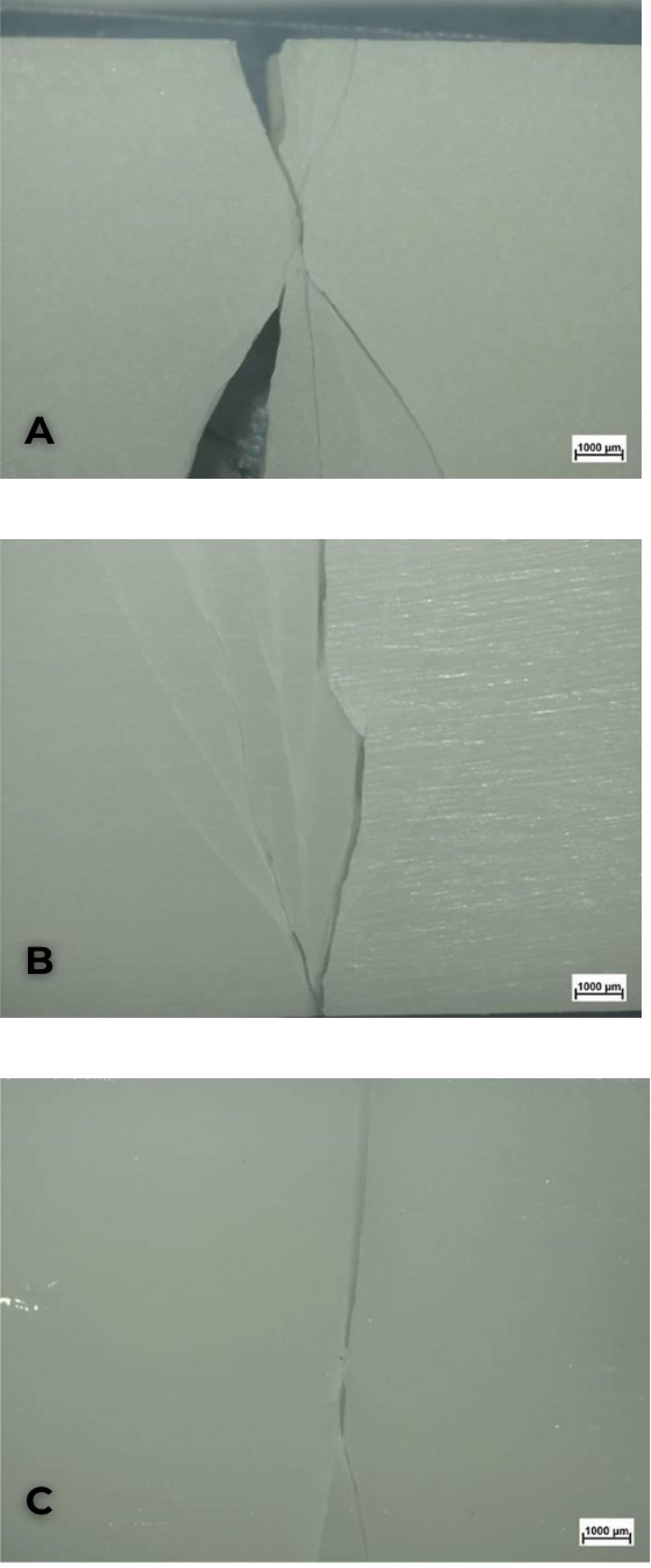
Based on the data obtained using the adopted methodology, the surfaces showed distinct characteristics after surface modification (figure 2). The AP group exhibited some surface irregularities after finishing, which were obliterated by the application of glaze in the PQ group. In contrast, the surface of the PM group presented polishing lines resulting from the action of rubber polishers and brushes. The use of polishing techniques promoted a reduction in surface roughness values across all analyzed parameters when compared with the finishing-only condition. The PQ group demonstrated the lowest surface roughness values among the groups in the study, and in some parameters showed statistically significant differences compared with the other surface treatments tested. Ra (p=0.003) and Rq (p=0.002) showed statistically significant differences among groups, whereas Rz values did not show significance (p=0.063) (table 1). The surface images obtained by confocal microscopy represent the AP, PM and PQ groups, as shown in (figure 3). Regarding flexural strength, the polishing technique showed a statistically significant effect (p=0.000) among the experimental groups. In descending order of mean flexural strength, the groups were ranked as PQ, PM and AP (table 1). With respect to the flexural modulus, a statistically significant difference among groups was also identified (p=0.002), with statistical similarity observed between the AP group and the other experimental groups (table 1). A higher number of fragments after fracture was observed in the AP and PM groups, whereas the PQ group showed a predominance of two fragments (figures 4 and 5).

Stereomicroscope images of the specimens: A) Stereomicroscope image at 16× magnification, AP group. B) Stereomicroscope image at 16× magnification, PM group. C) Stereomicroscope image at 16× magnification, PQ group.

Confocal microscope images of the specimens: A) Surface image of specimen 2 from the AP group (Ra = 1.695 µm, Rz = 23.989 µm, and Rq = 2.324 µm). B) Surface image of specimen 5 from the PM group (Ra = 1.344 µm, Rz = 10.845 µm, and Rq = 1.621 µm). C) Surface image of specimen 6 from the PQ group (Ra = 0.690 µm, Rz = 12.660 µm, and Rq = 0.860 µm).


Stereomicroscopic analysis of the fragments: A) Fragments after fracture in the AP group. Stereomicroscope image at 6.5× magnification. B) Fragments after fracture in the PM group. Stereomicroscope image at 6.5× magnification. C) Fragments after fracture in the PQ group. Stereomicroscope image at 6.5× magnification.
DISCUSSION
Additive technology has received significant interest from the dental research community, as it can expand the use of digital applications in clinical practice. Dental crowns and bridges are examples of devices that can be produced using 3D printing technology. Therefore, understanding the properties of provisional materials used in the fabrication of dental crowns is essential for the evaluation of new 3D printing materials, thus providing a clearer perspective on whether a material is suitable for clinical use, including long-term applications [8].
Based on the results obtained in this study, the null hypothesis that there would be no statistically significant differences among the polishing techniques tested with respect to mean surface roughness, flexural strength, and flexural modulus was rejected. This conclusion is supported by the fact that only the Rz values did not show significant differences among the experimental groups, whereas all other analyses in the study were influenced by the polishing technique.
The polishing techniques adopted in this study promoted surface changes in the 3D-printed resin. From a qualitative perspective, stereomicroscope images demonstrated the distinct effects produced by the surface treatments in each experimental group, which is consistent with the confocal microscope findings, showing a color map indicative of lower surface roughness compared with the AP and PM groups. It is worth highlighting that the PQ group exhibited the least surface alteration, as the glazing procedure applied to restorations provides improved aesthetics and smoother surfaces by filling microcracks and porosities [2].
Regarding surface roughness analysis, the results demonstrated statistically significant differences for the Ra and Rq parameters, whereas the Rz data showed similarity among the groups. This finding is consistent with the study by Mârțu et al. [1], which also reported statistically significant differences before and after mechanical polishing of 3D-printed resin specimens for the same roughness parameters evaluated in the present study. Likewise, the results reported by Mazurek-Popczyk et al. [2] showed that the use of mechanical or chemical polishing on different 3D-printed resins reduced Ra, Rz, and Rq values, with statistical significance observed for the NextDent resin. Consequently, biofilm formation was attenuated on resin surfaces treated with glaze for most of the microorganisms tested, thereby reducing potential final restoration failures. In contrast, the absence of surface polishing and the presence of mechanical polishing alone resulted in a dense biofilm layer rich in microorganisms.
Similar studies have reported results that differ from those of the present research regarding surface roughness. Wadhwani et al. [6], in their analysis of surface roughness and different printing techniques, did not identify significant differences in Ra values between resins printed by DLP and SLA. The Ra values on the buccal and lingual surfaces of bridges produced by both printing methods showed similar mean values among the experimental groups. In the study by Santos et al. [3], the 3D-printed resin did not show statistically significant differences in Ra when compared with autopolymerizing and heat-polymerized resins, however, it exhibited the lowest roughness values among the tested resin materials after finishing the specimen surfaces with 600-grit water sandpaper. Additionally, the study by Wadhwani et al. [6] comparing different printing orientations (0°, 45°, and 90°) of a 3D resin with a composite resin also did not identify differences among experimental groups in terms of surface roughness values. In that study, the 3D-printed specimens were subjected to a sequence of sanding and polishing using the same resin as in the present research. The divergent findings among these studies may reflect differences in experimental variables and study designs.
The acceptable Ra value regarding the threshold for bacterial plaque accumulation on a hard surface in the oral environment after polishing is 0.2 µm [9]. In the present study, all Ra, Rq, and Rz values after mechanical or chemical polishing exceeded the limit recommended in the literature. However, the mean Ra and Rq values remained below the clinically unacceptable threshold of 10 µm [9]. Therefore, the material evaluated in this study may be considered suitable for clinical use, and further analyses involving accelerated aging are suggested to investigate the polishing techniques tested, thereby expanding the indication of this material as a long-term provisional restoration.
Regarding surface roughness parameters, Ra, Rz, and Rq were adopted in this study. Although Ra is the parameter most commonly used to quantify surface roughness, it is recommended to use different parameters, including amplitude or spacing parameters that influence bacterial adhesion, optical characteristics, or other Properties [2].
The mechanical strength findings of the 3D-printed resin for provisional prostheses demonstrated a significant influence of the polishing techniques adopted. The literature indicates that the tensile strength of a 3D-printed resin, after being subjected to a mechanical polishing sequence, reached acceptable mechanical strength values when compared with conventional and CAD-CAM–milled samples; therefore, these materials should be indicated for the fabrication of long-term temporary bridges, allowing proper restoration of function and oral rehabilitation [1]. In contrast, the findings of Santos et al. [3] reported lower mechanical strength values for 3D-printed resin after finishing with 600-grit water sandpaper when compared with heat-polymerized and autopolymerizing resins, with the material factor being statistically significant.
Factors such as printing angulation, printing layer thickness, water storage, curing time, and printing technique may influence the flexural strength of 3D-printed materials [8,10]. Possibly, because the DLP printing method promotes the formation of rougher layers, in which interlayer bonding is weaker, fracture may occur more rapidly, resulting in multiple fragments. In addition, the chemical composition of this acrylate-based 3D-printed resin exhibits good surface hardness, however, due to its chemical structure, it tends to be brittle. A high number of fragments from a fractured 3D-printed resin may compromise patient safety and cause injuries [11], as observed in the AP and PM groups, which showed a greater number of fragments after fracture. The fact that the printed samples in this study were obtained exclusively using the DLP method may represent a limitation. Future studies should also focus on the behavior of specimens produced by stereolithography or other printing techniques [1]. Therefore, further research is needed on the manufacturing process and post-printing conditions, as well as their effects on the mechanical properties of 3D-printed dental materials [8].
Therefore, the printed layers affected by the polishing process, mainly mechanical polishing, resulted in reduced MPa values compared with the finishing-only group, due to the greater number of rubber and brush sequences promoting more extensive surface polishing of the material. Regarding the reduction in flexural strength values after chemical polishing, this may be explained by the premature fracture of the glaze layer, leading to lower values recorded by the testing machine. The PQ group exhibited a higher flexural modulus, possibly due to a higher degree of polymer conversion promoted by increased specimen exposure in the curing chamber and reduced fragmentation after fracture. Nevertheless, the flexural strength results of all groups were higher than the minimum flexural strength of 50 MPa permitted for temporary crown materials, thus, although flexural strength values were influenced by the polishing techniques adopted in this study, the MPa values obtained exceeded the limit reported in the literature [8].
Regarding the flexural modulus, a reduction in values was identified, below those reported by the manufacturer, in the PM group, with statistically significant differences compared with the other experimental groups. This finding may be explained by the mechanical polishing performed using a sequence of three rubber points and two brushes, which may have affected the printed layers, thereby promoting material weakening. No studies were found in the literature analyzing the flexural modulus of 3D-printed provisional resins, therefore, further investigations are necessary to more comprehensively evaluate the performance of this material so that its use can be extrapolated to daily clinical practice. Mechanical properties play a fundamental role in restorative materials, as they must withstand functional and parafunctional occlusal forces while maintaining their anatomical characteristics and surface polish [10]. Understanding how the mechanical properties of printed materials are affected by different parameters can help improve the quality of dental restorations and their performance in daily practice [8].
Provisional prostheses protect tooth preparations during treatment and are available through both conventional and digital fabrication methods. Although 3D-printed resins show potential in terms of durability and mechanical properties, further research is required to clarify the clinical use of this material [1,3]. The results of this study may be useful for dentists in the selection and indication of materials for 3D printing, and the tested material may be cautiously indicated as a long-term temporary material, especially for individuals with limited economic resources, for whom ceramic materials cannot be used due to their higher cost [6].
Based on the results of the present study, the limitations of the research include the lack of long-term evaluation of the specimens in relation to the polishing techniques, as well as the absence of an association between mechanical and chemical polishing for surface and mechanical analyses. Therefore, further studies on this topic should be conducted, focusing on variables such as accelerated aging of the specimens, fabrication of real anatomical forms such as crowns and bridges, identification of the microbiological effects of polishing techniques, and evaluation of whether glaze removal occurs after wear testing. Studies that simulate in vivo oral cavity conditions are essential to assess the longevity and maintenance of the characteristics of these new 3D-printed materials.
CONCLUSION
The polishing techniques tested on a 3D-printed resin for provisional prostheses in this study demonstrated a significant influence on Ra and Rq values, as well as on flexural strength and flexural modulus. However, they did not show a significant effect on Rz values.
-
Article aligned with the Good Health and well-being goal of the Sustainable Development Goals (SDGs).
-
How to cite this article
Silva MP, Silva SMA, Tôrres Neto AJ, Figueiredo VMG. Effect of polishing techniques on a 3D printed resin for provisional dentures: an in vitro study on surface roughness and mechanical strength. RGO, Rev Gaúch Odontol. 2026;74:e20260019. http://dx.doi.org/10.1590/1981-86372026001920250091
Data Availability
The research data are available in the body of the document.
REFERENCES
-
1 Mârțu I, Murariu A, Baciu ER, Savin CN, Foia I, Tatarciuc M, et al. An interdisciplinary study regarding the characteristics of dental resins used for temporary bridges. Medicina (Kaunas). 2022;58(6):811. doi: https://doi.org/10.3390/medicina58060811
» https://doi.org/10.3390/medicina58060811 -
2 Mazurek-Popczyk J, Nowicki A, Arkusz K, Pałka Ł, Zimoch-Korzycka A, Baldy-Chudzik K. Evaluation of biofilm formation on acrylic resins used to fabricate dental temporary restorations with the use of 3D printing technology. BMC Oral Health. 2022;22(1):442. doi: https://doi.org/10.1186/s12903-022-02526-9
» https://doi.org/10.1186/s12903-022-02526-9 -
3 Santos HÉS, Nascimento MDL, Penafort KLS, Tôrres Neto AJ, Barreto LAL, Grangeiro MTV, et al. Performance of conventional acrylic resin vs. 3D printed resin in surface roughness, hardness, and mechanical resistance. Rev Odontol UNESP. 2024;53:e20240017. doi: https://doi.org/10.1590/rouensp.20240017
» https://doi.org/10.1590/rouensp.20240017 -
4 Saratti CM, Rocca GT, Krejci I. The potential of three-dimensional printing technologies to unlock the development of new “bio-inspired” dental materials: an overview and research roadmap. J Prosthodont Res. 2019;63(2):131-9. doi: https://doi.org/10.1016/j.jpor.2018.08.001
» https://doi.org/10.1016/j.jpor.2018.08.001 -
5 Alghazzawi TF. Advancements in CAD/CAM technology: options for practical implementation. J Prosthodont Res. 2016;60(2):72-84. doi: https://doi.org/10.1016/j.jpor.2016.01.002
» https://doi.org/10.1016/j.jpor.2016.01.002 -
6 Wadhwani V, Sivaswamy V, Rajaraman V. Surface roughness and marginal adaptation of stereolithography versus digital light processing three-dimensional printed resins: an in-vitro study. J Indian Prosthodont Soc. 2022;22(4):377-81. doi: https://doi.org/10.1007/s13191-021-02010-3
» https://doi.org/10.1007/s13191-021-02010-3 -
7 Folwaczny M, Ahantab R, Kessler A, Ern C, Frasheri I. Cytotoxicity of 3D printed resin materials for temporary restorations on human periodontal ligament (PDL-hTERT) cells. Dent Mater. 2023;39(5):529-37. doi: https://doi.org/10.1016/j.dental.2023.01.009
» https://doi.org/10.1016/j.dental.2023.01.009 -
8 Alshamrani AA, Raju R, Ellakwa A. Effect of printing layer thickness and post-printing conditions on the flexural strength and hardness of a 3D-printed resin. Biomed Res Int. 2022;2022:8353137. doi: https://doi.org/10.1155/2022/8353137
» https://doi.org/10.1155/2022/8353137 -
9 Ozer NE, Sahin Z, Yikici C, Duyan S, Kilicarslan MA. Bacterial adhesion to composite resins produced by additive and subtractive manufacturing. Odontology. 2024;112(2):460-71. doi: https://doi.org/10.1007/s10266-023-00862-5
» https://doi.org/10.1007/s10266-023-00862-5 -
10 Araújo LV, Siqueira FSF, Macedo RFC, Gomes FS, Castro GG, Dibai DB, et al. Analysis of mechanical properties and printing orientation influence of composite resin for 3D printing compared to conventional resin. Materials (Basel). 2024;17(22):5626. doi: https://doi.org/10.3390/ma17225626
» https://doi.org/10.3390/ma17225626 -
11 Park SM, Park JM, Kim SK, Heo SJ, Koak JY. Flexural strength of 3D-printing resin materials for provisional fixed dental prostheses. Materials (Basel). 2020;13(18):3970. doi: https://doi.org/10.3390/ma13183970
» https://doi.org/10.3390/ma13183970
Edited by
-
Assistant editor
Luciana Butini Oliveira


 Note: Experimental groups that do not share the same letter are significantly different in each column, according to the Tukey’s test.