 Minimally Invasive Repair of Rectus Diastasis (MILA) with Preservation of the Umbilical Ligament: A Technical Description
ABSTRACT
Diastasis rectus abdominis (DRA) is a frequent condition of the anterior abdominal wall, with functional and aesthetic impact, whose surgical treatment has progressively evolved to less invasive approaches. Among these, MILA (Minimally Invasive Lipoabdominoplasty) stands out for allowing the correction of diastasis endoscopically in the supra-aponeurotic plane. The present article details the MILA technique with umbilical ligament preservation, emphasizing its technical and anatomical aspects, as well as the selection criteria and the fundamental surgical steps for its reproducibility. This is a technical description, without analysis of clinical outcomes or comparison between methods. The preservation of the umbilical ligament is a differential of the technique, contributing to the maintenance of the umbilical anatomy and the natural abdominal contour. The systematized description of MILA can help surgeons standardize the technique and serve as a basis for future comparative clinical studies.
Keywords:
Diastasis Rectus Abdominis; Minimally Invasive Surgery; Laparoscopy; Abdominal Wall; Navel
RESUMO
A diástase do reto abdominal (DRA) é uma condição frequente da parede abdominal anterior, com impacto funcional e estético, cujo tratamento cirúrgico tem evoluído progressivamente para abordagens menos invasivas. Entre essas, a técnica MILA (Minimally Invasive Lipoabdominoplasty) destaca-se por permitir a correção da diástase por via endoscópica no plano supra-aponeurótico. O presente artigo tem como objetivo descrever detalhadamente a técnica MILA com preservação do ligamento umbilical, enfatizando seus aspectos técnicos e anatômicos, bem como os critérios de seleção e os passos cirúrgicos fundamentais para sua reprodutibilidade. Trata-se de uma descrição técnica, sem análise de desfechos clínicos ou comparação entre métodos. A preservação do ligamento umbilical constitui um diferencial da técnica, contribuindo para a manutenção da anatomia umbilical e do contorno abdominal natural. A descrição sistematizada da MILA pode auxiliar cirurgiões na padronização da técnica e servir de base para futuros estudos clínicos comparativos.
Palavras-chave:
Diástase do Reto Abdominal; Cirurgia Minimamente Invasiva; Laparoscopia; Parede Abdominal; Umbigo
INTRODUCTION
Diastasis rectus abdominis (DRA) is characterized by excessive distancing of the rectus abdominis muscles along the linea alba, resulting from changes in the strength and integrity of its connective tissue. It is a condition often associated with pregnancy, but it can also occur in individuals with significant weight gain, previous abdominal surgeries, or constitutional factors1,2. Epidemiological studies have shown high prevalence of DRA during pregnancy and in the postpartum period, which may persist in a sizable portion of patients1.
Although sometimes considered an aesthetic alteration, DRA may be associated with relevant functional repercussions, including low back pain, sensation of abdominal wall instability, decreased core strength, and negative impact on quality-of-life2,3. From an anatomical point of view, linea alba plays a fundamental role in the transmission of forces from the abdominal muscles, and the pathological increase in its width is related to anterior abdominal wall dysfunction4,5.
Therapeutic options for DRA include conservative and surgical approaches. Conservative treatment, based on physical therapy and muscle strengthening, has variable results and limited efficacy in cases of moderate to severe diastasis6. In these situations, surgical correction is the most effective alternative for restoring the abdominal wall’s anatomy and function7,8.
Traditionally, surgical correction of DRA has been performed using open techniques, such as conventional abdominoplasty. Although effective, these procedures are associated with greater morbidity, prolonged recovery time, and extensive scarring9. With the advancement of minimally invasive surgery, endoscopic and laparoscopic techniques have emerged that allow diastasis correction with less tissue aggression, lower complication rate, and faster recovery10,12.
Among the minimally invasive approaches described in the literature, laparoscopic subcutaneous repair (SCOLA), endoscopic preaponeurotic repair (REPA), and endoscopic reconstruction of the linea alba stand out, all of which with promising results in the treatment of DRA, whether isolated or in association with midline defects10,13. However, despite the increasing diffusion of these techniques, there is still heterogeneity in the nomenclature, indication criteria, and technical details described12,14.
The MILA (Minimally Invasive Lipoabdominoplasty) technique has been proposed as an endoscopic approach in the supra-aponeurotic plane, allowing plication of the rectus muscles associated with complementary aesthetic procedures15. However, the systematized preservation of the umbilical ligament, especially in patients without associated umbilical hernia, still lacks a standardized technical description in the literature. Considering the anatomical importance of the umbilical ligament for the maintenance of the contour and viability of the umbilicus, its preservation may represent a relevant technical differential16.
This context justifies a detailed technical description of MILA with umbilical ligament preservation, emphasizing its anatomical foundations, selection criteria, and surgical steps, with the objective of contributing to the standardization of the technique and serving as a basis for future clinical investigations.
MATERIALS AND METHODS
Nature of the study
This is a descriptive study of surgical technique, whose objective is to systematically present the technical principles of minimally invasive correction of diastasis abdominis by the MILA technique, with emphasis on the preservation of the umbilical ligament. There was no collection of individual clinical data, statistical analysis, or systematized follow-up of patients, and it was not configured as a case series or comparative study, in accordance with technical descriptions previously published in the minimally invasive surgical literature10,12.
Ethical issues
As it is exclusively a technical description, there was no direct participation of patients or animals, nor collection of identifiable information. The images used have a strictly didactic and illustrative character, not allowing individual identification. Thus, the study does not fit the criteria that require submission and approval by the Ethics in Research Committee, according to practices adopted in similar publications of technical description in abdominal wall surgery10,11.
Inclusion and exclusion criteria
The criteria described below refer to the technical indication of MILA with umbilical ligament preservation, and not to the definition of a clinical sample
Inclusion criteria
Body mass index (BMI) ≤ 27 kg/m2; diastasis rectus abdominis of up to 4 cm, measured by physical examination and/or imaging methods; absence of associated umbilical hernia; good skin quality and adequate thickness of the subcutaneous tissue, allowing the in-plane, supra-aponeurotic approach.
These criteria agree with recommendations described for minimally invasive approaches to diastasis rectus abdominis8,15,17.
Exclusion criteria
Presence of umbilical hernia or other midline hernias; previous complex abdominal surgeries involving the midline; high BMI; severe skin flaccidity that contraindicates the minimally invasive approach.
Patient Preparation
The preoperative evaluation includes detailed anamnesis, careful physical examination of the abdominal wall, and imaging tests, such as ultrasound or computed tomography, to confirm diastasis extent and exclude associated hernias17. Discontinuation of anticoagulants and non-steroidal anti-inflammatory drugs is recommended, according to institutional protocols.
Before the procedure, the patient is instructed about the technique characteristics, potential risks and limitations of the approach, and signs an informed consent for the surgical procedure. The preoperative demarcation is performed with the patient in the orthostatic position, identifying the abdominal midline, the projection of the umbilicus, and the endoscopic access points, following principles described for minimally invasive surgeries of the abdominal wall10,11.
Detailed Surgical Technique
First stage:
Liposuction of the anterior abdomen is previously performed by the plastic surgery team, with the aim of facilitating the creation of the supra-aponeurotic space and optimizing the abdominal contour. The procedure is conducted using 3 mm cannulas and negative pressure less than 0.5 atm, strictly respecting the limits established by the current standards of the Federal Council of Medicine for combined procedures. The incisions used for liposuction, including those in the upper abdomen and periumbilical region, are reviewed and closed, allowing the proper construction of the supra-aponeurotic workspace.
Second stage:
Endoscopic Plication of DRA
-
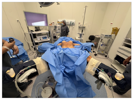
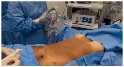
1. Positioning and Antisepsis:
Position: Supine position with arms in abduction at 90° and legs open (French position).
Antisepsis of the abdominal wall: 2% alcoholic chlorhexidine (or according to the service’s protocol).
Sterile surgical drape with xiphopubic exposure.
-
2. Minimally Invasive Access:
Optic portal (11 mm): A 1.5-cm transverse incision in the suprapubic sulcus, with blunt dissection up to the supra-aponeurotic space in the midline.
Working portals (5 mm): Right and left, using previous liposuction cannula incisions.
-
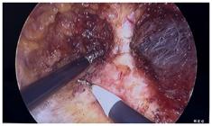
3. Workspace Creation:
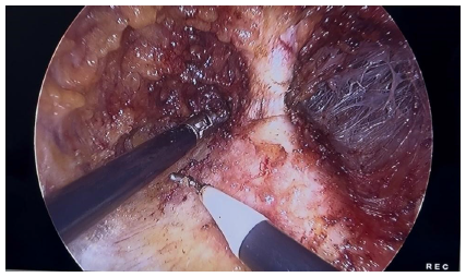
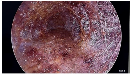
CO2 insufflation at 10 mmHg from the previously dissected space, allowing endoscopic visualization of the supra-aponeurotic space and the trabecular network after liposuction.
Conical dissection in the cranial direction of the subcutaneous cellular tissue, careful identification of the umbilical ligament, which is fully preserved in cases without associated umbilical hernia. In the presence of an umbilical hernia, the ligament can be released as technically needed.
-
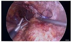
4. Plication of Rectus Abdominis:
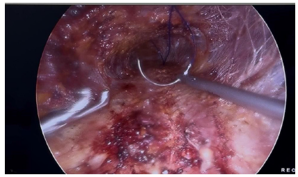
Continuous craniocaudal suture with bidirectional monofilament barbed suture without knot (preferably 0 or 1.0), in an inverted “U” pattern, with a spacing of 1 cm between stitches (usually two units for complete coverage of the defect along the midline).
Correction of the umbilical defect (when associated with hernia).
-
5. Finishing:
Removal and cutting of the needle under the sight of the 11-mm portal.
Deflation of the supra-aponeurotic space.
Skin synthesis with absorbable intradermal suture or surgical glue on the portals.




Third stage:
In the final stage, adjuvant skin retraction technologies can be used, such as radiofrequency, ultrasound, laser, or others available at the service. The choice of technology is based on clinical criteria, skin characteristics, and institutional availability, and does not interfere with the execution or reproducibility of the MILA technique itself.


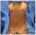
At the end of the procedure, the drain is positioned and fixed with a vacuum system, followed by sterile dressing and placement of a compressive modeling band.
DISCUSSION
The MILA technique was originally developed as a minimally invasive approach in the supra-aponeurotic plane for correction of diastasis rectus abdominis, combining principles of endoscopic surgery of the abdominal wall with complementary aesthetic objectives15. The present description proposes the systematized preservation of the umbilical ligament as a differential in selected patients without associated umbilical hernia, an aspect that is still poorly detailed in the literature.
In the context of minimally invasive approaches, several techniques have been described for the treatment of diastasis of the rectus abdominis, including laparoscopic subcutaneous repair (SCOLA), endoscopic preaponeurotic repair (REPA), and endoscopic reconstruction of the linea alba10,13. Although they share similar technical principles, these approaches present variations in plane dissection, plication systematization, and umbilicus management. The preservation of the umbilical ligament is not addressed in a standardized manner in these techniques, and its release is often performed when necessary for surgical exposure.
Umbilical ligament preservation, as described in this technique, is based on well-established anatomical principles. The umbilical ligament plays a key role in maintaining vascularization and the anatomical position of the umbilicus, and its preservation can contribute to the maintenance of the natural abdominal contour and to the reduction of umbilical complications described in open techniques and in some endoscopic approaches18. Previous studies suggest that anatomical integrity of the umbilicus is associated with better aesthetic outcomes, although comparative clinical evidence is still limited16.
Compared to traditional open techniques such as conventional abdominoplasty, minimally invasive approaches to DRA have well-documented theoretical advantages, including less tissue aggression, faster recovery, and lower incidence of wound-related complications9. In this scenario, MILA is an alternative technique that allows endoscopic correction of diastasis, maintaining principles of functional reconstruction of the linea alba described in other minimally invasive techniques10,11.
It is important to emphasize that the present manuscript is limited to the technical description, and does not present its own clinical data, casuistry, or analysis of functional and aesthetic outcomes. This characteristic is an inherent limitation of the study, but it is compatible with previous publications of technical description in abdominal wall surgery10,12. The absence of clinical follow-up and comparative evaluation prevents inferences as to the superiority of the technique in relation to other approaches, and these aspects should be explored in future studies.
Thus, prospective studies, case series, and multicenter investigations with medium- and long-term follow-up are necessary to objectively evaluate the functional and aesthetic outcomes, and the complication profile of the MILA technique with umbilical ligament preservation, as well as to compare it with other currently available minimally invasive approaches8,12,15.
CONCLUSIONS
The MILA technique represents a minimally invasive approach for the correction of diastasis rectus abdominis in the supra-aponeurotic plane, allowing endoscopic plication of the linea alba. The preservation of the umbilical ligament, as described in this article, constitutes a technical differential that aims to maintain the anatomical integrity of the umbilicus in selected patients without associated umbilical hernia.
The systematized description of the technique, with a clear definition of the surgical steps, indication criteria, and technical care contributes to its reproducibility and standardization, and can help surgeons in the planning and execution of the procedure. As this is a technical description, the present study does not allow inferences regarding clinical, functional, or aesthetic outcomes, which should be evaluated in future studies.
REFERENCES
-
1 Sperstad JB, Tennfjord MK, Hilde G, Ellström-Engh M, Bø K. Diastasis recti abdominis during pregnancy and 12 months after childbirth: prevalence, risk factors, and report of lumbopelvic pain. Br J Sports Med. 2016;50(17):1092-1096. doi:10.1136/bjsports-2016-096065.
» https://doi.org/10.1136/bjsports-2016-096065 -
2 Akram J, Matzen SH. Rectus abdominis diastasis. J Plast Surg Hand Surg. 2014;48(3):163-169. doi:10.3109/2000656X.2013.859145.
» https://doi.org/10.3109/2000656X.2013.859145 -
3 Emanuelsson P, Gunnarsson U, Dahlstrand U, Strigård K, Stark B. Operative correction of abdominal rectus diastasis reduces pain and improves abdominal wall muscle strength: a randomized prospective trial. Surgery. 2016;160(5):1367-1375. doi:10.1016/j.surg.2016.05.035.
» https://doi.org/10.1016/j.surg.2016.05.035 -
4 Beer GM, Schuster A, Seifert B, Manestar M, Mihic-Probst D, Weber SA. Normal width of the linea alba in nulliparous women. Clin Anat. 2009;22(6):706-711. doi:10.1002/ca.20836.
» https://doi.org/10.1002/ca.20836 -
5 Van de Water AT, Benjamin DR. Measurement methods to assess diastasis of the rectus abdominis muscle: a systematic review. Man Ther. 2016;21:41-53. doi:10.1016/j.math.2015.09.013.
» https://doi.org/10.1016/j.math.2015.09.013 -
6 Gormely E, Srai J, Pathak S, Gagliardi F, Shiwcharan A, Mok E. Impact of rectus diastasis repair on abdominal strength and function: a systematic review. Cureus. 2020;12(12):e12358. doi:10.7759/cureus.12358.
» https://doi.org/10.7759/cureus.12358 -
7 Hickey F, Finch JG, Khanna A. A systematic review on the outcomes of correction of diastasis of the recti. Hernia. 2011;15(6):607-614. doi:10.1007/s10029-011-0839-4.
» https://doi.org/10.1007/s10029-011-0839-4 -
8 Mommers EHH, Ponten JEH, Al Omar AK, de Vries Reilingh TS, Bouvy ND, Nienhuijs SW. The general surgeon's perspective of rectus diastasis: a systematic review of treatment options. Surg Endosc. 2017;31(12):4934-4949. doi:10.1007/s00464-017-5607-9.
» https://doi.org/10.1007/s00464-017-5607-9 -
9 ElHawary H, Chartier C, Alam P, Janis JE. Open versus laparoscopic surgical management of rectus diastasis: systematic review and pooled analysis. World J Surg. 2022;46(8):1878-1885. doi:10.1007/s00268-022-06550-9.
» https://doi.org/10.1007/s00268-022-06550-9 -
10 Claus CMP, Malcher F, Cavazzola LT, Furtado M, Morrell A, Azevedo M, et al. Subcutaneous onlay laparoscopic approach (SCOLA) for ventral hernia and rectus abdominis diastasis repair: technical description and initial results. Arq Bras Cir Dig. 2018;31(4):e1407. doi:10.1590/0102-672020180001e1407.
» https://doi.org/10.1590/0102-672020180001e1407 -
11 Köckerling F, Botsinis MD, Rohde C, Reinpold W, Schug-Pass C. Endoscopic-assisted linea alba reconstruction: new technique for treatment of symptomatic umbilical, trocar, and/or epigastric hernias with concomitant rectus abdominis diastasis. Eur Surg. 2017;49(2):71-75. doi:10.1007/s10353-017-0463-3.
» https://doi.org/10.1007/s10353-017-0463-3 -
12 Malcher F, Lima DL, Lima RNCL, Cavazzola LT, Claus CMP, Dong CT, et al. Endoscopic onlay repair for ventral hernia and rectus abdominis diastasis repair: why so many different names for the same procedure? Surg Endosc. 2021;35(10):5414-5421. doi:10.1007/s00464-021-08560-5.
» https://doi.org/10.1007/s00464-021-08560-5 - 13 Juárez Muas DM, Palmisano E, Pou Santonja G, Cuccomarino S, González Higuera G, Mayo P, et al. Reparación endoscópica preaponeurótica (REPA) como tratamiento de la diástasis de los músculos rectos asociada o no a hernias de la línea media. Rev Hispanoam Hernia. 2019;7(2):59-65.
- 14 Palmisano EM. Proposal for a single name in the endoscopic treatment of abdominal diastasis: a scientific need. Rev Hispanoam Hernia. 2023;11(1):1-3.
-
15 Claus CMP, Di-Biasio GA, Ribeiro RD, Corrêa MAMF, Pagnoncelli B, Palmisano E. Minimally invasive lipoabdominoplasty (MILA) tactic. Rev Col Bras Cir. 2024;51:e20243692. doi:10.1590/0100-6991e-20243692-en.
» https://doi.org/10.1590/0100-6991e-20243692-en -
16 Van Uchelen JH, Kon M, Werker PM. Long-term follow-up of umbilical complications after diastasis repair. Plast Reconstr Surg Glob Open. 2022;10(1):e4031. doi:10.1097/GOX.0000000000004031.
» https://doi.org/10.1097/GOX.0000000000004031 -
17 Hernández-Granados P, Henriksen NA, Berrevoet F, Cuccurullo D, López-Cano M, Nienhuijs S, et al. Management of rectus diastasis: a consensus statement. Br J Surg. 2021;108(10):1189-1191. doi:10.1093/bjs/znab128.
» https://doi.org/10.1093/bjs/znab128 -
18 Köhler G, Luketina RR, Emmanuel K. Sutured repair of primary small umbilical and epigastric hernias: concomitant rectus diastasis is a significant risk factor for recurrence. World J Surg. 2015;39(1):121-126. doi:10.1007/s00268-014-2765-y.
» https://doi.org/10.1007/s00268-014-2765-y -
19 Corrêa MA. Técnicas subcutâneas videoendoscópicas para cirurgia plástica estética e reconstrutiva. Plast Reconstr Surg. 1995;96(2):446-453. doi:10.1097/00006534-199508000-00023.
» https://doi.org/10.1097/00006534-199508000-00023 -
20 Champault GG, Catheline JM, Barrat C. Traitement pariétoscopique des défauts de la paroi abdominale: à propos de 15 cas. Hernia. 1999;3(1):15-18. doi:10.1007/BF01194634.
» https://doi.org/10.1007/BF01194634 -
21 Brucchi F, Boni L, Cassinotti E, Baldari L. Short-term outcomes of minimally invasive endoscopic onlay repair for diastasis recti and ventral hernia repair: a systematic review and meta-analysis. Surg Endosc. 2025;39(3):1490-1500. doi:10.1007/s00464-025-11555-1.
» https://doi.org/10.1007/s00464-025-11555-1 -
22 Forester E, Sadiq A. Comparative analysis of the efficacy and functionality of abdominoplasty versus minimally invasive techniques in the surgical treatment of diastasis rectus abdominis in postpartum women. Surg Endosc. 2023;37(12):9052-9061. doi:10.1007/s00464-023-10540-w.
» https://doi.org/10.1007/s00464-023-10540-w -
23 Henriksen NA, Kaufmann R, Simons MP, et al. EHS and AHS guidelines for treatment of primary ventral hernias in rare locations or special circumstances. BJS Open. 2020;4(2):342-353. doi:10.1002/bjs5.50252.
» https://doi.org/10.1002/bjs5.50252 -
24 Manetti G, Lolli MG, Belloni E, Nigri G. A new minimally invasive technique for the repair of diastasis recti: a pilot study. Surg Endosc. 2021;35(7):4028-4034. doi:10.1007/s00464-021-08393-2.
» https://doi.org/10.1007/s00464-021-08393-2 -
25 Carrara A, Catarci M, Fabris L, et al. Prospective observational study of abdominal wall reconstruction with THT technique in primary midline defects with diastasis recti. Surg Endosc. 2021;35(9):5104-5114. doi:10.1007/s00464-020-07997-4.
» https://doi.org/10.1007/s00464-020-07997-4 -
26 Ferrara F, Fiori F. Laparoendoscopic extraperitoneal surgical techniques for ventral hernias and diastasis recti repair: a systematic review. Hernia. 2024;28(6):2111-2124. doi:10.1007/s10029-024-03144-3.
» https://doi.org/10.1007/s10029-024-03144-3 -
27 Ngo P, Cossa JP, Gueroult S, Pélissier E. Minimally invasive bilayer suturing technique for the repair of concomitant ventral hernias and diastasis recti. Surg Endosc. 2023;37(7):5326-5334. doi:10.1007/s00464-023-10034-9.
» https://doi.org/10.1007/s00464-023-10034-9