 Morphological and histopathological description of Calyptospora sp. parasitism in Cichla monoculus Spix, 1929 (Osteichthyes, Cichlidae) from the lake region of Pracuúba-Amapá, Brazil
Abstract
The tucunaré (Cichla sp.) is an Amazonian fish that is heavily commercialized in the state of Amapá, and it can be infected by a variety of parasites, including coccidia of the genus Calyptospora, which are identified at the genus level by analyzing the structures that comprise its morphology. This study aimed to describe the morphology and histopathology of Calyptospora sp. parasitism in Cichla monoculus Spix, 1929 in the Municipality of Pracuúba, Amapá, Brazil. Nine specimens were acquired from the Lake Sacaizal by artisanal fishermen and transported in isothermal boxes to the Integrated Morpho-molecular and Technologies Laboratory (LIMT) of the Federal Rural University of the Amazon in Belém, Pará, where they were necropsied. Fragments of the liver were removed to visualize cysts using light microscopy and processed for scanning electron microscopy and histology analyses. The analysis revealed that 66.6% of the fish examined had clusters of oocysts in the hepatic region, resulting in the formation of melanomacrophagic centers. The oocysts were sphere-like, with a diameter of 21 µm. They contained four pyriform sporocysts, 8.7 µm long and 4.9 µm wide, with sporopods in the posterior region.
Keywords:
Coccidia; Tucunaré; Amazon; SEM
Resumo
O tucunaré (Cichla sp.) é um peixe amazônico de elevada comercialização no Estado do Amapá e que pode estar sujeito a infecção por diferentes parasitos, dentre eles os coccídeos do gênero Calyptospora sp. que são identificados quanto ao gênero, por meio da análise das estruturas que compõem sua morfologia. Este trabalho tem o objetivo de realizar a descrição morfológica e histopatológica do parasitismo por Calyptospora sp. em Cichla monoculus Spix, 1929, oriundos do Município de Pracuúba, Amapá, Brasil. Os 9 espécimes mortos foram adquiridos do lago Sacaizal por pescadores artesanais, em seguida transportados em caixas isotérmicas até o Laboratório de Integração Morfo-molecular e Tecnologias (LIMT) da Universidade Federal Rural da Amazônia, em Belém, Pará, aonde foram necropsiados e retirados fragmentos do fígado para visualização de cistos em microscopia de luz (ML) e processados para microscopia eletrônica de Varredura (MEV) e histologia. Por meio das análises, foi possível confirmar que 66,6% dos peixes analisados apresentavam aglomerados de oocistos na região hepática com a formação de centros melanomacrofágicos. Os oocistos eram arredondados, com diâmetro de 21 µm, contendo quatro esporocistos piriformes de 8.7 µm de comprimento e 4.9 µm de largura, que apresentavam esporopódes na região posterior.
Palavras-chave:
Coccídeos; Tucunaré; Amazônia; MEV
Introduction
The state of Amapá in the Municipality of Pracuúba has five primary fishing locations, including the “lake region.” This area is known for its rivers and streams, which are home to a diverse range of fish (Silva & Dias, 2010; Zacardi et al., 2021). In this location, fish of the genus Cichla Schneider, 1801 are among the most commonly caught by extractive fishing. Species of the genus are generally known in Brazilian territory as “tucunarés.” They are native to the Amazon basin and belong to the family Cichlidae within the order Cichliformes (Kullander, 2003; Kullander & Ferreira, 2006; Zacardi et al., 2021).
Cichla monoculus Spix, 1929, is resident in floodplain lakes and other ecotopes in Brazil and Peru. Their diet typically consisted of shrimp and fish. This species is characterized by a horizontal spot on the surface of the pectoral fin and a discontinuous lateral line on both sides (Kullander, 1987; Rabelo & Araújo-Lima, 2002; Neto et al., 2017; Kullander & Ferreira, 2006).
Apicomplexa Levine, 1970, is a protist phylum that includes obligate intracellular parasites found in both vertebrates and invertebrates. The class Coccidia Leuckart, 1897 comprises species that infect the gastrointestinal systems of their hosts. In freshwater fish, the genus Calyptospora is notable as the cause of highly prevalent liver infections that can be evident as a change in the color of the liver to whitish (Bonar et al., 2006; Acosta et al., 2016; Eiras et al., 2016; Votýpka et al., 2016; Ramos et al., 2018).
In general, parasites from Calyptospora are distinguished by the formation of oocysts or parasitophorous vacuoles that contain four structures known as sporocysts and are covered by a membrane veil. Each sporocyst contained two sporozoites. Scanning electron microscopy (SEM) can be used to view structural details such as sutures and sporopods, which are useful for determining the similarities and differences between morphological structures and are essential for identification (Azevedo et al., 1995; Matos et al., 1999; Oliveira et al., 2021). In addition, histopathological analyses are effective in detecting parasitic damage to tissues (Videira et al., 2013).
The purpose of this study was to describe the morphology and histopathology of Calyptospora sp. parasitizing C. monoculus in the lake region of the Municipality of Pracuúba-Amapá, Brazil.
Material and Methods
Collection of hosts and parasites
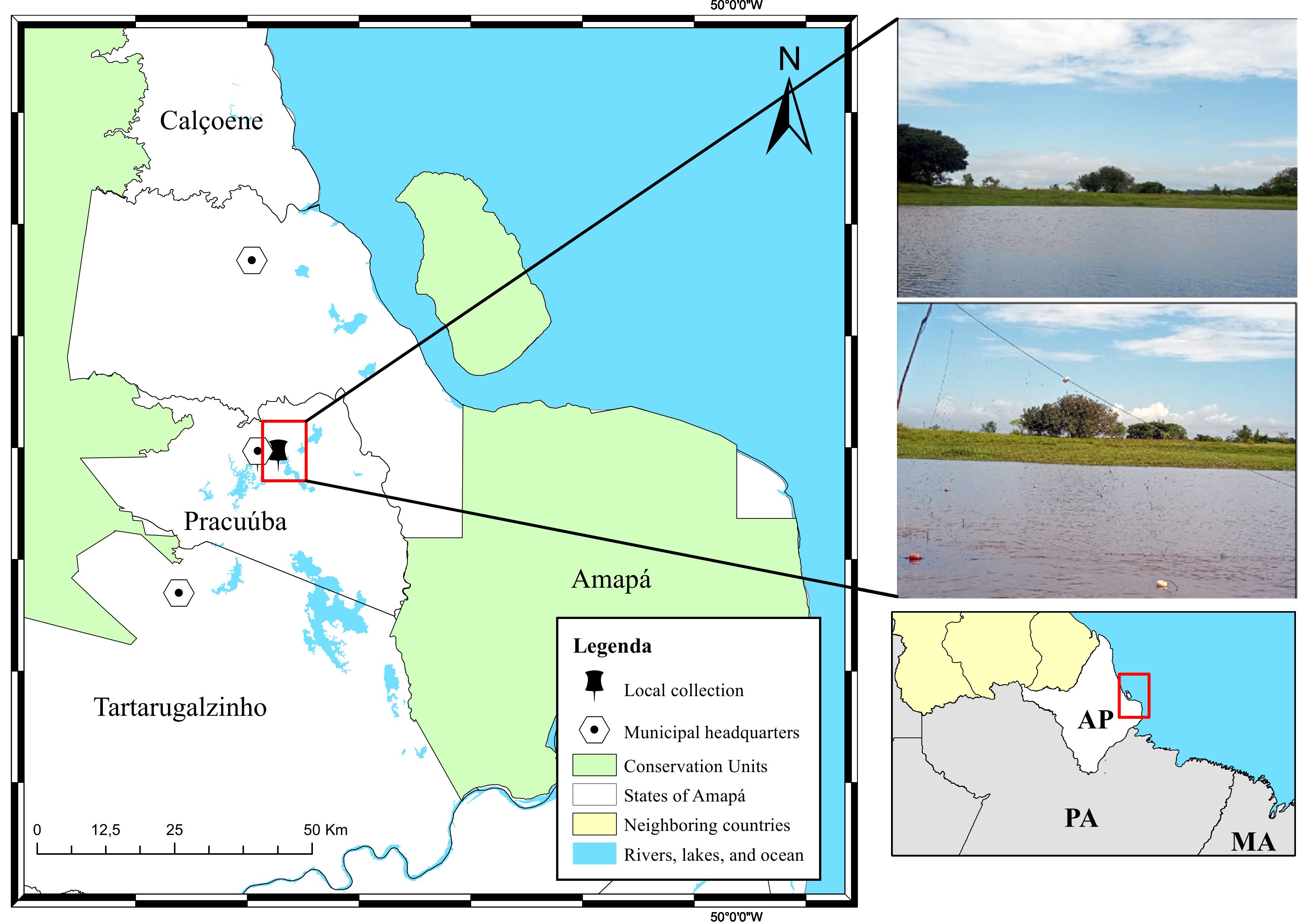
A total of nine dead specimens of peacock bass (C. monoculus) were acquired from artisanal fishermen in the Municipality of Pracuúba-Amapá (1°42'.79”N 50°43'17.5”W), which is close to the towns of Calçoene, Amapá, and Tartarugualzinho (Figure 1). Fish were caught from Lake Sacaizal, packed in isothermal boxes with ice, and transported to the Integrated Morpho-Molecular and Technologies Laboratory (LIMT) of the Federal Rural University of the Amazon (UFRA), Belém campus Pará. In the laboratory, specimen biometrics and identification were performed using the dichotomous key proposed by Kullander & Ferreira (2006). The fish were necropsied to inspect the entire body surface, including internal organs, using a Zeiss Stemi DCR stereoscope. Small chunks of the liver with oocysts were extracted, placed in the center of a slide containing a drop of water, softly compressed with a coverslip, and examined using light microscopy.
Geographic location map of the fish collection area, in the municipality of Pracuúba, state of Amapá in Brazil.
Morphometric analysis of oocysts
The morphometric parameters of oocysts (n=17), including oocyst diameter (OD), sporocyst length (SL), and sporocyst width (SW), were measured using ImageJ version 3.2 software as previously described (Schneider et al., 2012). The data were compiled with data from other Calyptospora sp. using principal component analysis (PCA) with PAST 3.0 software (Hammer et al., 2001).
Histopathological analysis
For histological processing, tissue fragments were fixed in Davidson’s solution (95% alcohol, formaldehyde, acetic acid, and distilled water) for 24 h before being dehydrated in an increasing series of alcohol solutions (70%, 80%, 90%, absolute I, absolute II, and absolute III) for 1 h each. After clearing with absolute alcohol–xylol for 30 min, absolute xylol (Xylol I and Xylol II) was successively added for 15 min each. The material was impregnated with paraffin, and the solid blocks that formed were used to obtain 5 μm-thick sections with a model HM315 rotary microtome (Microm). The sections were extruded onto a water bath, collected on glass slides, kept in the oven for 24 h at 60˚C, and stained using hematoxylin-eosin (Luna, 1968).
SEM
Liver fragments were fixed in 5% glutaraldehyde with 0.2 M sodium cacodylate buffer at pH 7.2 for 3 h at 4°C, washed in the same solution for 2 to 4 h, then fixed in 2% osmium tetroxide in the buffer for 2 h at 4°C. Following the dehydration step, increasing amounts of ethanol were added, followed by critical point drying and coating with gold. Photomicrographs of parasites were acquired at the Scanning Electron Microscopy Laboratory, Museu Paraense Emílio Goeldi. Structural details were observed, and morphometric parameters were measured in mature oocysts (n = 17) using ImageJ 3.2 software. OD, SL, and SW were measured, and average values were determined using Excel 2013 software. The data were compiled for comparison with those of published studies on the morphometry of Calyptospora sp.
Results and Discussion
The fish displayed an average weight of 298.0±1.8 g and a length of 21.0±1.2 cm. The prevalence of parasitism by Calyptospora sp. was 66.6%, and its morphology was consistent with that of coccidia of the genus. Several mature and rounded oocysts contained pyriform sporocysts in the inner region (Figures 2A-B). SEM of sporocysts revealed externally scattered sporopods (Figure 2C). These are regarded as another crucial characteristic for identification, as documented by Oliveira et al. (2021) in Serrasalmus rhombeus (Linnaeus, 1776).
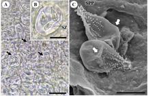
Infection of Cichla monoculus Spix, 1929 liver by Calyptospora sp. A- Groups of Calyptospora sp. oocysts, observed under light microscopy (ML) in the hepatic region, highlighting the sporocysts (arrows), Scale Bar 20 μm. B- Oocyst with sporocysts, indicating the spozoites internally (SZ), Scale Bar 10 μm. C- Sporocysts visualized in greater detail in Scanning Electron Microscopy (SEM), where structures on their surface, the sporopodia (SPP) and the apical and lateral suture lines (arrow) can be observed, Scale Bar 5 μm.
The prevalence of this study was higher than that of 40% of Calyptospora sp. in the hepatopancreas of Cichla temensis (Humboldt, 1821), reported by Velasco et al. (2012), although there were morphological similarities with the rounded oocysts, pyriform sporocysts and two sporozoites. Santiago et al. (2012) observed parasitism in 56% of C. temensis liver specimens from Marajó Island, with a higher incidence during the wet season. Another study reported that fish from the municipality of Vigia in the state of Pará, approximately 60% of Brachyplatystoma vaillantii (Valenciennes, 1940) displayed several clustered oocysts that replaced the liver parenchyma region (Silva et al., 2012). Negrão et al. (2019) identified parasitic forms of Calyptospora sp. in Crenicichla lepidota (Heckel 1840), Cichla ocelaris (Bloch & Schneider 1801), Hoplias malabaricus (Bloch, 1794), Mesonauta festivus (Heckel 1840), Hoplerythrinus unitaeniatus (Spix & Agassiz, 1829), Astronotus ocellatus (Agassiz, 1831), Geophagus proximus (Castelnau, 1855), Pterophyllum scalare (Schultze 1823), Satanoperca jurupari (Heckel 1840) and Heros efasciatus (Heckel, 1840), from the Curiaú-Amapá River floodplain, which infected the gallbladder, liver, and heart. Morphologically, the oocysts were scarcely recognizable.
The oocysts had an average diameter of 21±2.7 μm, while the pyriform-shaped sporocysts had an average length of 8.7±1.3 μm and a width of 4.9±0.7 μm. This Calyptospora presented oocyst diameter, shape, length and width of sporocysts similar to those found by Békési & Molnar (1991), Bonar et al. (2006), Santiago et al. (2012) and Silva et al. (2012, 2019, 2020) (Table 1). Furthermore, this oocyst differed from those reported by Albuquerque & Brasil-Sato (2010), who reported ellipsoidal sporocysts and greater oocyst diameter (24.5 μm) and sporocyst length (11.5 μm). Fournie et al. (1985) also reported this sporocyst form; however, the structural morphometric differences did not differ significantly from the morphotype of this study.
Morphometric comparison of Calyptospora spp. described in fish species, with data on oocyst diameter, shape, length and width of sporocysts, host, infection site and location. All data are in micrometers (µm).
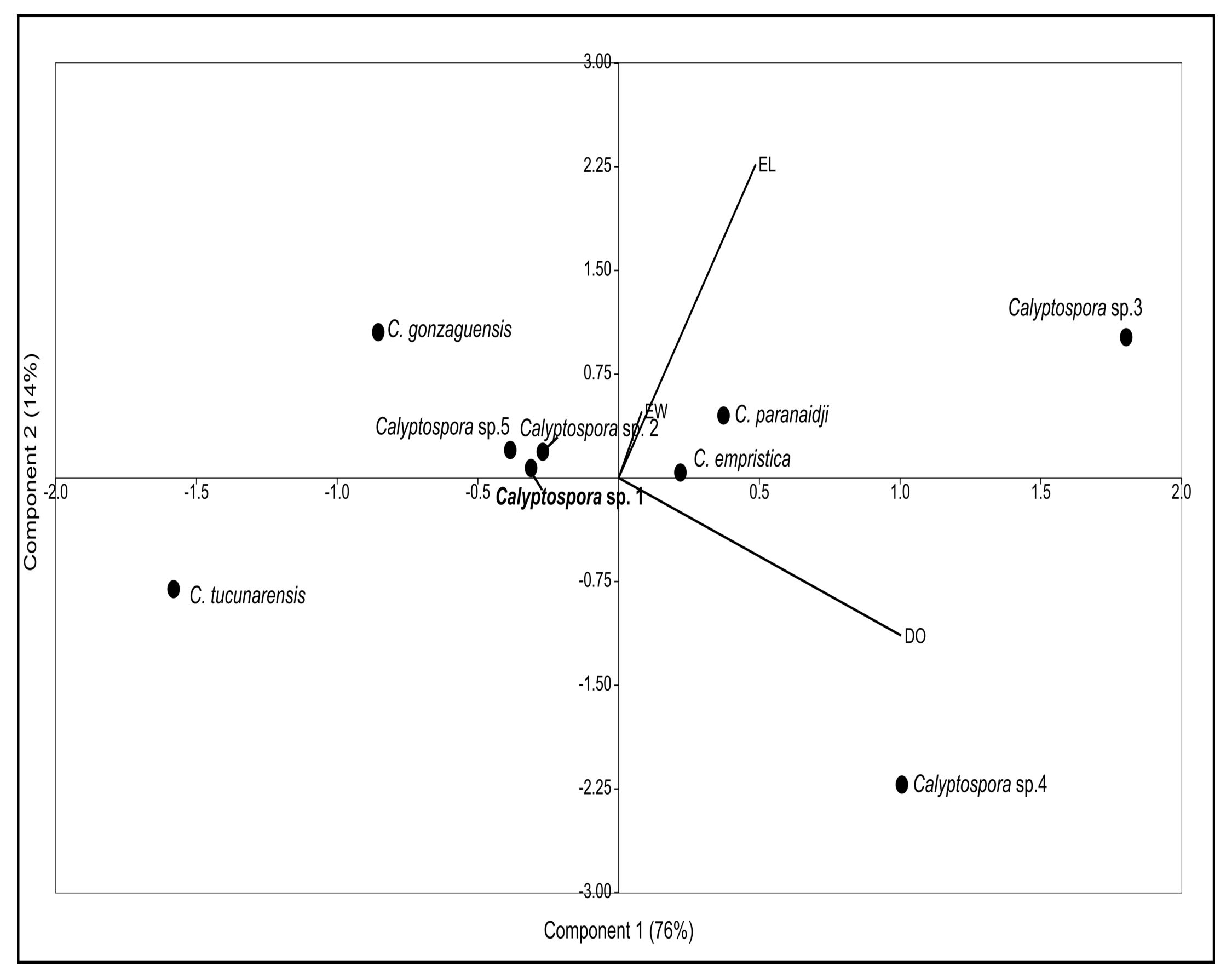
The morphometric PCA of Calyptospora sp. (Figure 3) revealed that component 1 (OD) accounted for approximately 76% of the variance, whereas components 2 and 3 (SL and SW, respectively) accounted for 14% and 10% of the variance, respectively. The findings imply that components 1 and 2 account for 89% of the correlation of variables. The Calyptospora sp. oocysts observed in the current study were similar to Calyptospora sp. 2 reported in C. temensis, with an oocyst diameter of 21.0 vs. 21.2 μm, but differed in sporocyst length of 8.7 vs. 9.2 μm and sporocyst width 4.9 vs. 3.1 μm (Table 1). Although they still demonstrate small morphological variations from Calyptospora sp. to Cichla sp., it remains uncertain whether they belong to the same taxonomic group. Further molecular data, specifically markers of small ribosomal subunit DNA data, are required (Berto et al., 2014).
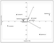
The Principal component analysis (PCA) of oocysts of Calyptospora spp. in Cichla monoculus from Pracaúuba, Amapá. The oocyst diameter (OD), sporocyst length (SL), and sporocyst width (SW) components were combined. The components 1 and 2, on each axis of the graph, display the variables that vary the most and are in charge of the morphometric ordering of the species.
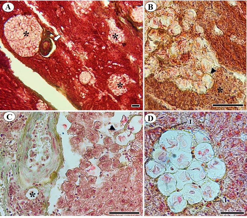
According to histopathological analysis, there were many oocysts that matured into hepatocytes and hepatopancreas region; some of these were found near blood arteries with a higher blood supply, which may have been related to the sporocysts' chemotaxis or nutrition (Figure 4 A-C). Furthermore, infiltrates of mononuclear cells were noted, suggesting the presence of an inflammatory process (Figure 4D). Oocysts in the liver parenchyma close to blood vessels were also observed by Oliveira et al. (2021). This location is related to the development to the oocyst stage, where the sporocysts are transported by the blood vessels and mature into oocysts in the hepatocytes, as described in the C. funduli life cycle (Fournie et al., 2000). Other studies of Calyptospora spp. have shown the presence of inflammatory cells and, in some occasions, necrosis (Fournie & Overstreet, 1993; Békési & Molnár, 1991; Whipps et al., 2012; Videira et al., 2013; Silva et al., 2019; Negrão et al., 2019). This demonstrates that the parasitic intensity found causes damage to the liver tissue of C. monoculus.
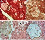
Gomori-stained photomicrographs of Calyptospora sp. oocysts in the liver parenchyma of Cichla monoculus Spix, 1929. A- Oocysts (*) adjacent to the hepatic artery (arrow), Scale Bar 50 μm. B- Adjacent oocytes (arrowhead) in a region of hepatopancreas (*), Scale Bar 50 μm. C- Oocysts interspersed between hepatocytes with loss of cellular architecture and collagen deposition (arrowhead), perivascular oocysts (*), Scale Bar 50 μm. D- Oocysts surrounded by inflammatory infiltrate (I), Scale Bar 20 μm.
This is the first report of Calyptospora sp. in C. monoculus. Morphological and morphometric analyses indicated that parasitism occurred only in the liver tissue of this host. Additional research is required to understand the action of the immunological response and to verify the causes for the formation of melanomacrophagic bodies, the parasite's life cycle, and the forms of prophylaxis and treatment to promote the conservation of the species in its natural habitat.
Acknowledgements
We acknowledge the assistance of the Aquatic Organisms Research Group of the Federal Rural University of the Amazon (UFRA) and the Scanning Electron Microscopy Laboratory of the Museum Paraense Emílio Goeldi.
-
How to cite: Amoras ES, Eduard J, Progene MPS, Silva JFBR, Videira MN, Sindeaux-Neto JL, et al. Morphological and histopathological description of Calyptospora sp. parasitism in Cichla monoculus Spix, 1929 (Osteichthyes, Cichlidae) from the lake region of Pracuúba-Amapá, Brazil. Braz J Vet Parasitol 2024; 33(4): e012324. https://doi.org/10.1590/S1984-29612024078
References
- Acosta AA, Godoy AT, Yamada FH, Brandão H, Paes JVK, Bongiovani MF, et al. Aspectos parasitológicos dos peixes. In: Silva RJ. Integridade ambiental da represa de Jurumirim: ictiofauna e relações ecológicas São Paulo: Editora UNESP; 2016. p. 115-192.
-
Albuquerque MC, Brasil-Sato MC. First report of Calyptospora sp. (Apicomplexa, Calyptosporidae) in forage characid fish from the Três Marias Reservoir, São Francisco Basin, Brazil. Eur J Protistol 2010; 46(2): 150-152. http://doi.org/10.1016/j.ejop.2009.12.005 PMid:20163938.
» http://doi.org/10.1016/j.ejop.2009.12.005 -
Azevedo C, Matos E, Matos P. Ultrastructural data on sporogony of the coccidian parasite Calyptospora spinosa from the liver of the Amazonian fish, Crenicichla lepidota Heckel. J Fish Dis 1995; 18(6): 475-479. http://doi.org/10.1111/j.1365-2761.1995.tb00351.x
» http://doi.org/10.1111/j.1365-2761.1995.tb00351.x -
Békési L, Molnár K. Calyptospora tucunarensis n. sp. (Apicomplexa: Sporozoea) from the liver of tucunare Cichla ocellaris in Brazil. Syst Parasitol 1991; 18(2): 127-132. http://doi.org/10.1007/BF00017665
» http://doi.org/10.1007/BF00017665 -
Berto BP, Mclntosh D, Lopes CWG. Studies on coccidian oocysts (Apicomplexa: eucoccidiorida). Rev Bras Parasitol Vet 2014; 23(1): 1-15. http://doi.org/10.1590/S1984-29612014001 PMid:24728354.
» http://doi.org/10.1590/S1984-29612014001 -
Bonar CJ, Poynton SL, Schulman FY, Rietcheck RL, Garner MM. Hepatic Calyptospora sp. (Apicomplexa) infection in a wild-born, aquarium-held clutch of juvenile arapaima Arapaima gigas (Osteoglossidae). Dis Aquat Organ 2006; 70(1-2): 81-92. http://doi.org/10.3354/dao070081 PMid:16875394.
» http://doi.org/10.3354/dao070081 - Eiras JC, Veloso AL, Pereira JJR. Parasitos de peixes marinhos da América do Sul. Rio Grande: Editora Furg; 2016.
-
Fournie JW, Hawkins WE, Overstreet RM. Calyptospora empristica n. sp. (Eimeriorina: Calyptosporidae) from the liver of the starhead topminnow, Fundulus notti. J Protozool 1985; 32(3): 542-547. http://doi.org/10.1111/j.1550-7408.1985.tb04058.x
» http://doi.org/10.1111/j.1550-7408.1985.tb04058.x -
Fournie JW, Overstreet RM. Host Specificity of Calyptospora funduli (Apicomplexa: Calyptosporidae) in Atheriniform Fishes. J Parasitol 1993; 79(5): 720-727. http://doi.org/10.2307/3283611 PMid:8410544.
» http://doi.org/10.2307/3283611 -
Fournie JW, Vogelbein WK, Overstreet RM, Hawkins WE. Life cycle of Calyptospora funduli (Apicomplexa: calyptosporidae). J Parasitol 2000; 86(3): 501-505. http://doi.org/10.1645/0022-3395(2000)086[0501:LCOCFA]2.0.CO;2 PMid:10864246.
» http://doi.org/10.1645/0022-3395(2000)086[0501:LCOCFA]2.0.CO;2 - Hammer Ø, Harper DAT, Ryan PD. Past: paleontological statistics software package for education and data analysis. Palaeontol Electronica 2001; 4(1): 4.
-
Kullander S. Cichlid fishes of the Amazon River drainage of Peru. Copeia 1987; 4(4): 1078-1080. http://doi.org/10.2307/1445586
» http://doi.org/10.2307/1445586 - Kullander SO, Ferreira EJG. A review of the South American cichlid genus Cichla, with descriptions of nine new species (Teleostei: cichlidae). Ichthyol Explor Freshwat 2006; 17(4): 289-398.
- Kullander SO. Family Cichlidae. In: Reis RE, Kullander SO, Ferraris CJ,editors, Checklist of the Freshwater Fishes of South America Porto Alegre: Edipucrs; 2003. p. 605–654.
- Luna LG. Manual of histologic staining methods of the Armed Forces Institute of Pathology New York: McGraw-Hill; 1968.
-
Matos E, Azevedo C, Corral L, Casal G, Matos P. Ultraestrutura de protozoários parasitas de peixes da região amazônica. Acta Amaz 1999; 29(4): 575-585. http://doi.org/10.1590/1809-43921999294585
» http://doi.org/10.1590/1809-43921999294585 -
Negrão MCS, Silva MRL, Videira MN, Viana LA. Prevalence and molecular characterisation of Calyptospora parasites Overstreet, Hawkins and Fournié, 1984 (Apicomplexa: Calyptosporidae) in fishes from the Eastern Amazon, Brazil. Parasitol Int 2019; 73: 101975. http://doi.org/10.1016/j.parint.2019.101975 PMid:31421267.
» http://doi.org/10.1016/j.parint.2019.101975 -
Neto CF, Alves WS, Junior HNM. Dieta do tucunaré Cichla monoculus Agassiz, 1831- açude Thomaz Osterne de Alencar semiárido cearense. Cad Cult Cien 2017; 16(1): 60-73. http://doi.org/10.14295/cad.cult.cienc.v16i1.1466
» http://doi.org/10.14295/cad.cult.cienc.v16i1.1466 -
Oliveira JEF, Figueredo RTA, Sindeaux-Neto JL, Vilhena MPSP, Berrêdo JF, Matos E, et al. Hepatic coccidiosis in Serrasalmus rhombeus Linnaeus, 1766 from the Amazon basin: morphological and histopathological aspects. Rev Bras Parasitol Vet 2021; 30(1): e026120. http://doi.org/10.1590/s1984-29612021007
» http://doi.org/10.1590/s1984-29612021007 -
Rabelo H, Araújo-lima CARM. A dieta e o consumo diário de alimento de Cichla monoculus na Amazônia central. Acta Amazon 2002; 32(4): 707-724. http://doi.org/10.1590/1809-43922002324724
» http://doi.org/10.1590/1809-43922002324724 -
Ramos ABMA, Santos PFS, Hemeterio LCA, Santos WJP, Araújo RS, Matos E. Coccidiose hepática em Aspistor quadriscutis, Valenciennes, 1840 (Teleostei: Ariidae) capturados no Estuário do nordeste paraense, Amazônia, Brasil. Arch Vet Sci 2018; 23(3): 63-67. http://doi.org/10.5380/avs.v23i3.52244
» http://doi.org/10.5380/avs.v23i3.52244 -
Santiago H, Corrêa JL, Tortelly R, Menezes RC, Matos P, Matos E. Morphology and histopathology of Calyptospora sp. (Apicomplexa: Calyptosporidae) in speckled peacock bass, Cichla temensis Humboldt, 1821 (Perciformes: Cichlidae), from the Marajó-Açu River, Marajó Island, Brazil. Parasitol Res 2012; 110(6): 2569-2572. http://doi.org/10.1007/s00436-011-2770-0 PMid:22200958.
» http://doi.org/10.1007/s00436-011-2770-0 -
Schneider CA, Rasband WS, Eliceiri KW. NIH Image to ImageJ: 25 years of image analysis. Nat Methods 2012; 9(7): 671-675. http://doi.org/10.1038/nmeth.2089 PMid:22930834.
» http://doi.org/10.1038/nmeth.2089 -
Silva LMA, Dias MT. A pesca artesanal no estado do Amapá: estado atual e desafios. Bol Tec-Cient CEPNOR 2010; 10(1): 43-53. http://doi.org/10.17080/1676-5664/btcc.v10n1p43-53
» http://doi.org/10.17080/1676-5664/btcc.v10n1p43-53 -
Silva MC, São Clemente SC, Picanço Júnior JA, Silva MVO, Matos ER. Calyptospora sp in Brachyplatystoma vaillantii trapped at the Vigia, State of Pará, Brazil. Rev Bras Parasitol Vet 2012; 21(2): 176-178. http://doi.org/10.1590/S1984-29612012000200020
» http://doi.org/10.1590/S1984-29612012000200020 -
Silva MF, Orlanda JFF, Araújo-Costa MJ, Hamoy I, Matos E. Hepatic Coccidiosis in Triportheus angulatus Spix & Agassiz, 1829 (Characiformes: Triportheidae), a Tropical Fish from the Eastern Brazilian Amazon, with the Description of a New Species of Calyptospora (Apicomplexa: Calyptosporidae). J Eukaryot Microbiol 2020; 67(3): 352-358. http://doi.org/10.1111/jeu.12785 PMid:31943507.
» http://doi.org/10.1111/jeu.12785 -
Silva MF, Silva DT, Giese EG, Hamoy IG, Matos ER. Morphology and Phylogeny of Calyptospora paranaidji n. sp. (Eimeriorina: Calyptosporidae), an Apicomplexan Parasite of the Hepatic Tissue of Cichla piquiti Kullander & Ferreira, 2006, From a Reservoir in the Brazilian Amazon Region. J Eukaryot Microbiol 2019; 66(4): 608-616. http://doi.org/10.1111/jeu.12707 PMid:30548108.
» http://doi.org/10.1111/jeu.12707 -
Velasco M, Videira M, Matos P, São Clemente SC, Carmona S, Sanches O, et al. Morfologia e nova ocorrência de um coccídio hepatopancreático parasita de peixe amazônico. Rev Cienc Agrar (Belem) 2012; 55(3): 231-235. http://doi.org/10.4322/rca.2012.063
» http://doi.org/10.4322/rca.2012.063 -
Videira M, Velasco M, Tortelly R, Menezes RC, São Clemente SC, Matos E. An anatomopathological study of hepatic coccidiosis (Calyptospora sp.) in the Acará-pixuna, Aequidens plagiozonatus Kullander, 1984 from the Brazilian state of Pará. Arq Bras Med Vet Zootec 2013; 65(1): 91-94. http://doi.org/10.1590/S0102-09352013000100014
» http://doi.org/10.1590/S0102-09352013000100014 -
Votýpka J, Modrý D, Oborník M, Slapeta J, Lukes J. Apicomplexa. In: Archibald JM, Simpson AGB, Slamovits CH, editors. Handbook of the Protists 2nd ed. New York: Springer; 2016. p. 1-58. http://doi.org/10.1007/978-3-319-32669-6_20-1
» http://doi.org/10.1007/978-3-319-32669-6_20-1 -
Whipps CM, Fournie JW, Morrison DA, Azevedo C, Matos E, Thebo P, et al. Phylogeny of fish- infecting Calyptospora species (Apicomplexa: eimeriorina). Parasitol Res 2012; 111(3): 1331-1342. http://doi.org/10.1007/s00436-012-2969-8 PMid:22645034.
» http://doi.org/10.1007/s00436-012-2969-8 -
Zacardi DM, Passos LS, Silva TC. A pesca artesanal na região dos lagos: o caso do município de Pracuúba, Amapá. Rev Desafios 2021; 08(1): 85-94. http://doi.org/10.20873/uftv8-9552
» http://doi.org/10.20873/uftv8-9552