 Molecular detection of hemoplasmas in domestic cats from different Brazilian regions
Abstract
Domestic cats can be parasitized by Mycoplasma haemofelis, ‘Candidatus Mycoplasma haemominutum’, and ‘Candidatus Mycoplasma turicensis’. Although the molecular occurrence of hemoplasmas in domestic cats has been investigated in the five geographical regions of Brazil, no studies have been conducted in the states of Rondônia and Minas Gerais to date. The present work aimed to investigate the molecular occurrence of hemoplasmas in cats from four Brazilian states in the Northern, Central-Western and Southeastern regions of the country. Among 486 blood samples – 80 from Rondônia (RO), 100 from Goiás (GO), 155 from Minas Gerais (MG), and 151 from São Paulo (SP) – submitted to the endogenous gapdh gene-based PCR, 94.44% (459/486) were positive, of which 1.96% (9/459; 1 from RO, 2 from GO, 5 from MG, and 1 from SP) were positive in the qPCR assay for hemoplasmas based on the 16S rRNA gene. In the phylogenetic analyses, the obtained 16S rRNA (two genotypes) and 23S rRNA (three genotypes) sequences were positioned together with ‘Candidatus Mycoplasma haemominutum’. The present study showed the first molecular evidence of infection by hemoplasmas in cats from MG and RO, contributing to a better understanding of the epidemiology of feline hemoplasmosis agents in Brazil.
Keywords:
'Candidatus Mycoplasma haemominutum'; Mycoplasma spp.; qPCR; feline hemoplasmosis
Resumo
Gatos domésticos podem ser parasitados por Mycoplasma haemofelis, ‘Candidatus Mycoplasma haemominutum’ e ‘Candidatus Mycoplasma turicensis’. Embora a ocorrência molecular de hemoplasmas em gatos domésticos tenha sido investigada nas cinco regiões geográficas do Brasil, nenhum estudo foi conduzido nos estados de Rondônia e Minas Gerais até o momento. O presente trabalho teve como objetivo investigar a ocorrência molecular de hemoplasmas em gatos de quatro estados brasileiros nas regiões Norte, Centro-Oeste e Sudeste do país. Dentre 486 amostras de sangue – 80 de Rondônia (RO), 100 de Goiás (GO), 155 de Minas Gerais (MG) e 151 de São Paulo (SP) – submetidas à PCR baseada no gene endógeno gapdh, 94,44% (459/486) foram positivas, das quais 1,96% (9/459; 1 de RO, 2 de GO, 5 de MG e 1 de SP) foram positivas no ensaio de qPCR para hemoplasmas baseado no gene 16S rRNA. Nas análises filogenéticas, as sequências 16S rRNA (2 genótipos) e 23S rRNA (3 genótipos) obtidas foram posicionadas juntamente com ‘Candidatus Mycoplasma haemominutum’. O presente estudo mostrou a primeira evidência molecular de infecção por hemoplasmas em gatos dos estados de Minas Gerais e Rondônia, contribuindo para um melhor entendimento da epidemiologia dos agentes da hemoplasmose felina no Brasil.
Palavras-chave:
'Candidatus Mycoplasma haemominutum'; Mycoplasma spp.; qPCR; hemoplasmose felina
Introduction
Hemoplasmas (Mollicutes: Mycoplasmataceae) are cell wall-lacking small epicellular bacteria that infect the surface of erythrocytes of vertebrate hosts (Razin et al., 1998). These bacteria have already been reported in different species of domestic and wild mammals, including pigs, cattle, sheep, bears, bats, rodents, dogs, and cats (Harasawa et al., 2015; Mesquita et al., 2021; Tasker, 2022). While the transmission of such agents via aggressive interactions and blood transfusion is well documented, vector transmission by fleas, ticks and mosquitoes is still poorly elucidated, being restricted to studies that detected DNA from hemoplasmas resulting from the blood meal of these ectoparasites (Tasker, 2022; Moore et al., 2024). Furthermore, hemoplasma infections are important causes of anemia in felines (Tasker, 2022; Moore et al., 2024).
Domestic cats can be parasitized by three hemoplasma species, namely Mycoplasma haemofelis (Mhf), ‘Candidatus Mycoplasma haemominutum’ (CMhm) and ‘Candidatus Mycoplasma turicensis’ (CMt), the former considered the most pathogenic species (Tasker, 2022; Moore et al., 2024). Mhf is responsible for causing severe extravascular and occasionally intravascular hemolytic anemia in immunocompetent cats without other comorbidities (Willi et al., 2005; Tasker, 2022; Hoseinpoor et al., 2024). CMhm, in its turn, causes clinical anemia in cats with neoplasms, immunosuppressed, or infected with the feline leukemia virus (FeLV) (Rosa Maciel et al., 2023). On the other hand, CMt infection can result in a small reduction in the number of erythrocytes experimentally, but rarely causes clinical anemia (Willi et al., 2005; Tasker, 2022).
Feline hemoplasmosis is often associated with old, male, mixed-breed cats with outdoor access (Kellner et al., 2018; Tasker, 2022). Díaz-Regañón et al. (2018) showed a significant relationship between some species of hemoplasma and retroviruses, especially the feline immunodeficiency virus (FIV) (Attipa et al., 2017; Rosa Maciel et al., 2023). Due to failures in the in vitro cultivation of hemotropic Mycoplasma spp., as well as the low sensitivity of cytological tests and blood smears for detecting these agents, molecular diagnosis by Polymerase Chain Reaction (PCR) is the most suitable technique for diagnosing hemoplasmosis (Tasker, 2022).
Different research groups have molecularly detected hemoplasmas in domestic cats in various countries, such as Iran (CMhm: 10.5%; Mhf: 2.2%; CMt: 0.6%) (Hoseinpoor et al., 2024), China (CMhm: 3.4%; Mhf: 0.9%; CMt: 1.2%) (Zhang et al., 2021), Germany (CMhm: 8.76%; Mhf: 0.41%) (Bergmann et al., 2016), Portugal (CMhm: 41.56%; Mhf: 12.81%; CMt: 1.25%) (Martínez-Díaz et al., 2013), Albania (CMhm: 21.9%; Mhf: 10.3%; CMt: 5.5%) (Silaghi et al., 2014), Spain (CMhm: 9.9%; Mhf: 3.7%; CMt: 0.5%) (Díaz-Regañón et al., 2018), Romania (CMhm: 72.7%; Mhf: 27.3%) (Imre et al., 2020), Italy (CMhm: 12.3%; Mhf: 9.4%; CMt: 4.8%) (Ravagnan et al., 2017), South Africa (CMhm: 38%; Mhf: 15%; CMt: 26%) (Willi et al., 2006), Angola (CMhm: 7.5%; Mhf: 1.5%) (Mesquita et al., 2021) and the United States (Mycoplasma spp.: 13.69%) (Manvell et al., 2021). In Brazil, the molecular occurrence of hemotropic Mycoplasma spp. in domestic cats has been investigated in the five geographical regions of the country (Bortoli et al., 2012; Braga et al., 2012; Miceli et al., 2013; André et al., 2014; Santis et al., 2014; Santos et al., 2009; Raimundo et al., 2016; Munhoz et al., 2018; Pedrassani et al., 2019; Santos et al., 2021; Carvalho et al., 2024). However, no studies have been conducted in the states of Rondônia and Minas Gerais to date, and few studies have been conducted in the states of São Paulo and Goiás.
Although traditionally recognized for their veterinary significance, the zoonotic potential of hemoplasmas species has gained increasing attention in recent years, particularly in immunocompromised humans (Sykes et al., 2010). ‘Candidatus M. haematohominis’, ‘Candidatus M. haematoparvum’, M. haemofelis, Mycoplasma haemocanis, Mycoplasma suis, and Mycoplasma ovis have been molecularly detected in humans (Yuan et al., 2009; Sykes et al., 2010; Steer et al., 2011; Maggi et al., 2013, Hattori et al., 2020; Kmetiuk et al., 2025). For instance, Mhf has already been reported in a patient coinfected with HIV virus and Bartonella henselae in Brazil (Santos et al., 2008). CMt has been described in a patient with hemolytic anemia and pyrexia in the United States (Steer et al., 2011). Such reports imply constant epidemiological surveillance of feline hemoplasmosis in domestic cats and a One Health approach to the zoonotic risks of these agents. The present work aimed to investigate the molecular occurrence and genetic diversity of hemoplasmas in blood samples from cats from four Brazilian states in the Northern, Central-Western and Southeastern regions of the country.
Material and Methods
Study area and sampling
A total of 486 cat blood samples were collected in the states of Goiás (n=100), Minas Gerais (n=155), Rondônia (n=80) and São Paulo (n=151) (Figure 1) through partnerships with veterinary clinics and autonomous animal protection shelters, and during castration campaigns promoted by the Centro de Castração da Associação Protetora dos Animais, located in the Faculdade de Ciências Agrárias e Veterinárias (FCAV/UNESP, Jaboticabal, São Paulo), as well as the Projeto de Controle Populacional Animal developed by the Universidade Federal de Uberlândia (UFU, Uberlândia, Minas Gerais) and the Centro de Zoonoses in the same city (Furquim et al., 2021; André et al., 2022). The procedures and management involving domestic cats were approved by the “Comissão de Ética no Uso de Animais” (CEUA) from FCAV/UNESP, under protocol numbers 012017/17 and 5439/20.

Map generated by QGIS 2.18.2.8, showing the states of Brazil (in different colors) and the location of cities by state where cats were sampled in the present study.
Approximately 500 μL of blood from each feline was collected in sterilized tubes with EDTA anticoagulant (ethylenediaminetetraacetic acid). Then, samples were stored in cryotubes, free of RNAse and DNAse, and transported in a liquid nitrogen cylinder to the Vector Borne-Bioagents Laboratory (VBBL) (UNESP/FCAV – Campus Jaboticabal, SP). The samples were kept at -70 °C until further processing.
DNA extraction
DNA extraction from cat blood samples was performed using the Biopur Mini Spin Plus kit (Mobius Life Sciences, Pinhais, Paraná, Brazil), following the manufacturer's recommendations. The 260/280 and 260/230 ratios and DNA concentration were measured using a NanoDrop spectrophotometer device (Thermo Scientific, Waltham, MA, USA).
Conventional PCR (PCR) for the endogenous mammalian glyceraldehyde-3-phosphate dehydrogenase (gapdh) gene
PCR assays were performed to verify the absence of PCR inhibitors in DNA samples extracted from feline blood through amplification of the endogenous mammalian gapdh gene (Birkenheuer et al., 2003). Samples negative in this protocol were not used in further molecular analyzes.
Quantitative real-time PCR (qPCR) based on the 16S rRNA gene from hemoplasmas
DNA samples positive for the endogenous gene were subjected to a qPCR to detect and quantify the number of copies of a fragment of the 16S rRNA gene from hemoplasmas per microliter, as described by Willi et al. (2009) (Table 1). To construct the standard curve for each reaction, serial dilutions were performed at different concentrations (1.0 × 10⁷ to 1.0 × 100 copies) of a GBLOCK that encodes a 259 bp fragment of the 16S rRNA gene from Mycoplasma haemofelis (pIDTSMART; Integrated DNA Technologies, Coralville, Iowa, USA). DNA samples were evaluated in duplicates and, when the differences in Cq values were greater than 0.5, they were re-tested in triplicates. Ultrapure water free of DNAses and RNAses (Promega, Madison, Wisconsin, USA) was used as a negative control in all reactions. The qPCR assays were conducted in the C1000-CFX96 thermocycler (BioRad®, Hercules, California, USA). The amplification efficiency (E) was calculated according to the slope of the standard curve using the formula (E= 10-1/slope) (Bustin et al., 2009). The data generated were visualized and stored in the CFX manager program (BioRad®, Hercules, California, USA).
Description of the PCR assays used in this study based on the mammalian gapdh gene, and 16S rRNA and 23S rRNA of hemoplasmas.
Molecular characterization of hemoplasmas based on the 16S rRNA gene
DNA samples positive in qPCR were subjected to a semi-nested PCR (snPCR) assay amplifying an 1,107 bp fragment of the 16S rRNA gene described by Di Cataldo et al. (2020) to additional molecular characterization (Table 1). In all reactions, Mycoplasma ovis DNA was used as a positive control (Mongruel et al., 2020), while ultra-pure water free of DNAses and RNAses (Promega, Madison, Wisconsin, USA) was used as a negative control.
Molecular characterization of hemoplasmas based on the 23S rRNA gene
Samples positive in qPCR were subjected to a conventional PCR assay (PCR) described by Mongruel et al. (2020), amplifying an 800 bp fragment of the 23S rRNA gene (Table 1). In all reactions, a sample positive for Mycoplasma ovis (Mongruel et al., 2020) was used as a positive control, while ultra-pure water free of DNAses and RNAses (Promega, Madison, Wisconsin, USA) was used as a negative control.
Agarose gel electrophoresis
The products amplified in the snPCR and PCR assays were subjected to horizontal electrophoresis in a 1.0% agarose gel stained with Ethidium Bromide (0.5 μL/mL) in TBE pH 8.0 running buffer (44.58 M Tris-base; 0.44 M boric acid; 12.9 mM EDTA), being subjected to 90 V/ 150 mA for 60 minutes. A 100 base pair molecular marker (Life Technologies - Invitrogen, Carlsbad, CA, USA) was used and the results were visualized and analyzed using an ultraviolet light transilluminator (ChemiDoc MP Imaging System, BioRad). The data generated was stored in the Image lab program, BioRad.
Purification, sequencing and phylogenetic analysis
The amplicons obtained in the snPCR and PCR assays for the 16S rRNA and 23S rRNA genes from hemoplasmas were purified using the Wizard SV Gel and PCR Clean-Up System kit (Promega, Madison, Wisconsin, USA). The purified products were sequenced using an automated technique based on the Sanger method (Sanger et al., 1977). Sequencing was conducted by the Centro de Recursos Biológicos (CREBIO) at FCAV/UNESP.
The Phred-Phrap version 23 program (Ewing & Green, 1998; Ewing et al., 1998) was used to evaluate the quality of the electropherograms. Only sequences presenting Phred quality equal or higher than 20 were selected for generation of consensus sequences and downstream analyses (Ewing et al., 1998).
Next, the consensus sequences were subjected to BLASTn analysis to designate the molecular identity of the samples by comparing them with sequences previously deposited in the GenBank database (Benson et al., 2018). The sequences were aligned with other homologous sequences of the same gene using the Clustal W software via Bioedit v. 7.0.5.3 (Hall, 1999). The best nucleotide substitution model was determined by the software jModeltest v.2.1.10 (Darriba et al., 2012) through the PAUP* Version 4c software (Swofford, 2003). For Bayesian Inference, the software MrBayes 3.2.2 on XSEDE (Ronquist & Huelsenbeck, 2003) was used via the CIPRES (2025) portal.
Genetic diversity analysis
The analysis of genetic diversity was performed using the sequences obtained from both the 16S rRNA and 23S rRNA genes. The calculation of nucleotide diversity (π), number of variable sites (VS), number of genotypes (g), genotype diversity (gd) and the average number of nucleotide differences (K) among the sequences obtained and closely related sequences previously deposited in GenBank (Benson et al., 2018) was done using the DnaSP v5 software (Librado & Rozas, 2009). A genotype network was constructed using the TCS v.1.21 (Clement et al., 2001), with figures generated using Population Analysis with Reticulate Trees software (PopART) (Leigh & Bryant, 2015). Additionally, a distance-based analysis was performed using SplitsTree V6.4.13 (University of Tubingen, Tubingen, Germany) and NeighborNet method (Huson & Bryant, 2006; Bryant & Huson, 2023) to investigate the genetic relationship among sequences in the present study and sequences previously deposited in GenBank and used in the genetic diversity analysis.
Results
PCR for the endogenous mammalian gapdh gene
Among 486 samples submitted to the mammalian endogenous gapdh gene, 94.44% (459/486) were positive: 80/80 from RO, 100/100 from GO, 147/155 from MG, and 132/151 from SP. The extracted DNA samples had a mean concentration and absorbance ratio (260/280 nm) of 30.3 ng/uL (ranging from 1.0 to 567.8 ng/μl, standard deviation [SD] ± 100.4) and 2.0 (ranging from 0.78 to 3.9, SD ± 0.42).
Molecular detection of hemoplasmas using qPCR assays based on the 16S rRNA gene
In the qPCR assays, a positivity of 1.96% (9/459) was detected for hemoplasmas based on the 16S rRNA gene, previously positive in the endogenous gapdh gene: 1/80 from RO, 2/100 from GO, 5/147 from MG, and 1/132 from SP. The identification and origin of positive samples in the qPCR assays are shown in Table 2. The Efficiency, R2, Y-intercept and slope values of the qPCR assays ranged from 97.9% to 103.8%, 0.983 to 0.997, 31.328 to 34.574, and -3.375 to -3.234, respectively, according to MIQE recommendations (Bustin et al., 2009). In the Melting temperature analysis, the positive samples corresponded to the Melting temperature (Tm) for ‘Candidatus Mycoplasma turicensis’ (Tm= 76 – 77.5 °C) (Willi et al., 2009) (Table 1).
Positive samples according to the Melting temperature (Tm) obtained in the qPCR assay based on a fragment of the 16S rRNA gene with the respective quantification (number of DNA copies per microliter), and the Quantification Cycle (Cq) value.
Molecular characterization of hemoplasmas targeting the 16S rRNA and 23S rRNA genes
Of the nine samples submitted to molecular characterization, two (22.2% or 2/9) readable 16S rRNA gene sequences were obtained (both with 100% identity to ‘Candidatus Mycoplasma haemominutum’ [KR905457; EU839985.1]) from cats sampled in the states of Goiás and São Paulo, and three (33.3% or 3/9) sequences of the 23S rRNA gene were obtained in the states of Minas Gerais (99.85% identity to ‘Candidatus Mycoplasma haemominutum’ [HE613254.1]), São Paulo (99.85% of identity to ‘Candidatus Mycoplasma haemominutum’ [HE613254.1]) and Goiás (99.21% identity to ‘Candidatus Mycoplasma haemominutum’ [HE613254.1]). All sequences have been deposited in GenBank under accession numbers OR381484, PP809395, OR738640, PP691102 and PP669273.
Phylogenetic analyzes based on the 16S rRNA and 23S rRNA genes
The Bayesian inference (GTR+I+G evolutionary model) of 35 homologous sequences of the 16S rRNA gene (989 bp alignment) showed the formation of two large distinct clades, which are subdivided into seven smaller clades (Figure 2). The eight subclades were numbered from one to seven in the phylogenetic tree represented in Figure 2. In the seventh clade, five sequences from ‘Candidatus Mycoplasma haemominutum’ were grouped, together with the sequences obtained in the present study obtained from two domestic cats in Brazil (PP809395 from São Paulo; OR381484 from Goiás). Among the ‘Candidatus Mycoplasma haemominutum’ sequences allocated in subclade 7, one sequence was detected in a dog (Canis lupus familiaris) from China (AM691834) and another in a dog from the United States (AY297712), as well as a sequence detected in a margay cat (Leopardus tigrinus) from Switzerland (DQ825439). Two sequences of ‘Candidatus Mycoplasma haematoparvum’ obtained in dogs (Canis lupus familiaris) were also grouped, one from the United States (AY383241) and one from Switzerland (GQ129113) in the subclade 6. The Mycoplasma haemofelis and ‘Candidatus Mycoplasma turicensis’ sequences were positioned in subclades 2 and 3, respectively, remaining distant from the sequences obtained in the present study.

Phylogenetic tree based on an alignment of 989 bp length of Mycoplasma spp. 16S rRNA sequences, created using the Bayesian method, and GTR+I+G as the evolutionary model. The sequences obtained in this study are highlighted in red. Mycoplasmoides pneumoniae, Bacillus subtilis and Bacillus mycoides were used as an outgroup.
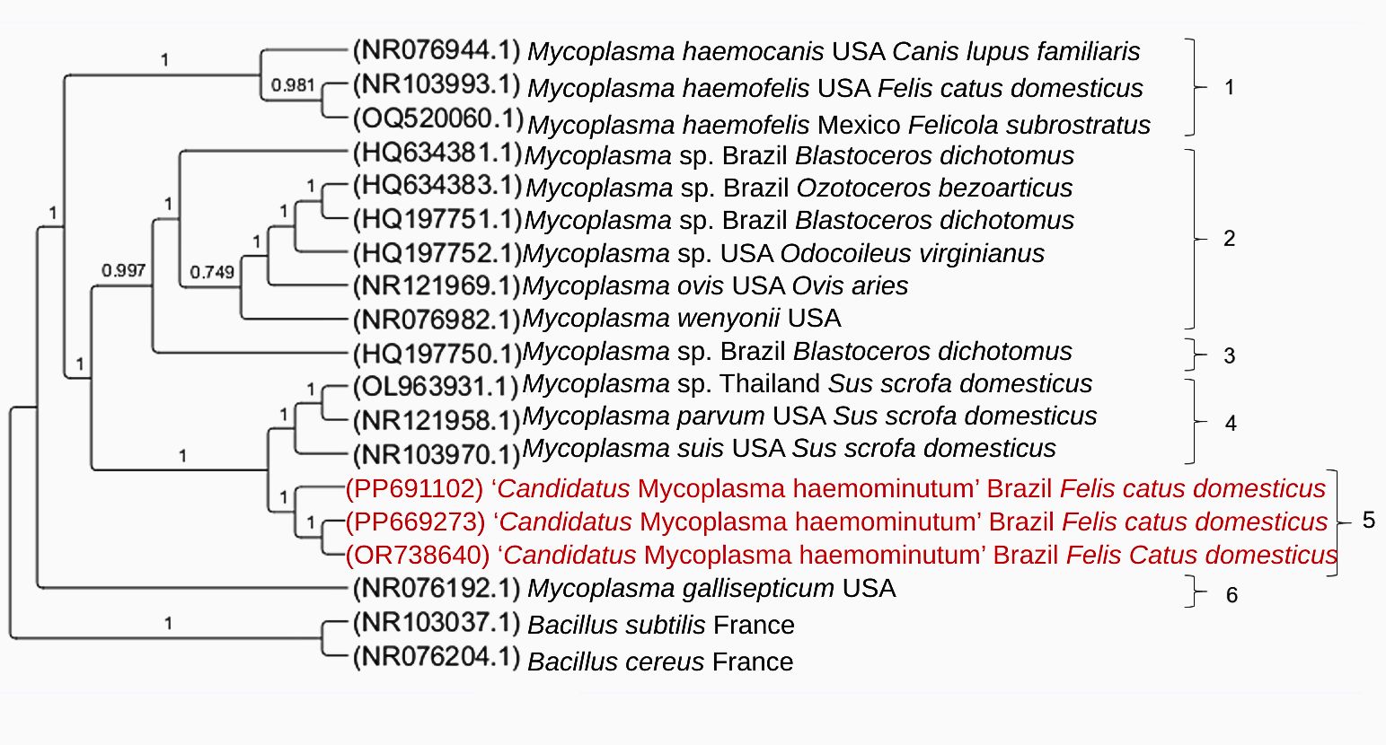
The Bayesian inference (GTR+I+G evolutionary model) of 21 homologous sequences of the 23S rRNA gene (759 bp alignment) showed the formation of two large distinct clades, which were subdivided into six smaller clades (Figure 3). The six subclades were numbered from one to six in the phylogenetic tree represented in Figure 3. In the subclade 5, the three sequences obtained in the present study (PP669273 from Minas Gerais, PP691102 from São Paulo, and OR738640 from Goiás) of ‘Candidatus Mycoplasma haemominutum’ from naturally infected domestic cats in Brazil were positioned together in a sister subclade to sequences of hemoplasma species detected in pigs (OL963931 [Mycoplasma sp], NR121958 [Mycoplasma parvum], and NR103970 [Mycoplasma suis]) from Thailand and the United States of America).

Phylogenetic tree based on an alignment of 759 bp length of Mycoplasma spp. 23S rRNA sequences, created using the Bayesian method and GTR+G as the evolutionary model. The sequences obtained in this study are highlighted in red. Bacillus cereus and Bacillus subtilis were used as an outgroup.
Genetic diversity analysis
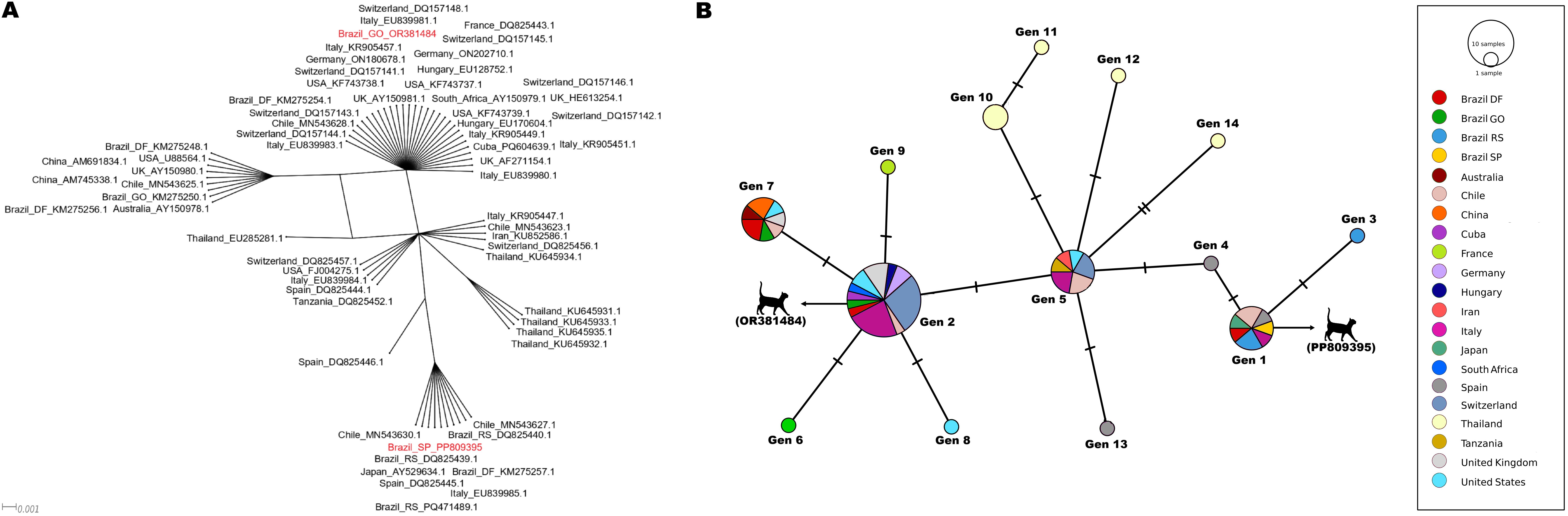
The SplitsTree distance-based analyses were constructed based on the same alignments used in the genetic diversity analyses for the 16S rRNA (469 bp) and 23S rRNA (671 bp) genes, containing the sequences detected in the present study and closely related ‘Candidatus Mycoplasma haemominutum’ sequences from GenBank (Figures 4A and 5A).

(A) ‘Candidatus Mycoplasma haemominutum’ split-network distance analysis of the 16S rRNA sequences. The analysis was performed applying the NeighborNet method based on a 469 bp alignment. The sequences obtained in the present study are highlighted in red; (B) Genotype network obtained from 16S rRNA sequences (469 bp) of 'Candidatus Mycoplasma haemominutum' detected in domestic cats, wild felids and a dog worldwide. The size of the circles varies according to the number of sequences comprising each genotype. Each color represents the location where each sequence was sampled. The black vertical lines represent mutational events between each genotype. The 16S rRNA genotypes obtained in the present study are represented by an illustration of a cat (
 ), followed by the accession number on GenBank.

(A) ‘Candidatus Mycoplasma haemominutum’ split-network distance analysis of the 23S rRNA sequences. The analysis was performed applying the NeighborNet method based on a 671 bp alignment. The sequences obtained in the present study are highlighted in red; (B) Genotype network obtained from 23S rRNA sequences (671 bp) of 'Candidatus Mycoplasma haemominutum' detected in domestic cats and humans worldwide. The size of the circles varies according to the number of sequences from each genotype. Each color represents the location where each sequence was sampled. The black vertical lines represent mutational events between each genotype. The 23S rRNA genotypes obtained in the present study are represented by an illustration of a cat (
), followed by the accession number on GenBank.
16S rRNA sequences detected in domestic cats from the states of GO and SP represented two different genotypes (Figure 4): while the sequence OR381484 (GO) corresponded to genotype #1, the sequence PP809395 (SP) corresponded to genotype #2. The number of sequences analyzed, variable sites number, G+C content, genotypes number, genotype diversity, standard deviation, nucleotide diversity (per site) and nucleotide differences number are shown in Table 3.
23S rRNA sequences detected in domestic cats from the states of GO, SP, and MG represented three different genotypes (Figure 5): while the sequence PP669273 (GO) corresponded to genotype #6, the sequence PP691102 (SP) corresponded to genotype #2, and OR738640 (MG) corresponded to genotype #1. The number of sequences analyzed, variable sites number, G+C content, genotypes number, genotype diversity, standard deviation, nucleotide diversity (per site) and nucleotide differences number are shown in Table 3.
Discussion
In the present study, molecular techniques were used to detect the occurrence of hemoplasmas in domestic cats from different veterinary clinics, autonomous animal protection shelters and castration campaigns in the states of Goiás, São Paulo, Minas Gerais and Rondônia. Among the 459 samples submitted to qPCR assays based on the 16S rRNA gene for detection of hemotropic mycoplasmas, 1.96% (9/459) were positive, making this the first study to describe the occurrence of such agents in the states of Rondônia and Minas Gerais, to the best of our knowledge.
The molecular occurrence of hemoplasmas varies between regions of Brazil. In the South, detection varies between 6.6 and 21.4% (Pedrassani et al., 2019; Santis et al., 2014), 6.5 to 32% in the Southeast (André et al., 2014; Bortoli et al., 2012; Raimundo et al., 2016), 8.4 to 36.4% in the Central-West (Miceli et al., 2013; Santis et al., 2014, Carvalho et al., 2024), 12 to 35.5% in the Northeast (Braga et al., 2012; Munhoz et al., 2018), and 44% in the North (Santos et al., 2021). Although the present study used qPCR to detect hemoplasmas, which is a more sensitive technique compared to PCR, the frequency of hemoplasma DNA detected was considered low, but similar to other studies carried out in Brazil. Miceli et al. (2013) detected 8.4% positivity for hemoplasmas in blood samples from naturally infected cats in the city of Cuiabá (MT), using conventional PCR assays. Likewise, Bortoli et al. (2012) and Pedrassani et al. (2019) reported frequencies corresponding to 6.5% and 6.6% in the states of São Paulo and Santa Catarina, respectively. The variation in the molecular occurrence of hemoplasmas in domestic cats between different regions of the country can be influenced by environmental and climatic conditions, veterinary care, characteristics of the sampled feline population (housed versus non-housed cats), immune status of the sampled animal, sample size, and sensitivity of the molecular assay used in the diagnosis of infection by such agents (Imre et al., 2020; Mesquita et al., 2021; Hoseinpoor et al., 2024).
Although there are reports of co-infections between two or three hemoplasma species, the present work detected only CMhm in nine (9/459) naturally infected cats (Tasker, 2022; Hoseinpoor et al., 2024). Furthermore, CMhm is the species most frequently detected in domestic cats in Brazil and other countries, corroborating the findings of this study (Santos et al., 2021; Munhoz et al., 2018; Santis et al., 2014; Imre et al., 2020; Mesquita et al., 2021; Hoseinpoor et al., 2024). Such observations are associated with CMhm’s high efficiency in infecting and multiplying in cats compared with Mhf and CMt (Tanahara et al., 2010; Hoseinpoor et al., 2024).
Although the 16S rRNA gene is highly conserved, bacterial species present considerable variations in this molecular marker, allowing differentiation between them. Based on this, the 16S rRNA gene has been extensively used in PCR assays to detect and differentiate hemoplasma species through different primer pairs, including those used in the present study (Martinez-Porchas et al., 2017; Citti et al., 2020). Although the Melting curve analysis of the positive samples in this study corresponded molecularly to CMt (Tm= 76.5 °C and 77.5 °C), the semi-nested PCR characterization assays based on the 16S rRNA gene and conventional PCR based on the 23S rRNA gene, followed by sequencing and BLASTn analysis, confirmed the molecular identity of CMhm, indicating a possible mistake in the Melting temperatures established in the protocol by Willi et al. (2009). These findings were corroborated with phylogenetic analyses based on both 16S rRNA and 23S rRNA, which positioned the sequences detected in this study with others from CMhm and separately from CMt sequences.
In the phylogenetic analysis, the 16S rRNA sequences obtained in this study (OR381484 and PP809395) were positioned within the clade of ‘Candidatus Mycoplasma haemominutum’ (CMhm), clustering with sequences from different geographic regions, such as Brazil, Switzerland, China, and the United States, supported by high statistical values. This result reinforces the low genetic diversity commonly reported for CMhm worldwide when using this molecular marker and suggests the existence of genotypes with a wide geographic distribution (Imre et al., 2020). In addition, the genotype network analysis demonstrated that PP809395 and OR381484 were represented by two distinct genotypes (Genotypes #1 and #2, respectively), distant from each other by a few mutational events and two genotypes (#4 and #5). The grouping of these genotypes with others from different countries may evidence limited global 16S rRNA gene flow of CMhm. Nonetheless, the presence of exclusive genotypes in Brazilian samples (genotypes #1 and #2) suggests local variations and possible regional adaptations, despite the overall low diversity observed. These findings might suggest recent population expansion or transmission of CMhm among domestic cats, characterized by low selective pressure and limited accumulated genetic diversity. Although CMhm is classically associated with subclinical infections in immunocompetent cats, the results of the present study are epidemiologically and clinically relevant, as this agent can cause hemolytic anemia, particularly in immunosuppressed animals or those co-infected with other pathogens, such as feline immunodeficiency virus (FIV), feline leukemia virus (FeLV) or other hemoplasma and hemoparasite species (Rosa Maciel et al., 2023).
A robust genetic diversity analysis of CMhm 23S rRNA was not feasible due to the limited number of sequences available in public databases. Another limitation of this study was the lack of information related to the clinical history of the sampled animals, especially if they had been recently treated with antibiotics capable of controlling hemoplasma infection, such as doxycycline and enrofloxacin. The inclusion of hematological and biochemical profiles, as well as the investigation of the association between hemoplasma infection and feline immunodeficiency virus (FIV) and feline leukemia virus (FeLV) infections should be included in future studies. Additionally, considering that sampling was performed by convenience, the real prevalence of these hemotropic agents should be cautiously interpreted.
Conclusion
This is the first molecular detection of hemoplasmas in cat blood samples in the states of Minas Gerais and Rondônia. Furthermore, the identification of two genotypes with wide global distribution circulating in Brazil contributes to a better understanding of the molecular epidemiology of feline hemoplasmosis agents in Brazil. The presence of exclusive genotypes in Brazilian samples suggests local variations and possible regional adaptations, despite the overall low diversity observed.
Data availability
Data will be made available on request.
Acknowledgements
This research was financed by FAPESP (Fundação de Amparo à Pesquisa do Estado de São Paulo – Processos #2022/06616-2, #2022/08543-2, #2022/16321-0, #2023/06168-2), CAPES (Coordenação de Aperfeiçoamento de Pessoal de Nível Superior), and CNPq (National Council for Scientific and Technological Development; Productivity Grant to MRA - Process #303701/2021-8).
-
How to cite:
Dias CM, Francisco KP, Oliveira GV, Silva JVSA, Franco EO, Lee DAB, et al. Molecular detection of hemoplasmas in domestic cats from different Brazilian regions. Braz J Vet Parasitol 2025; 34(3): e003925. https://doi.org/10.1590/S1984-29612025035
-
Ethics declaration
The procedures and management involving domestic cats were approved by the “Comissão de Ética no Uso de Animais” (CEUA) from FCAV/UNESP, under protocol numbers 012017/17 and 5439/20.
References
-
André MR, Calchi AC, Furquim MEC, de Andrade I, Arantes PVC, de Melo Lopes LC, et al. Molecular detection of tick-borne agents in cats from southeastern and northern Brazil. Pathogens 2022; 11(1): 106. http://doi.org/10.3390/pathogens11010106 PMid:35056054.
» http://doi.org/10.3390/pathogens11010106 -
André MR, Denardi NCB, Sousa KCM, Gonçalves LR, Henrique PC, Ontivero CRGR, et al. Arthropod-borne pathogens circulating in free-roaming domestic cats in a zoo environment in Brazil. Ticks Tick Borne Dis 2014; 5(5): 545-551. http://doi.org/10.1016/j.ttbdis.2014.03.011 PMid:24889035.
» http://doi.org/10.1016/j.ttbdis.2014.03.011 -
Attipa C, Papasouliotis K, Solano-Gallego L, Baneth G, Nachum-Biala Y, Sarvani E, et al. Prevalence study and risk factor analysis of selected bacterial, protozoal and viral, including vector-borne, pathogens in cats from Cyprus. Parasit Vectors 2017; 10(1): 130. http://doi.org/10.1186/s13071-017-2063-2 PMid:28285597.
» http://doi.org/10.1186/s13071-017-2063-2 -
Benson DA, Cavanaugh M, Clark K, Karsch-Mizrachi I, Ostell J, Pruitt KD, et al. GenBank. Nucleic Acids Res 2018; 46(D1): D41-D47. http://doi.org/10.1093/nar/gkx1094 PMid:29140468.
» http://doi.org/10.1093/nar/gkx1094 -
Bergmann M, Englert T, Stuetzer B, Hawley JR, Lappin MR, Hartmann K. Risk factors of different hemoplasma species infections in cats. BMC Vet Res 2016; 13(1): 52. http://doi.org/10.1186/s12917-017-0953-3 PMid:28202016.
» http://doi.org/10.1186/s12917-017-0953-3 -
Birkenheuer AJ, Levy MG, Breitschwerdt EB. Development and evaluation of a seminested PCR for detection and differentiation of Babesia gibsoni (Asian genotype) and B. canis DNA in canine blood samples. J Clin Microbiol 2003; 41(9): 4172-4177. http://doi.org/10.1128/JCM.41.9.4172-4177.2003 PMid:12958243.
» http://doi.org/10.1128/JCM.41.9.4172-4177.2003 -
Bortoli CP, André MR, Seki MC, Pinto AA, Machado STZ, Machado RZ. Detection of hemoplasma and Bartonella species and co-infection with retroviruses in cats subjected to a spaying/neutering program in Jaboticabal, SP, Brazil. Rev Bras Parasitol Vet 2012; 21(3): 219-223. http://doi.org/10.1590/S1984-29612012000300008 PMid:23070430.
» http://doi.org/10.1590/S1984-29612012000300008 -
Braga MSCO, André MR, Freschi CR, Teixeira MCA, Machado RZ. Molecular detection of hemoplasma infection among cats from São Luís island, Maranhão, Brazil. Braz J Microbiol 2012; 43(2): 569-575. http://doi.org/10.1590/S1517-83822012000200018 PMid:24031865.
» http://doi.org/10.1590/S1517-83822012000200018 -
Bryant D, Huson DH. NeighborNet: improved algorithms and implementation. Front Bioinform 2023; 3: 1178600. http://doi.org/10.3389/fbinf.2023.1178600 PMid:37799982.
» http://doi.org/10.3389/fbinf.2023.1178600 -
Bustin SA, Benes V, Garson JA, Hellemans J, Huggett J, Kubista M, et al. The MIQE guidelines: minimum information for publication of quantitative real-time PCR experiments. Clin Chem 2009; 55(4): 611-622. http://doi.org/10.1373/clinchem.2008.112797 PMid:19246619.
» http://doi.org/10.1373/clinchem.2008.112797 -
Carvalho SF, Pádua GT, Paula WVF, Tavares MA, Neves LC, Pereira BG, et al. Feline vector-borne diseases and their possible association with hematological abnormalities in cats from midwestern Brazil. Microorganisms 2024; 12(11): 2171. http://doi.org/10.3390/microorganisms12112171 PMid:39597560.
» http://doi.org/10.3390/microorganisms12112171 -
CIPRES [online]. 2025 [cited 2025 Mar 15]. Available from: https://www.phylo.org/
» https://www.phylo.org/ -
Citti C, Baranowski E, Dordet-Frisoni E, Faucher M, Nouvel LX. Genomic Islands in Mycoplasmas. Genes (Basel) 2020; 11(8): 836. http://doi.org/10.3390/genes11080836 PMid:32707922.
» http://doi.org/10.3390/genes11080836 - Clement M, Derington J, Posada D. TCS: estimating gene genealogies. Version 1.13 Provo, UT: Brigham Young University; 2001.
-
Darriba D, Taboada GL, Doallo R, Posada D. jModelTest 2: more models, new heuristics and parallel computing. Nat Methods 2012; 9(8): 772. http://doi.org/10.1038/nmeth.2109 PMid:22847109.
» http://doi.org/10.1038/nmeth.2109 -
Di Cataldo S, Kamani J, Cevidanes A, Msheliza EG, Millán J. Hemotropic mycoplasmas in bats captured near human settlements in Nigeria. Comp Immunol Microbiol Infect Dis 2020; 70: 101448. http://doi.org/10.1016/j.cimid.2020.101448 PMid:32109761.
» http://doi.org/10.1016/j.cimid.2020.101448 -
Díaz-Regañón D, Villaescusa A, Ayllón T, Rodríguez-Franco F, García-Sancho M, Agulla B, et al. Epidemiological study of hemotropic mycoplasmas (hemoplasmas) in cats from central Spain. Parasit Vectors 2018; 11(1): 140. http://doi.org/10.1186/s13071-018-2740-9 PMid:29554981.
» http://doi.org/10.1186/s13071-018-2740-9 -
Ewing B, Green P. Base-calling of automated sequencer traces using phred II. Error probabilities. Genome Res 1998; 8(3): 186-194. http://doi.org/10.1101/gr.8.3.186 PMid:9521922.
» http://doi.org/10.1101/gr.8.3.186 -
Ewing B, Hillier L, Wendl MC, Green P. Base-calling of automated sequencer traces using phred I. Accuracy assessment. Genome Res 1998; 8(3): 175-185. http://doi.org/10.1101/gr.8.3.175 PMid:9521921.
» http://doi.org/10.1101/gr.8.3.175 -
Furquim MEC, do Amaral R, Dias CM, Gonçalves LR, Perles L, Lima CAP, et al. Genetic diversity and Multilocus Sequence Typing Analysis of Bartonella henselae in domestic cats from Southeastern Brazil. Acta Trop 2021; 222: 106037. http://doi.org/10.1016/j.actatropica.2021.106037 PMid:34224716.
» http://doi.org/10.1016/j.actatropica.2021.106037 - Hall TA. BioEdit: a user-friendly biological sequence alignment editor and analysis program for Windows 95/98/NT. Nucleic Acids Symp Ser 1999; 41(1): 95-98.
-
Harasawa R, Fujita H, Kadosaka T, Ando S, Rikihisa Y. Proposal for ’Candidatus Mycoplasma haemomuris subsp. musculi’ in mice, and ’Candidatus Mycoplasma haemomuris subsp. ratti’ in rats. Int J Syst Evol Microbiol 2015; 65(Pt 2): 734-737. http://doi.org/10.1099/ijs.0.069856-0 PMid:25406232.
» http://doi.org/10.1099/ijs.0.069856-0 -
Hattori N, Kuroda M, Katano H, Takuma T, Ito T, Arai N, et al. Candidatus Mycoplasma haemohominis in human, Japan. Emerg Infect Dis 2020; 26(1): 11-19. http://doi.org/10.3201/eid2601.190983 PMid:31855136.
» http://doi.org/10.3201/eid2601.190983 -
Hoseinpoor E, Goudarztalejerdi A, Sazmand A. Molecular prevalence and phylogenetic analysis of hemotropic Mycoplasma species in cats in different regions of Iran. BMC Microbiol 2024; 24(1): 198. http://doi.org/10.1186/s12866-024-03356-8 PMid:38849724.
» http://doi.org/10.1186/s12866-024-03356-8 -
Huson DH, Bryant D. Application of phylogenetic networks in evolutionary studies. Mol Biol Evol 2006; 23(2): 254-267. http://doi.org/10.1093/molbev/msj030 PMid:16221896.
» http://doi.org/10.1093/molbev/msj030 -
Imre M, Văduva C, Dărăbuș G, Morariu S, Herman V, Plutzer J, et al. Molecular detection of hemotropic mycoplasmas (hemoplasmas) in domestic cats (Felis catus) in Romania. BMC Vet Res 2020; 16(1): 399. http://doi.org/10.1186/s12917-020-02626-7 PMid:33092590.
» http://doi.org/10.1186/s12917-020-02626-7 -
Kellner A, Carver S, Scorza V, McKee CD, Lappin M, Crooks KR, et al. Transmission pathways and spillover of an erythrocytic bacterial pathogen from domestic cats to wild felids. Ecol Evol 2018; 8(19): 9779-9792. http://doi.org/10.1002/ece3.4451 PMid:30386574.
» http://doi.org/10.1002/ece3.4451 -
Kmetiuk LB, Shaw P, Wallington A, Kattoor JJ, Johnson M, Wilkes RP, et al. Hemotropic mycoplasmas (hemoplasmas) in indigenous populations and their dogs living in reservation areas, Brazil. Sci Rep 2025; 15(1): 7973. http://doi.org/10.1038/s41598-025-87938-0 PMid:40055404.
» http://doi.org/10.1038/s41598-025-87938-0 -
Leigh JW, Bryant D. POPART: full-feature software for haplotype network construction. Methods Ecol Evol 2015; 6(9): 1110-1116. http://doi.org/10.1111/2041-210X.12410
» http://doi.org/10.1111/2041-210X.12410 -
Librado P, Rozas J. DnaSP v5: a software for comprehensive analysis of DNA polymorphism data. Bioinformatics 2009; 25(11): 1451-1452. http://doi.org/10.1093/bioinformatics/btp187 PMid:19346325.
» http://doi.org/10.1093/bioinformatics/btp187 -
Maggi RG, Compton SM, Trull CL, Mascarelli PE, Mozayeni BR, Breitschwerdt EB. Infection with hemotropic Mycoplasma species in patients with or without extensive arthropod or animal contact. J Clin Microbiol 2013; 51(1): 3237-3241. http://doi.org/10.1128/JCM.01125-13 PMid:23863574.
» http://doi.org/10.1128/JCM.01125-13 -
Manvell C, Ferris K, Maggi R, Breitschwerdt EB, Lashnits E. Prevalence of Vector-Borne Pathogens in Reproductive and Non-Reproductive Tissue Samples from Free-Roaming Domestic Cats in the South Atlantic USA. Pathogens 2021; 10(9): 1221. http://doi.org/10.3390/pathogens10091221 PMid:34578253.
» http://doi.org/10.3390/pathogens10091221 -
Martínez-Díaz VL, Silvestre-Ferreira AC, Vilhena H, Pastor J, Francino O, Altet L. Prevalence and co-infection of haemotropic mycoplasmas in Portuguese cats by real-time polymerase chain reaction. J Feline Med Surg 2013; 15(10): 879-885. http://doi.org/10.1177/1098612X13480985 PMid:23482254.
» http://doi.org/10.1177/1098612X13480985 -
Martinez-Porchas M, Villalpando-Canchola E, Ortiz Suarez LE, Vargas-Albores F. How conserved are the conserved 16S-rRNA regions? PeerJ 2017; 5: e3036. http://doi.org/10.7717/peerj.3036 PMid:28265511.
» http://doi.org/10.7717/peerj.3036 -
Mesquita JR, Oliveira AC, Neves F, Mendoza JR, Luz MF, Crespo I, et al. Hemotropic Mycoplasma and Bartonella Species Diversity in Free-Roaming Canine and Feline from Luanda, Angola. Pathogens 2021; 10(6): 735. http://doi.org/10.3390/pathogens10060735 PMid:34200906.
» http://doi.org/10.3390/pathogens10060735 -
Miceli NG, Gavioli FA, Gonçalves LR, André MR, Sousa VRF, Sousa KCM, et al. Molecular detection of feline arthropod-borne pathogens in cats in Cuiabá, state of Mato Grosso, central-western region of Brazil. Rev Bras Parasitol Vet 2013; 22(3): 385-390. http://doi.org/10.1590/S1984-29612013000300011 PMid:24142170.
» http://doi.org/10.1590/S1984-29612013000300011 -
Millán J, Travaini A, Cevidanes A, Sacristán I, Rodríguez A. Assessing the natural circulation of canine vector-borne pathogens in foxes, ticks and fleas in protected areas of Argentine Patagonia with negligible dog participation. Int J Parasitol Parasites Wildl 2019; 8: 63-70. http://doi.org/10.1016/j.ijppaw.2018.11.007 PMid:30622893.
» http://doi.org/10.1016/j.ijppaw.2018.11.007 -
Mongruel ACB, Spanhol VC, Valente JDM, Porto PP, Ogawa L, Otomura F, et al. Survey of vector-borne and nematode parasites involved in the etiology of anemic syndrome in sheep from Southern Brazil. Rev Bras Parasitol Vet 2020; 29(3): e007320. http://doi.org/10.1590/s1984-29612020062 PMid:32935770.
» http://doi.org/10.1590/s1984-29612020062 -
Moore CO, Lashnits E, Lappin M, Hawley J, Breitschwerdt EB. A case of mistaken identity: a systematic review, meta-analysis, and reinvestigation of hemotropic Mycoplasma spp. infection in Ctenocephalides felis (cat flea). Parasit Vectors 2024; 17(1): 209. http://doi.org/10.1186/s13071-024-06292-8 PMid:38720359.
» http://doi.org/10.1186/s13071-024-06292-8 -
Munhoz AD, Simões IGPC, Calazans APF, Macedo LS, Cruz RDS, Lacerda LC, et al. Hemotropic mycoplasmas in naturally infected cats in Northeastern Brazil. Rev Bras Parasitol Vet 2018; 27(4): 446-454. http://doi.org/10.1590/s1984-296120180074 PMid:30462823.
» http://doi.org/10.1590/s1984-296120180074 -
Pedrassani D, Biolchi J, Gonçalves LR, Mendes NS, Zanatto DCS, Calchi AC, et al. Molecular detection of vector-borne agents in cats in Southern Brazil. Rev Bras Parasitol Vet 2019; 28(4): 632-643. http://doi.org/10.1590/s1984-29612019077 PMid:31596318.
» http://doi.org/10.1590/s1984-29612019077 -
Raimundo JM, Guimarães A, Rodrigues RB, Botelho CFM, Peixoto MP, Pires MS, et al. Hematological changes associated with hemoplasma infection in cats in Rio de Janeiro, Brazil. Rev Bras Parasitol Vet 2016; 25(4): 441-449. http://doi.org/10.1590/s1984-29612016086 PMid:27982300.
» http://doi.org/10.1590/s1984-29612016086 -
Ravagnan S, Carli E, Piseddu E, Da Rold G, Porcellato E, Zanardello C, et al. Prevalence and molecular characterization of canine and feline hemotropic mycoplasmas (hemoplasmas) in northern Italy. Parasit Vectors 2017; 10(1): 132. http://doi.org/10.1186/s13071-017-2069-9 PMid:28285588.
» http://doi.org/10.1186/s13071-017-2069-9 -
Razin S, Yogev D, Naot Y. Molecular Biology and Pathogenicity of Mycoplasmas. Microbiol Mol Biol Rev 1998; 62(4): 1094-1156. http://doi.org/10.1128/MMBR.62.4.1094-1156.1998 PMid:9841667.
» http://doi.org/10.1128/MMBR.62.4.1094-1156.1998 -
Ronquist F, Huelsenbeck JP. MrBayes 3: bayesian phylogenetic inference under mixed models. Bioinformatics 2003; 19(12): 1572-1574. http://doi.org/10.1093/bioinformatics/btg180 PMid:12912839.
» http://doi.org/10.1093/bioinformatics/btg180 -
Rosa Maciel A, Biezus G, de Cristo TG, Miletti LC, Maciel UC, Medeiros ALV, et al. Mycoplasma haemofelis infection and its correlation with feline leukemia virus (FeLV) and feline immunodeficiency virus (FIV) in cats in Southern Brazil. Comp Immunol Microbiol Infect Dis 2023; 93: 101941. http://doi.org/10.1016/j.cimid.2022.101941 PMid:36630814.
» http://doi.org/10.1016/j.cimid.2022.101941 -
Sanger F, Nicklen S, Coulson AR. DNA sequencing with chain-terminating inhibitors. Proc Natl Acad Sci USA 1977; 74(12): 5463-5467. http://doi.org/10.1073/pnas.74.12.5463 PMid:271968.
» http://doi.org/10.1073/pnas.74.12.5463 -
Santis ACGA, Herrera HM, Sousa KCM, Gonçalves LR, Denardi NCB, Domingos IH, et al. Molecular detection of hemotrophic mycoplasmas among domiciled and free-roaming cats in Campo Grande, state of Mato Grosso do Sul, Brazil. Rev Bras Parasitol Vet 2014; 23(2): 231-236. http://doi.org/10.1590/S1984-29612014039 PMid:25054503.
» http://doi.org/10.1590/S1984-29612014039 -
Santos AP, Santos RP, Biondo AW, Dora JM, Goldani LZ, Oliveira ST, et al. Hemoplasma infection in HIV-positive patient, Brazil. Emerg Infect Dis 2008; 14(12): 1922-1924. http://doi.org/10.3201/eid1412.080964 PMid:19046522.
» http://doi.org/10.3201/eid1412.080964 -
Santos AP, Messick JB, Biondo AW, Oliveira ST, Pedralli V, Lasta SC, et al. Design, optimization, and application of a conventional PCR assay with an internal control for detection of ‘Candidatus Mycoplasma turicensis’ 16S rDNA in domestic cats from Brazil. Vet Clin Pathol 2009; 38(4): 443-452. http://doi.org/10.1111/j.1939-165X.2009.00158.x PMid:19548972.
» http://doi.org/10.1111/j.1939-165X.2009.00158.x -
Santos CMP, Barra ECM, Almeida JCF, Jesus IS, Cruz AV, Costa JC, et al. Occurrence of hemotropic mycoplasmas in domestic cats (Felis catus) from Belém, Brazil. Int J Dev Res 2021; 11(3): 44949-44952. http://doi.org/10.37118/ijdr.21256.03.2021
» http://doi.org/10.37118/ijdr.21256.03.2021 -
Silaghi C, Knaus M, Rapti D, Kusi I, Shukullari E, Hamel D, et al. Survey of Toxoplasma gondii and Neospora caninum, haemotropic mycoplasmas and other arthropod-borne pathogens in cats from Albania. Parasit Vectors 2014; 7(1): 62. http://doi.org/10.1186/1756-3305-7-62 PMid:24517118.
» http://doi.org/10.1186/1756-3305-7-62 -
Steer JA, Tasker S, Barker EN, Jensen J, Mitchell J, Stocki T, et al. A novel hemotropic Mycoplasma (hemoplasma) in a patient with hemolytic anemia and pyrexia. Clin Infect Dis 2011; 53(11): e147-e151. http://doi.org/10.1093/cid/cir666 PMid:22021921.
» http://doi.org/10.1093/cid/cir666 - Swofford DL. PAUP: Phylogenetic Analysis Using Parsimony (and othermethods). Version 4.10b Sunderland: Sinauer Associates, 2003.
-
Sykes JE, Lindsay LL, Maggi RG, Breitschwerdt EB. Human coinfection with Bartonella henselae and two hemotropic mycoplasma variants resembling Mycoplasma ovis. J Clin Microbiol 2010; 48(10): 3782-3785. http://doi.org/10.1128/JCM.01029-10 PMid:20702675.
» http://doi.org/10.1128/JCM.01029-10 -
Tanahara M, Miyamoto S, Nishio T, Yoshii Y, Sakuma M, Sakata Y, et al. An epidemiological survey of feline hemoplasma infection in Japan. J Vet Med Sci 2010; 72(12): 1575-1581. http://doi.org/10.1292/jvms.10-0143 PMid:20686353.
» http://doi.org/10.1292/jvms.10-0143 -
Tasker S. Hemotropic Mycoplasma Vet Clin North Am Small Anim Pract 2022; 52(6): 1319-1340. http://doi.org/10.1016/j.cvsm.2022.06.010 PMid:36336423.
» http://doi.org/10.1016/j.cvsm.2022.06.010 -
Willi B, Boretti FS, Cattori V, Tasker S, Meli ML, Reush C, et al. Identification, molecular characterization and experimental transmission of a new hemoplasma isolate from a cat with hemolytic anemia in Switzerland. J Clin Microbiol 2005; 43(6): 2581-2585. http://doi.org/10.1128/JCM.43.6.2581-2585.2005 PMid:15956367.
» http://doi.org/10.1128/JCM.43.6.2581-2585.2005 -
Willi B, Tasker S, Boretti FS, Doherr MG, Cattori V, Meli ML, et al. Phylogenetic analysis of ‘Candidatus Mycoplasma turicensis’ isolates from pet cats in the United Kingdom, Australia, and South Africa, with analysis of risk factors for infection. J Clin Microbiol 2006; 44(12): 4430-4435. http://doi.org/10.1128/JCM.00987-06 PMid:17035497.
» http://doi.org/10.1128/JCM.00987-06 -
Willi B, Meli ML, Luthy R, Honegger H, Wengi N, Hoelzle LE, et al. Development and application of a universal hemoplasma screening assay based on the SYBR green PCR principle. J Clin Microbiol 2009; 47(12): 4049-4054. http://doi.org/10.1128/JCM.01478-09 PMid:19828748.
» http://doi.org/10.1128/JCM.01478-09 -
Yuan CL, Liang AB, Yao CB, Yang ZB, Zhu JG, Cui L, et al. Prevalence of Mycoplasma suis (Eperythrozoon suis) infection in swine and swine-farm workers in Shanghai, China. Am J Vet Res 2009; 70(7): 890-894. http://doi.org/10.2460/ajvr.70.7.890 PMid:19566474.
» http://doi.org/10.2460/ajvr.70.7.890 -
Zhang Y, Zhang Z, Lou Y, Yu Y. Prevalence of hemoplasmas and Bartonella species in client-owned cats in Beijing and Shanghai, China. J Vet Med Sci 2021; 83(5): 793-797. http://doi.org/10.1292/jvms.20-0681 PMid:33731498.
» http://doi.org/10.1292/jvms.20-0681
Edited by
-
Assistant Scientific Editor:
Maristela Peckle Peixoto