Ocular Whipple's disease: a very rare clinical presentation
ABSTRACT
Whipple's disease is a rare multisystemic condition caused by Tropheryma whipplei. In its early stages, it typically presents with joint symptoms, progressing in later stages primarily to digestive disorders. Although ocular involvement is uncommon, it may be one of the first manifestations of the disease, presenting a diagnostic challenge. Polymerase chain reaction testing or periodic acid Schiff staining of affected tissues can assist in early diagnosis when symptoms are non-specific, which is crucial for initiating early antibiotic therapy and thus modifying the progressive course of the disease, preventing irreversible ocular damage.
Keywords:
Whipple's disease, Tropheryma; Periodic acid-Schiff reaction; Ceftriaxone; Trimethoprim; Sulfamethoxazole
RESUMO
A doença de Whipple é uma doença multissistêmica rara causada por Tropheryma whipplei. Nos estágios iniciais, ela geralmente se manifesta com sintomas articulares, levando, em estágios posteriores, principalmente a distúrbios digestivos, embora também possa afetar o sistema ocular, o cérebro, os pulmões e/ou o coração. Testes de diagnóstico, como reação em cadeia da polimerase ou coloração ácido periódico de Schiff nos tecidos afetados, podem nos ajudar a fazer um diagnóstico precoce nos estágios iniciais com sintomas muito inespecíficos, o que é crucial para estabelecer uma antibioterapia precoce e, assim, modificar o curso progressivo da doença.
Descritores:
Doença de Whipple; Tropheryma; Reação do ácido periódico de Schiff; Ceftriaxona; Trimetoprima; Sulfametoxazol
INTRODUCTION
Whipple's disease (WD) is a multisystemic infectious disease first described in 1907 and caused by Tropheryma whipplei, a Gram-positive bacterium commonly found in sewage and faecal matter.(1,2) Worldwide, WD is a rare disorder, with an incidence of one to three cases per 1.000.000 people, associated with the human leukocyte antigen (HLA) B27 haplotype. It is much more common in males and the development of symptoms usually occurs around the fifth decade of life. Its pathogenesis is unclear, but it has been shown that in diseased individuals there is impaired macrophage function with an abnormal T-cell response.(3)
In the course of WD, during the initial or prodromal phases, non-specific symptoms such as fever, arthralgia, and migratory arthritis appear. Subsequently, in the classic or advanced phase, gastrointestinal symptoms predominate, typical of a malabsorption process after involvement mainly of the small intestine. In the late or disseminated phase, other organs may be affected, leading to ocular (occurring in less than 5% of WD cases), central nervous system, pulmonary and cardiac manifestations. Other signs such as lymphadenopathy, hyperpigmentation, anaemia, splenomegaly and ascites can be found at any phase of WD.(4,5)
Due to the rarity of WD, associated with its heterogeneous pattern of presentation, hence known as "the great simulator", its identification is a diagnostic challenge. T. whipplei is detected by PCR testing of biological fluids or biopsied tissues, or by specific histological techniques such as PAS and haematoxylin-eosin.(6)
Its chronic and progressive nature inexorably leads to death if antibiotic therapy is not initiated in time, which is based on broad-spectrum antibiotics that penetrate the blood-brain barrier such as ceftriaxone, penicillin G and trimethoprim-sulfamethoxazole. Unfortunately, relapses can occur years after treatment, and evaluation of body fluids is important to ensure proper cure, which can take one to two years.(3)
CASE REPORT
A 47-year-old woman with a biopsy-confirmed diagnosis of lymph node sarcoidosis in 2018, presented to the rheumatology department, after two years without attending her follow-up appointments, reporting fever, dyspnea, arthromyalgia, constitutional syndrome and decreased visual acuity (VA) for 6 months. Ophthalmological examination revealed VA of 20/32 in the right eye and 20/50 in the left eye, as well as a normal anterior segment examination. A posterior segment examination of both eyes (OU) revealed vitritis with diffuse perivascular infiltrates, multiple retinal haemorrhages, Roths’ spots and discrete vascular dilatation and tortuosity (Figure 1). In view of these findings, fluorescein angiography (FFA) was performed, showing occlusive vasculitis with areas of peripheral ischaemia of both eyes (Figure 2).
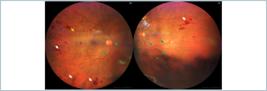
Fundus examination (both eyes): deep retinal haemorrhages (white arrows), perivascular infiltrates (yellow arrows), Roths’ spots (red arrows), dilatation and venous tortuosity (green arrows).
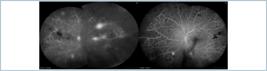
Fluorescein angiography (both eyes) showing large areas of peripheral ischaemia, extravasation, retinal neovessels and vasculitis.
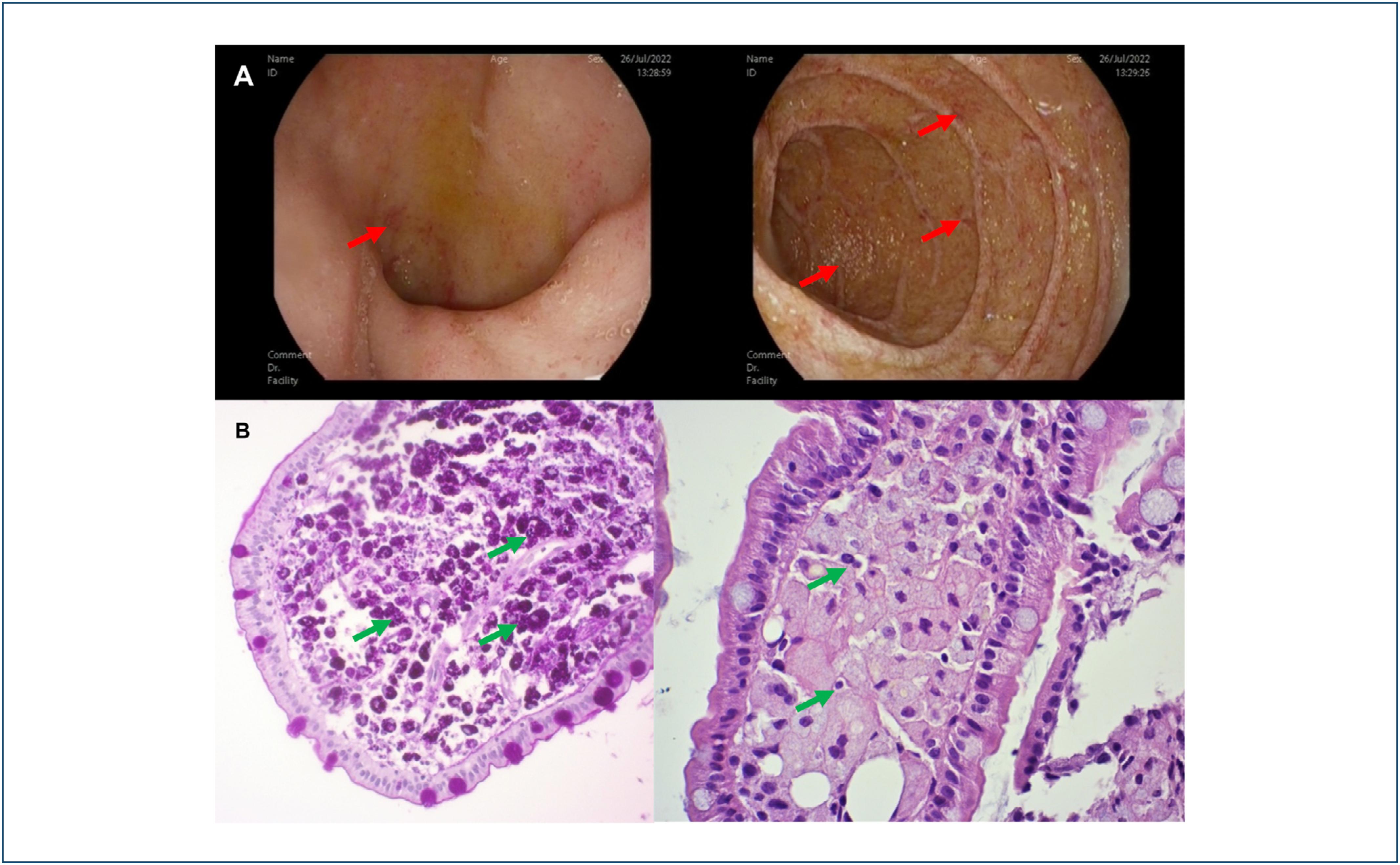
Simultaneously, the study of autoantibodies and serologies (human immunodeficiency virus, syphilis, toxoplasmosis, Mycobacterium tuberculosis, antinuclear antibodies, extractable nuclear antibodies, angiotensin-converting enzyme (ACE), rheumatoid factor, anti-transglutaminase and anti-endomysium antibodies) was negative, so a computed tomography (CT) scan was performed. This revealed splenomegaly, polyadenopathies, alteration of pulmonary vessels, which eventually resulted in severe pulmonary hypertension, and thickening of duodenal folds (Figure 3). A complete gastrointestinal endoscopic study was also performed, showing signs of non-erosive duodenitis. Duodenal biopsies were obtained and polymerase chain reaction (PCR) and periodic acid Schiff (PAS) staining were performed, with confirmatory results of T. whipplei infection (Figure 4).

Thoracoabdominal computerized tomography with multiple lymphadenopathies (red arrows), as well as splenomegaly (yellow arrow).

(A) Gastroscopy showing a hyperemic, oedematous and friable mucosa with whitish "rice-grain" stippling in the bulb and duodenum (red arrows). (B) Histological section of duodenum with periodic acid Schiff-positive foamy macrophages in lamina propria (green arrows).
Because of worsening ocular symptoms, aqueous humour and vitreous humour samples were taken for bacteriological, viral and cytological analysis with CD4/CD8 ratio to rule out a lymphoproliferative syndrome or ocular involvement as part of the WD. The results were inconclusive, so it was decided not to delay treatment and to start ceftriaxone 2 g once daily intravenously for four weeks. At the start of antibiotherapy, due to persistent intense vitritis, OU underwent pars plana vitrectomy (PPV) with endolaser, injection of intravitreal antiangiogenics and a new vitreous humour sampling for analysis, which this time yielded negative results for both T. whipplei and type B non-Hodgkin's lymphoma.
The patient is currently on oral maintenance treatment with trimethoprim 160 mg and sulfamethoxazole 800 mg twice a day until the completion of the year of treatment, with a clear improvement in systemic symptoms, but a guarded visual prognosis because of residual retinal atrophy and ischaemia. There have been periods of ocular reactivation of the lesions in the form of cystic macular oedema with optimal response to treatment with intravitreal dexamethasone implants.
DISCUSSION
The rarity of WD and the heterogeneous patterns of presentation lead, as described in the literature, to an erroneous diagnosis in up to 89% of patients, often with a presumptive diagnosis of sarcoidosis, as in our case, due to clinical similarities. This misdiagnosis frequently results in the administration of immunosuppressive drugs, such as corticosteroids, which suppress the immune response, worsen the symptoms and accelerate the progression of WD (7,8) Additionally, there are unusual presentations of the disease, such as in our case, with ophthalmological symptoms, which are usually preceded by gastrointestinal and/or neurological manifestations and can give rise to bilateral panuveitis, multifocal choroiditis, retinal vasculitis, myositis and optic neuropathy.(9)
The first-line antibiotic treatment initiated after the diagnosis of WD significantly improved systemic symptoms. However, unlike findings described in other studies, there was no ocular improvement due to the consequences of severe inflammation and established aggression.(6,10) Despite the negative result of the vitreous samples, possibly biased by the antibiotic therapy prior to collection, we can conclude that it was a disseminated WD due to the positive histopathological findings obtained from the duodenal biopsies.(6)
In summary, this case illustrates the need to include WD in the differential diagnosis of bilateral chronic uveitis, since its suspicion is crucial to initiate the search, just as a multidisciplinary approach is key, and the need to completely exclude the infectious etiology before considering immunosuppressive treatment.(10)
REFERENCES
- 1 Moreno García MS, Casorrán Berges M, del Río-Martínez PS, Bosque Peralta MT. Enfermedad de Whipple, la gran desconocida. Reumatol Clin. 2017;13(4):243-4.
- 2 Fernández Vecilla D, Ugalde Zárraga E, Tarín Nieto A, Díaz de Tuesta Del Arco JL. Enfermedad de Whipple con presentación múltiple en paciente reumatológico. Caso y revisión de la literatura [Whipple's disease with multiple presentation in a rheumatologic patient]. Rev Esp Quimioter. 2023;36(2):203-207. Spanish.
-
3 Antunes C, Singhal M. Whipple Disease [Updated 2023 Jul 4]. In: StatPearls. StatPearls Publishing; Treasure Island (FL): 2023 [cited 2025 Feb 5]. Available from: https://www.ncbi.nlm.nih.gov/books/NBK441937/
» https://www.ncbi.nlm.nih.gov/books/NBK441937/ - 4 Kucharz EJ, Kramza J, Grosicka A, Pieczyrak R. Clinical manifestations of Whipple's disease mimicking rheumatic disorders. Reumatologia. 2021;59(2):104-10.
-
5 Gomezjurado MG, Gomezjurado AG. Fisiopatología, diagnóstico y tratamiento de la enfermedad de whipple. Medwave; 2011 [Updated 2023 Nov 28]. Available from: https://www.medwave.cl/revisiones/revisionclinica/5112.html
» https://www.medwave.cl/revisiones/revisionclinica/5112.html - 6 Boumaza A, Ben Azzouz E, Arrindell J, Lepidi H, Mezouar S, Desnues B. Whipple's disease and Tropheryma whipplei infections: from bench to bedside. Lancet Infect Dis. 2022;22(10):e280-e291.
- 7 Meunier M, Puechal X, Hoppé E, Soubrier M, Dieudé P, Berthelot JM, et al. Rheumatic and musculoskeletal features of Whipple disease: a report of 29 cases. J Rheumatol. 2013;40(12):2061-6.
- 8 Kc P, Ayele FK, Karki S, Waleed MS. Whipple's disease mimicking sarcoidosis. Cureus. 2023;15(7):e41839.
- 9 Saint-Martin G, Urbanski G, Beucher AB, Ebran JM. An ophthalmologic complication of Whipple's disease: Case report. J Fr Ophtalmol. 2018;41(8):e387-e389.
- 10 Chan RY, Yannuzzi LA, Foster CS. Ocular Whipple's disease: earlier definitive diagnosis. Ophthalmology. 2001;108(12):2225-31.