Presence of retinal ischemic perivascular lesions prior to an ischemic stroke event
ABSTRACT
An 81-year-old male patient with systemic hypertension, diabetes mellitus, and dyslipidemia presented with sudden visual acuity loss in the right eye (RE) due to a branch retinal artery occlusion (BRAO) associated with macular edema. Despite treatment with intravitreal bevacizumab, the patient developed persistent inner retinal ischemia, evidenced by optical coherence tomography (OCT) findings of retinal inner plexiform layer (RIPL) alterations. Years later, he suffered an ischemic stroke, highlighting the association between retinal ischemic changes and systemic vascular risk. This case underscores the role of RIPLs as potential early markers for cerebrovascular and cardiovascular events, emphasizing the need for multidisciplinary management and vigilant cardiovascular monitoring in patients with ischemic retinal findings. The identification of RIPLs through noninvasive imaging may offer a valuable opportunity for early intervention, although further studies are necessary to establish their routine clinical use as predictive biomarkers.
Keywords:
Ischemic stroke; Diabetes mellitus; Dyslipidemias; Retinal artery occlusion; Biomarkers
RESUMO
Paciente do sexo masculino, de 81 anos, com hipertensão arterial sistêmica, diabetes mellitus e dislipidemia apresentou perda súbita da acuidade visual no olho direito devido à oclusão de um ramo da artéria retiniana (OAR) associada a edema macular. Apesar do tratamento com bevacizumabe intravítreo, o paciente desenvolveu isquemia retiniana interna persistente, evidenciada por achados de tomografia de coerência óptica (OCT) de alterações na camada plexiforme interna da retina (CIPIR). Anos depois, ele sofreu um acidente vascular cerebral isquêmico, destacando a associação entre alterações isquêmicas da retina e risco vascular sistêmico. Este caso ressalta o papel das camadas plexiformes internas da retina como potenciais marcadores precoces de eventos cerebrovasculares e cardiovasculares, enfatizando a necessidade de tratamento multidisciplinar e monitoramento cardiovascular vigilante em pacientes com achados retinianos isquêmicos. A identificação de camada plexiforme interna da retina por meio de exames de imagem não invasivos pode oferecer uma oportunidade valiosa para intervenção precoce, embora mais estudos sejam necessários para estabelecer seu uso clínico rotineiro como biomarcadores preditivos.
Descritores:
AVC isquêmico; Diabetes mellitus; Dislipidemia; Oclusão da artéria retiniana; Biomarcadores
INTRODUCTION
Branch retinal artery occlusion (BRAO) represents an ophthalmic emergency associated with significant visual morbidity and a close relationship with systemic vascular diseases. In particular, the detection of alterations in the inner plexiform layers of the retina, recently described as paracentral acute middle maculopathy (PAMM) or ischemic injury of the inner retina (CIPIR), has emerged as a marker of retinal microvascular ischemia and a potential indicator of cerebrovascular risk. These findings, obtained through optical coherence tomography (OCT), reinforce the interconnection between retinal and cerebral circulation, highlighting the importance of an integrated approach between ophthalmology and internal medicine. This report describes the case of an elderly patient with multiple cardiovascular risk factors who presented with BRAO associated with CIPIR and subsequently developed an ischemic stroke, illustrating the relevance of ischemic retinal changes as predictors of systemic events.(1–3)
CASE REPORT
An 81-year-old male patient with a medical history of systemic arterial hypertension, diabetes mellitus, and dyslipidemia sought ophthalmological care on April 12, 2018, presenting with sudden visual acuity loss in the right eye (RE). Examination revealed a visual acuity of 0.05 (logMAR 1.3) in the RE and 0.40 (logMAR 0.4) in the left eye (LE). Fundoscopy showed branch occlusion of the superior temporal retinal artery in the RE, accompanied by macular edema. Additionally, an epiretinal membrane was identified in the LE, along with nuclear cataracts graded 2+/4+ in both eyes during slit-lamp examination.
One month later, optical coherence tomography (OCT) confirmed persistent cystoid macular edema in the RE. The patient was treated with intravitreal injections of bevacizumab, resulting in partial resolution of intraretinal macular edema after three treatment cycles. Visual acuity improved to 0.63 (logMAR 0.2), although the patient retained an irreversible inferior quadrant hemianopia in the RE as a sequela.
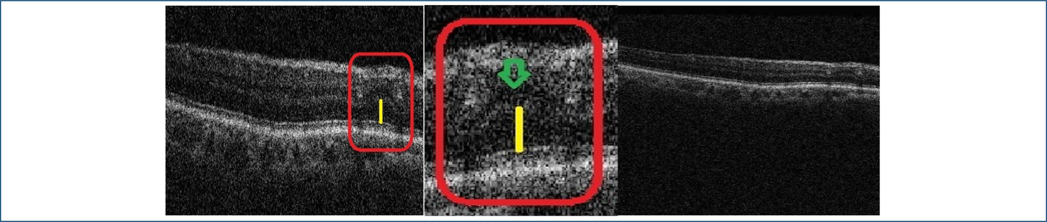
In 2019, the patient developed rhegmatogenous retinal detachment in the LE, requiring pars plana vitrectomy (PPV) with silicone oil implantation. During postoperative follow-up of the LE, the patient continued monitoring for chronic macular edema secondary to vascular occlusion in the RE. OCT imaging showed areas of inner retinal thinning and increased spacing in the outer nuclear layer, suggesting inner retinal ischemia. No new retinal occlusion episodes were observed after the initial treatment (Figure 1).

Optical coherence tomography imaging showed areas of inner retinal thinning and increased spacing in the outer nuclear layer, suggesting inner retinal ischemia.
On August 18, 2022, the patient was admitted to the emergency department with a decline in general condition and was diagnosed with an ischemic stroke.
This study was approved by the Research Ethics Committee Central Air Force Hospital CAAE: 77387924.3.0000.5250
DISCUSSION
Branch retinal artery occlusion (BRAO) is a vascular event commonly linked to systemic risk factors such as hypertension, diabetes, and dyslipidemia. Evidence suggests that ischemic events in the retina may act as predictors of an elevated risk for cerebrovascular complications.
In this context, the retinal inner plexiform layer (RIPL) biomarker identified in this case reflects ischemic injury to the inner retinal layer. Retinal ischemic perivascular lesions (RIPLs) are characterized by focal atrophy in the inner nuclear layer (INL), along with secondary expansion of the outer nuclear layer, giving the middle retinal layers an undulating appearance. These lesions are considered sequelae of paracentral acute middle maculopathy (PAMM), a condition thought to result from the inner nuclear layer's increased vulnerability to ischemic damage, especially at the level of the deep capillary plexus. Retinal hypoperfusion, which can occur due to reduced blood flow to the retinal capillary plexus, is commonly associated with conditions such as low ejection fraction, carotid artery stenosis, thrombus or embolus formation, and vascular stasis—all of which have been linked to PAMM. While PAMM has not yet been conclusively shown to increase cardiovascular risk, RIPLs appear to represent end-organ infarction due to vascular dysfunction and are associated with cardiovascular disease. As such, the eye may be one of the earliest organs to reveal signs of ischemia caused by systemic conditions, particularly in the middle retina, which is highly sensitive to even subtle changes in blood flow.(1)
This association underscores the critical role of identifying RIPLs in evaluating systemic vascular health, as they may serve as an early warning sign of broader vascular compromise. RIPLs are permanent, while hyperreflective PAMM lesions are transient and typically resolve within six weeks. However, they leave behind lasting changes in the inner nuclear layer, which can persist as residual damage.
Recent research suggests that retinal microinfarcts are strongly correlated with generalized microvascular dysfunction, often indicative of underlying atherosclerosis and systemic inflammation.(2)
The relationship between retinal vascular events and stroke risk has been further supported by studies like the Beaver Dam Eye Study. This research found that individuals with a history of retinal vascular occlusions have up to a twofold increased risk of stroke.(3) This case emphasizes the critical importance of continuous cardiovascular monitoring in patients with ischemic retinal changes, particularly those with classic cardiovascular risk factors.
Furthermore, the RIPL biomarker is increasingly recognized as a potential predictor of underdiagnosed systemic vascular diseases, such as atrial fibrillation.(4) It may also serve as an early indicator of severe vascular events, such as ischemic stroke or acute myocardial infarction (AMI). These findings underscore the need for rigorous clinical follow-up and vigilance for systemic vascular conditions in patients with ischemic retinal changes.(5)
Given that spectral-domain optical coherence tomography (SD-OCT) is a noninvasive and widely accessible imaging technique, the detection of retinal ischemic perivascular lesions (RIPLs) could serve as a crucial entry point for managing cardiovascular health. Identifying RIPLs may help pinpoint individuals who are at higher risk and who could benefit from further diagnostic evaluation and early therapeutic interventions.
The occurrence of retinal vascular occlusion, combined with the emergence of RIPLs, underscores their significance as potential early markers of systemic vascular issues and an elevated likelihood of cerebrovascular incidents. This case emphasizes the importance of a multidisciplinary approach involving ophthalmologists, neurologists, and cardiologists. Such collaboration enables a thorough assessment and facilitates the adoption of preventive measures, effectively minimizing the risk of serious complications.
Ongoing surveillance and proactive care are crucial for identifying early risks and implementing timely interventions that can greatly reduce the chances of negative outcomes. Leveraging RIPLs as a predictive marker allows healthcare professionals to customize strategies for cardiovascular health, leading to improved patient results. Nevertheless, additional studies and validation are required before RIPLs can be reliably integrated into routine clinical practice for screening and guiding decisions on further diagnostics and treatments.
Data Availability Statement:
The datasets generated and/or analyzed during the current study are included in the manuscript.
REFERENCES
- 1 Moura-Coelho N, Gaspar T, Ferreira JT, Dutra-Medeiros M, Cunha JP. Paracentral acute middle maculopathy-review of the literature. Graefes Arch Clin Exp Ophthalmol. 2020;258(12):2583-96.
- 2 Colcombe J, Mundae R, Kaiser A, Bijon J, Modi Y. Retinal findings and cardiovascular risk: prognostic conditions, novel biomarkers, and emerging image analysis techniques. J Pers Med. 2023;13(11):1564.
- 3 Klein R, Klein BE, Jensen SC, Moss SE, Meuer SM. Retinal emboli and stroke: the Beaver Dam Eye Study. Arch Ophthalmol. 1999 Aug;117(8):1063-8.
- 4 Bakhoum CY, Madala S, Lando L, Yarmohammadi A, Long CP, Miguez S, et al. Retinal ischemic perivascular lesions in individuals with atrial fibrillation. J Am Heart Assoc. 2023;12(16):e028853.
- 5 Drakopoulos M, Zhang DL, Cheng BT, Sadiq SA, Nadel A, Marchese A, et al. Swept-source optical coherence tomography angiography metrics of retinal ischaemic perivascular lesions in patients being evaluated for carotid artery stenosis and controls. BMJ Open Ophthalmol. 2023;8(1):e001226.
Edited by
-
Associate Editor:
Ricardo Augusto Paletta Guedes Universidade Federal de Juiz de Fora, Juiz de Fora, MG, Brazil https://orcid.org/0000-0002-9451-738X