Unilateral optic neuritis associated with MOG antibodies in a pediatric patient
ABSTRACT
Case report of a 14-year-old male presented with acute painless vision loss and left optic disc swelling. Extensive diagnostic investigations, including magnetic resonance imaging and serum MOG antibody testing, were conducted. Magnetic resonance imaging showed an extensive T2 hyperintense signal in the anterior portion of the left optic nerve without involving chiasm or retrochiasmatic areas. The management strategy involved intravenous methylprednisolone followed by azathioprine, resulting in complete vision recovery. This case highlights the importance of considering MOG antibody-associated optic neuritis in the differential diagnosis of acute vision loss in adolescents and discusses the clinical and imaging features that should prompt testing for anti-MOG antibodies.
Keywords:
Blindness; Vision loss; Methylprednisolone; Azathioprine; Adolescent
RESUMO
Relato de caso de paciente do sexo masculino de 14 anos que apresentou perda de visão aguda e indolor e inchaço do disco óptico esquerdo. Extensas investigações diagnósticas, incluindo ressonância magnética e testes séricos de anticorpos anti- MOG, foram realizadas. À ressonância magnética, evidenciou-se hipersinal em T2, extenso, na porção anterior do nervo óptico esquerdo, sem envolver quiasma, ou regiões retroquiasmáticas. A estratégia de manejo envolveu metilprednisolona intravenosa seguida de azatioprina, resultando em recuperação completa da visão. Este caso destaca a importância de considerar a neurite óptica associada a anticorpos anti-MOG no diagnóstico diferencial de perda aguda de visão em adolescentes e discute as características clínicas e de imagem que devem levar ao teste de anticorpos anti-MOG.
Descritores:
Cegueira; Neurite óptica; Metilprednisolona; Azatioprina; Adolescente
INTRODUCTION
Optic neuritis in pediatric patients can present with sudden, painless vision loss, posing a diagnostic challenge due to its broad differential diagnosis. Myelin oligodendrocyte glycoprotein (MOG) antibody-associated disease has emerged as an important consideration in such cases. MOG antibodies are associated with a spectrum of demyelinating diseases, and their presence necessitates distinct therapeutic strategies compared to other causes of optic neuritis, such as multiple sclerosis.(1) Recognizing the clinical and imaging characteristics that suggest MOG antibody involvement is crucial for timely diagnosis and management.
MOG antibody disease (MOGAD) is a rare autoimmune condition that affects the central nervous system, particularly the myelin. MOG antibody disease is characterized by recurrent inflammatory episodes, including optic neuritis, transverse myelitis, and encephalomyelitis.(2) Unlike multiple sclerosis (MS), another demyelinating disease, MOGAD presents a distinct clinical profile, with a better prognosis in terms of recovery after relapses, although patients may experience multiple recurrences over time. Additionally, the lesions observed on magnetic resonance imaging (MRI) can differ significantly, with a greater prevalence of central and hemispheric lesions in MOGAD, and in the pattern of optic nerve involvement.(3)
The diagnosis of MOGAD depends on the detection of specific antibodies against MOG (anti-MOG). These antibodies target MOG, a protein expressed on the surface of myelin cells, and their presence is an essential diagnostic marker.(4) The identification of anti-MOG antibodies in the patient's serum is crucial to distinguish MOGAD from other demyelinating diseases, such as MS and neuromyelitis optica (NMO), which have different treatments and prognoses.(5) The most commonly used assay technique for detecting these antibodies is cell-based immunofluorescence assay, which offers high sensitivity and specificity.
The management of MOGAD involves the acute treatment of inflammatory relapses, often with intravenous corticosteroids, and long-term strategies to prevent recurrences, which may include immunosuppressants such as azathioprine, mycophenolate mofetil, or rituximab. Although the response to treatment is generally positive, the recurrent nature of the disease requires continuous and personalized follow-up.(6) Early identification and appropriate management are key to minimizing associated morbidity, improving patient's quality of life. Ongoing research on MOGAD and the role of anti-MOG antibodies may provide further insights into pathogenic mechanisms and new therapeutic approaches.
REPORT CASE
A 14-year-old male with no noteworthy medical history was urgently admitted to the Emergency Department due to the sudden occurrence of painless vision loss in his left eye. Upon physical examination, the ophthalmologist noted significant swelling of the left optic disc. The rest of the neurological evaluation revealed no further abnormalities. Given the acute presentation and clinical findings, an immediate brain MRI and serum testing for MOG antibodies were ordered. The serum MOG antibody test, performed via flow cytometry (FACS), returned positive, confirming the suspicion of an autoimmune etiology. The MRI imaging provided further diagnostic clarity (Figure 1). Ethics Committee approved this study, and all subjects signed a consent form to participate (Research Ethical Committee Number: CAAE: 80164224.4.0000.5373.
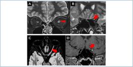
Magnetic resonance imaging of the orbits reveals increased signal intensity in left optic nerve on coronal T2-weighted images in intraorbital (A) and canalicular (B) segments (arrows). The axial T2 FLAIR-weighted image shows full extension of the nerve involvement (C) (arrow). Coronal T1-weighted image depicts the peripheric enhancement around the left optic nerve (arrow) (D).
The investigation was extensive and covered all the differential diagnosis. A brain, cervical, and dorsal spine MRI was performed, which showed no alterations. Regarding the serologies, anti-AQUAPORIN 4 in cerebrospinal fluid, oligoclonal bands, immunoglobin G (IgG), and polymerase chain reaction (PCR) tests were performed. Anti-MOG was performed by FACS with a titer of 1:40 (reference value titer: lower than 1:20). MOG-IgG has an overall specificity of 98%, but MOG-IgG with titers of 1:20 or 1:40 is considered weakly positive according to the 2023 diagnostic criteria for MOGAD and has a positive predictive value of approximately 50% for MOGAD.
Extensive rheumatological investigation was performed, including FAN assessment (screening for Hep-2 cell antibodies), anti-DSDNA antibodies, rheumatoid factor, anti-SS-A/RO antibodies, anti-SS-B/LA antibodies, anti-RNP/SM antibodies, anti-SCL-70 antibodies, anti-JO-1 antibodies, anti-chromatin antibodies, and anti-CPP antibodies). All results were within normal and non-reactive ranges.
Based on these findings, an aggressive treatment regimen was initiated. The acute phase of treatment consisted of high-dose intravenous methylprednisolone for 5 days. Following this, maintenance therapy with azathioprine was started to manage the underlying autoimmune response. The patient exhibited a remarkable recovery, with complete restoration of vision in the affected eye. Regular follow-ups ensured the stability of his condition, with no relapses reported during the maintenance therapy.
This case underscores the importance of rapid assessment and intervention in cases of acute vision loss associated with autoimmune optic neuritis, and it highlights the efficacy of immediate high-dose corticosteroid treatment followed by long-term immunosuppression for sustaining remission.
This case highlights unilateral optic neuritis with MOG antibody positivity, distinguishing it from other demyelinating diseases such as MS and aquaporin-4-positive NMO. The presentation of unilateral vision loss, longitudinal extensive optic neuritis sparing the chiasma, perineural enhancement, and disc edema were crucial in guiding the diagnosis. Early diagnosis and treatment are critical, as they can lead to excellent outcomes. MOG antibody-associated optic neuritis should be considered in adolescents presenting with sudden and painless vision loss. This case underscores the efficacy of timely steroid administration and the importance of maintenance therapy to prevent relapses.
DISCUSSION
MOG antibody-associated optic neuritis (MOG-ON) is increasingly recognized as a distinct clinical entity that can present with features overlapping with other demyelinating conditions such as MS and NMO spectrum disorder (NMOSD). However, there are key clinical and imaging features that should prompt the consideration of MOG antibody testing.(1)
Clinically, MOG-ON often presents with more severe optic disc swelling compared to MS-related optic neuritis, and the visual loss can be profound. Additionally, MOG-ON frequently presents as a unilateral process, though bilateral involvement can occur. Importantly, patients with MOG-ON tend to have a good visual prognosis if treated promptly with corticosteroids, but the risk of relapse is significant if long-term immunosuppression is not maintained.(2)
Imaging studies, particularly MRI, play a critical role in the differentiation of MOG-ON from other causes of optic neuritis. Typical MRI findings include longitudinally extensive optic nerve involvement, extending more than half the length of the optic nerve, and perineural enhancement, which are less commonly seen in MS-related optic neuritis. Additionally, the optic chiasm is usually spared, and there is often no involvement of other brain regions typically affected in MS.(3) Brain MRI is considered normal in 2/3 of cases or demonstrates nonspecific supratentorial subcortical or small, deep, white matter foci of hyperintensity on T2-weighted sequences, and these lesions are generally asymptomatic. Large lesions with blurred and hazy margins, referred to as ADEM-like lesions, predominantly in the supratentorial white matter, the basal ganglia, the thalamus, or the brainstem, were seen in almost one-third of MOG-IgG-positive patients.(4)
Given these distinctive features, MOG antibody testing should be strongly considered in the following scenarios: pediatric patients with unilateral optic neuritis and severe disc swelling; cases of optic neuritis with MRI findings of longitudinally extensive optic nerve involvement and perineural enhancement; patients who present with recurrent optic neuritis, especially if the clinical and imaging findings do not align with a typical MS diagnosis; situations in which the visual loss is more severe than expected for typical MS-associated optic neuritis.(5)
By recognizing these clinical and imaging characteristics, ophthalmologists can better identify patients who may benefit from anti-MOG antibody testing, leading to more tailored treatment strategies and improved patient outcomes.(6)
To sum up, this case underscores the importance of considering MOG antibody-associated optic neuritis in the differential diagnosis of acute vision loss in pediatric patients. Early treatment with high-dose corticosteroids, followed by immunosuppressive therapy, can lead to complete recovery, highlighting the need for prompt and accurate diagnosis. Recognizing the specific clinical and imaging features that indicate MOG antibody involvement is crucial for appropriate testing and management.
REFERENCES
- 1 Chen JJ, Flanagan EP, Jitprapaikulsan J, López-Chiriboga AS, Fryer JP, Leavitt JA, et al. Myelin oligodendrocyte glycoprotein antibody-positive optic neuritis: clinical characteristics, radiologic clues, and outcome. Am J Ophthalmol. 2018; 195:8-15.
- 2 Jurynczyk M, Messina S, Woodhall MR, Waters P, Kulakowska A, Chandratre S, et al. Clinical presentation and prognosis in mog-antibody disease: a european case series. Mult Scler. 2017;23(1):82-6.
- 3 Ramanathan S, Prelog K, Barnes EH, Tantsis EM, Reddel SW, Henderson AP, et al. Radiological differentiation of optic neuritis with myelin oligodendrocyte glycoprotein antibodies, aquaporin-4 antibodies, and multiple sclerosis. Mult Scler. 2016;22(4):470-82.
- 4 Narayan R, Simpson A, Fritsche K, Salama S, Pardo S, Mealy M, et al. MOG Antibody disease: a review of MOG Ant-IgG, its role in demyelinating diseases, and how mog antibody testing is changing practice. Curr Neurol Neurosci Rep. 2018;18(12):104.
- 5 Reindl M, Waters P. Reindl M, Waters P. Myelin oligodendrocyte glycoprotein antibodies in neurological disease. Nat Rev Neurol. 2019;15(2):89-102.
- 6 Pereira FV, Jarry VM, Castro JT, Appenzeller S, Reis F. Pediatric inflammatory demyelinating disorders and mimickers: how to differentiate with MRI? Autoimmun Rev. 2021;20(5):102801.