Pars plana vitrectomy and laser photocoagulation of vasoproliferative tumors: a new surgical approach to sight-threatening complications
ABSTRACT
Retinal vasoproliferative tumor is a rare disease associated with several sight-threatening complications. The optimum management of retinal vasoproliferative tumor is unknown and the need of surgery arises when there is diagnostic uncertainty or sight-threatening complications, such as 23G pars plana vitrectomy, perfluorooctane retinal flattening, fluid drainage through a peripheral retinotomy, fluid-air exchange, laser photocoagulation of the retinal vasoproliferative tumor, and 12.5% C3F8 tamponade. We described two cases of retinal vasoproliferative tumor with exudative retinal detachment threatening the macula, treated with the technique described above. Both eyes had complete tumor resolution, vanishing of lipid exudation, and remained stable in over three years of follow-up. Exudative retinal detachment can be a sight threatening complication of retinal vasoproliferative tumor. A surgical approach that does not involve the risks of tumor resection can provide successful treatment of the disease.
Keywords:
Vitrectomy; Retinal neoplasms; Retinal neovascularization; Rare diseases; Retinal detachment; Light coagulation
RESUMO
O tumor vasoproliferativo da retina é uma doença rara associada a várias complicações que ameaçam a visão. O tratamento ideal do tumor vasoproliferativo da retina é desconhecido, e a necessidade de cirurgia surge quando há incerteza diagnóstica ou complicações que ameaçam a visão, como vitrectomia pars plana 23G, achatamento da retina com perfluorooctano, drenagem de fluido por meio de uma retinotomia periférica, troca fluido-ar, fotocoagulação a laser do tumor vasoproliferativo da retina e tamponamento com C3F8 a 12,5%. Descrevemos dois casos de tumor vasoproliferativo da retina com descolamento exsudativo da retina ameaçando a mácula, tratados com a técnica descrita acima. Ambos os olhos apresentaram resolução completa do tumor, desaparecimento da exsudação lipídica e permaneceram estáveis em mais de três anos de acompanhamento. O descolamento de retina exsudativo pode ser uma complicação do tumor vasoproliferativo da retina que ameaça a visão. Uma abordagem cirúrgica que não envolva os riscos da ressecção tumoral pode proporcionar um tratamento bem-sucedido da doença.
Descritores:
Vitrectomia; Neoplasias da retina; Neovascularização retiniana; Doenças raras; Descolamento retiniano; Fotocoagulação
INTRODUCTION
Retinal vasoproliferative tumor (RVPT) is a rare disease considered to be of reactive nature, whose etiology is yet to be understood. It can be idiopathic or secondary to other ocular diseases such as uveitis, retinitis pigmentosa, previous surgery, and retinopathy of prematurity.(1,2)
Retinal vasoproliferative tumor affects patients of both genders and ages, although women are predominant, especially after the fifth decade.(2)
Vasoproliferative tumors are associated with several sight-threatening complications, most commonly cystoid macular edema, vitreous hemorrhage, macular epiretinal membranes, and progressive subretinal exudation threatening the macular region.(3) The optimum management of vasoproliferative tumors is unknown due to their rarity and uncertainty regarding their underlying pathobiology.(3,4) In the majority of cases, observation alone is required.(3) Medical management includes laser photocoagulation or photodynamic therapy for smaller tumors and anti-VEGF for associated macular edema.
The need for surgery arises when there is diagnostic uncertainty or sight-threatening complications are present.(3–5)
We described two cases of retinal vasoproliferative tumor with exudative retinal detachment threatening the macula.
CASE REPORT
A standard 3-port, 23-G vitrectomy (Constellation® System, Alcon, Geneva, Switzerland) with scleral depression-assisted was performed to ensure that all vitreous adhesions overlying the tumor were dissected. Perfluorooctane was injected and fluid was passively drained from a small peripheral retinotomy during a fluid-air exchange. The tumor and its immediate surroundings were treated directly with an endolaser photocoagulation until a complete whitish appearance was observed. Finally, surgery was completed with 12.5% C3F8 tamponade and postoperative face-down positioning for 1 day.
Case 1
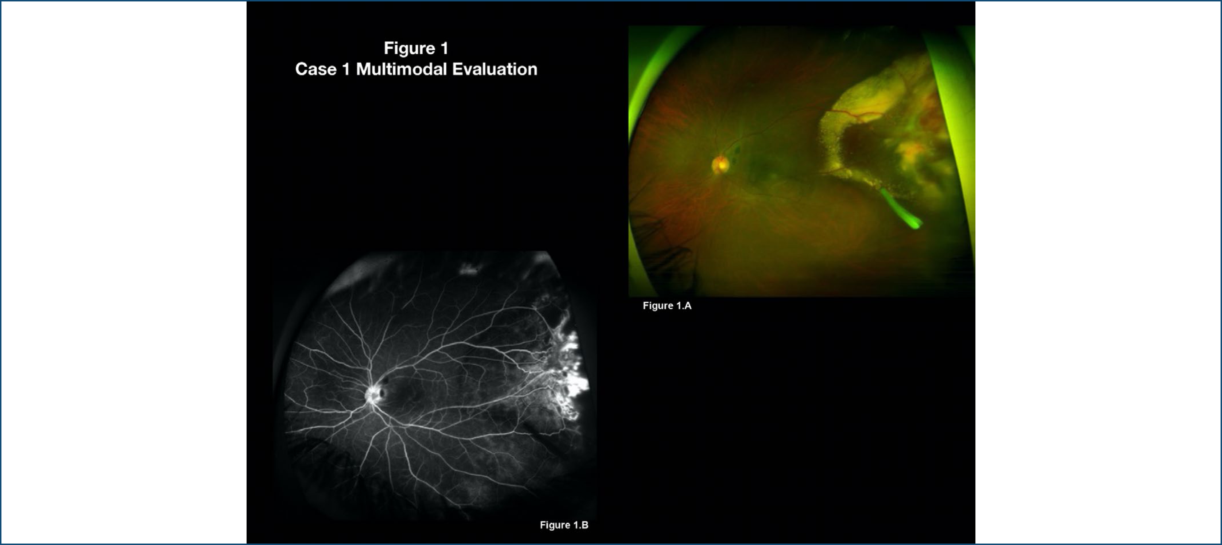
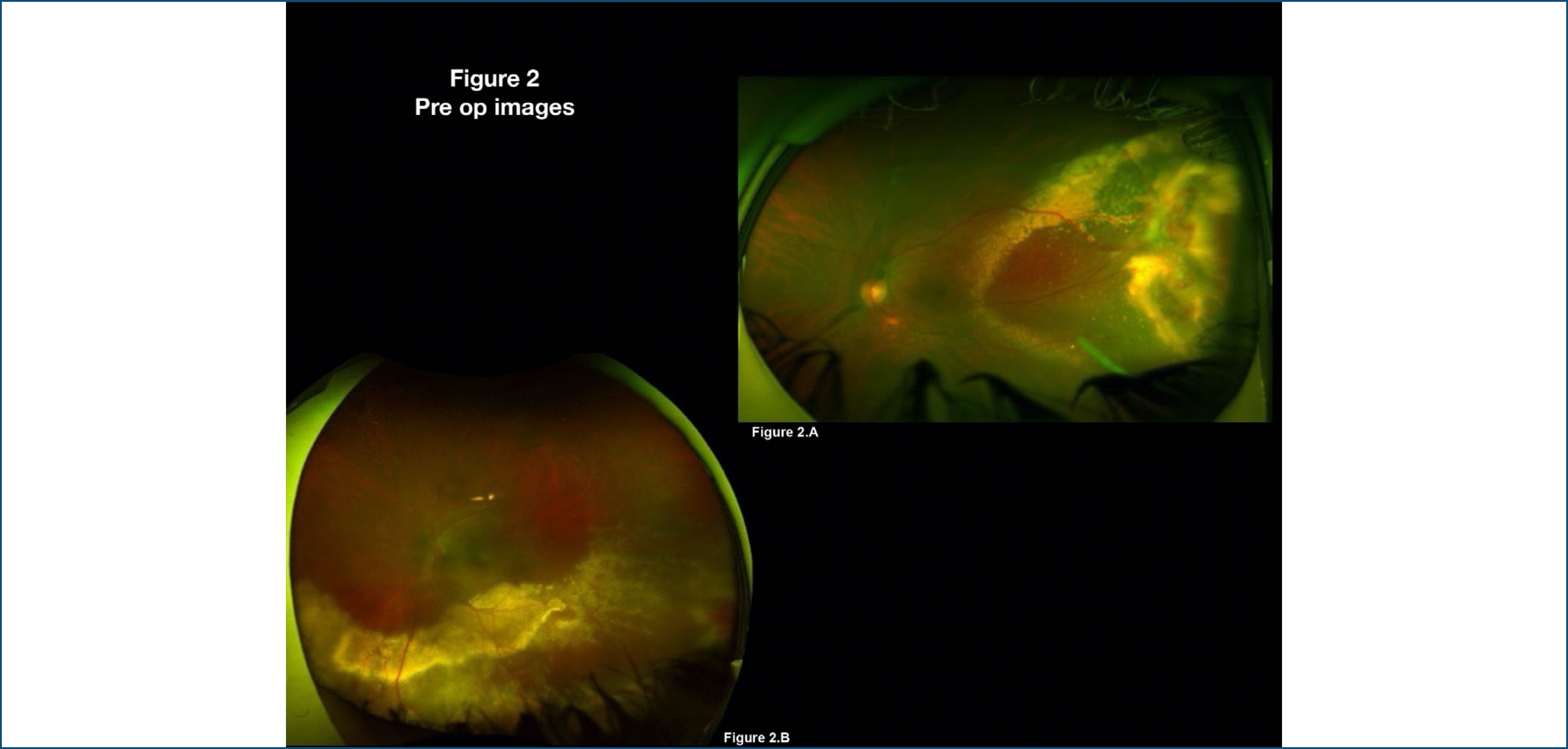
A 54-year-old Caucasian female was referred with a decreased vision in her left eye (OS) for 3 months. She had a previous history of retinal vasculitis due to suspected Behcet's disease. Visual acuity was 20/25 in the right eye (OD) and 20/50 in the OS. Intraocular pressure and anterior segment of both eyes (OU) were normal. Posterior segment was normal in OD, except for the presence of asteroid hyalosis, whereas OS revealed an ill-defined reddish yellow intraretinal mass in the temporal periphery with subretinal fluid and significant lipid exudation, along the temporal quadrant (Figures 1A and 1B). The patient had a dexamethasone implant injection 2 months before. Two months later, subretinal fluid increased and reached the posterior pole, threatening the macula (Figure 2A).

Case 1 multimodal evaluation. (A) Fundus widefield image of the left eye reveals a retinal vasoproliferative tumor in the temporal periphery with subretinal fluid and extensive lipid exudation. Dexamethasone implant is observed in the vitreous cavity. (B) Widefield fluorescein angiography of the left eye shows hyperfluorescence of the lesion and late leakage, consistent with retinal vasoproliferative tumor.
Pre op images. (A) Fundus widefield image of the left eye shows significant increase of the fluid towards the macula. (B) Fundus widefield image of the left eye (Case 2) partially shows an inferotemporal retinal vasoproliferative tumor associated with exudative retinal detachment and lipid exudation.
Case 2
A 29-year-old Caucasian female, was referred with the diagnosis of idiopathic intermediate uveitis for 12 years, having undergone treatment with corticosteroids, cataract surgery in OU and Ahmed Glaucoma Valve implant in OD. Visual acuity was 20/25 in OU. Fundus examination revealed an ill-defined reddish yellow intraretinal mass in the temporal inferior quadrant with significant subretinal fluid and lipid exudation over the inferotemporal vascular arcade, almost reaching the macular region (Figure 2B).
In both cases, the retinas remained fully attached. Retinal exudates progressively faded until disappeared, and only a laser photocoagulation pigmented scar could be seen in the previous RVPT area. The first patient developed a cataract, which was successfully treated. Both eyes had final 20/25 vision after over three years of follow-up.
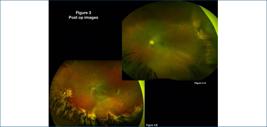
Post op images. (A) Fundus widefield image of the left eye (Case 1) shows that the retina is fully attached and pigmented laser photocoagulation scars are present at the previous site of the tumor and its surroundings. (B) Fundus widefield image of the left eye (Case 2) reveals an attached retina with pigmented laser photocoagulation scars in the inferior periphery. A moderate depigmentation of the inferotemporal quadrant is observed, consistent with previous chronic subretinal fluid.
DISCUSSION
Shields et al. described a series of 129 vasoproliferative tumors in 113 eyes of 103 patients and their ophthalmic disease associations, clinical features, and treatment modalities.(1) These tumors were found to develop in association with congenital, inflammatory, vascular, infectious, and traumatic ocular conditions. Compared with primary RVPTs, secondary tumors are more often bilateral (20%), multifocal (15%), and larger (mean, 7 mm), occurring at an earlier age and with poorer visual prognosis.(6,7)
Tumors < 2 mm could be treated with laser photocoagulation or cryotherapy.(8) Larger tumors with extensive associated subretinal fluid are less likely to absorb sufficient laser for adequate treatment.(9) Beyond that, cryotherapy causes significant chorioretinal disruption and atrophy, with reports of scleral thinning and potential concurrent increase in vascular endothelial growth factor (VEGF) production and vasoproliferative drive in adjacent tissue.(10) It may also increase the risk of further exudative changes and lead to complete exudative retinal detachment.
Photodynamic therapy has also been reported as an effective treatment in retinal and choroidal vascular tumors.(11,12) Its major limitation is the technical difficulties in reaching the typical peripheral location of RVPTs.(13)
There are reports of successful brachytherapy for RVPTs using either ruthenium106 or iode 125, reaching total tumor remission in most cases.(1,14, 15) It is also important to remember the risk of developing some adverse effects of eye irradiation: dry eyes, cataracts, actinic optic neuropathy, actinic retinopathy, and neovascular glaucoma.
The role of VEGF in the pathogenesis of RVPT is unclear. However, an upregulation of VEGF in the tissues surrounding such lesions has been shown positively after intravitreal Anti-VEGF in case reports. The effect seems short-lived with recurrence of exudation on cessation of treatment.
Despite all these therapeutic options, affected eyes lose vision owing to hemorrhagic and exudative effects and the formation of epiretinal membranes and cystoid macular edema in 50 to 60% of patients.(10) For this reason, the surgical approach could present itself as a definitive treatment for the tumor and its main vision-threatening complications, with good anatomic and functional results.(3,8)
The ideal treatment is yet to be established since most of what we have known is based on isolated case reports or small case series. We described two cases of successful pars plana vitrectomy and endolaser treatment of RVPT complicated by exudative retinal detachment threatening the central vision. The resolution of the subretinal exudates after 3 years of follow-up can be an indicator of tumor inactivity. The goal of laser photocoagulation of the tumor could be achieved with lesser risk than its resection.
REFERENCES
- 1 Shields CL, Shields JA, Barrett J, De Potter P. Vasoproliferative tumors of the ocular fundus. Classification and clinical manifestations in 103 patients. Arch Ophthalmol. 1995;113(5):615-23.
- 2 Marback EF, Guerra RL, Maia Junior OO, Marback RL. Retinal vasoproliferative tumor. Arq Bras Oftalmol. 2013;76(3):200-3.
- 3 Smith J, Steel D. The surgical management of vasoproliferative tumours. Ophthalmologica. 2011;226 Suppl 1:42-5.
- 4 Irvine F, O’Donnell N, Kemp E, Lee WR. Retinal vasoproliferative tumors: surgical management and histological findings. Arch Ophthalmol. 2000;118(4):563-9.
- 5 Castro-Navarro V, Saktanasate J, Say EA, Chiang A, Shields CL. Role of pars plana vitrectomy and membrane peel in vitreomacular traction associated with retinal vasoproliferative tumors. Oman J Ophthalmol. 2016;9(3):167-9.
- 6 Manjandavida FP, Shields CL, Kaliki S, Shields JA. Cryotherapy-induced release of epiretinal membrane associated with retinal vasoproliferative tumor: analysis of 16 cases. Retina. 2014;34(8):1644-50.
- 7 Zheng B, Chen Y, Chen L, Chen H, Zheng J, Chen F, et al. Comparative Study on the Efficacy and Safety of Tumor Resection in Vitrectomy for Retinal Vasoproliferative Tumors. J Ophthalmol. 2019;2019:7464123.
- 8 Krivosic V, Massin P, Desjardins L, Le Hoang P, Tadayoni R, Gaudric A. Management of idiopathic retinal vasoproliferative tumors by slit-lamp laser or endolaser photocoagulation. Am J Ophthalmol. 2014;158(1):154-61.e1.
- 9 Hussain RN, Jmor F, Damato B, Heimann H. Verteporfin photodynamic therapy for the treatment of retinal vasoproliferative tumors. Ophthalmology. 2015;122(11):2361-3.
- 10 Rodriguez-Coleman H, Spaide RF, Yannuzzi LA. Treatment of angiomatous lesions of the retina with photodynamic therapy. Retina. 2002;22(2):228-32.
- 11 Cohen VM, Shields CL, Demirci H, Shields JA. Iodine I 125 plaque radiotherapy for vasoproliferative tumors of the retina in 30 eyes. Arch Ophthalmol. 2008;126(9):1245-51.
- 12 Brockmann C, Rehak M, Heufelder J, Cordini D, Brockmann T, Corkhill C, et al. Predictors of treatment response of vasoproliferative retinal tumors to Ruthenium-106 brachytherapy. Retina. 2016;36(12):2384-90.
- 13 Saito W, Kase S, Fujiya A, Dong Z, Noda K, Ishida S. Expression of vascular endothelial growth factor and intravitreal anti-VEGF therapy with bevacizumab in vasoproliferative retinal tumors. Retina. 2013;33(9):1959-67.
- 14 Gibran SK. Trans-vitreal endoresection for vasoproliferative retinal tumours. Clin Exp Ophthalmol. 2008;36(8):712-6.
- 15 Yeh S, Wilson DJ. Pars plana vitrectomy and endoresection of a retinal vasoproliferative tumor. Arch Ophthalmol. 2010;128(9):1196-9.