
 Long-term results of intrastromal ring implantation for keratoconus
ABSTRACT
Objective: To evaluate refractive and topographic outcomes before and at least 7 years after intrastromal ring segment implantation surgery in patients with keratoconus.
Methods: Long-term retrospective longitudinal study carried out with 35 patients (62 eyes) aged 10 to 30 who underwent intrastromal ring segment implantation using a manual technique. Different arc rings were analyzed across various keratoconus types.
Results: Significant improvements in topographic astigmatism (p = 0.001) and corrected distance visual acuity (p < 0.001) were observed, with no significant changes in keratometry (p = 0.099) and a slight decrease in pachymetry (p = 0.033). Statistically significant improvements in K1 were noted with two 160° arc rings. K2 showed improvements with all arc rings except for one 210° ring. Topographic astigmatism improved notably with two 160° arc rings. No significant changes occurred between six months after surgery and final visits.
Conclusion: Long-term follow-up demonstrated favorable outcomes post-intrastromal ring segment implantation, particularly in manifest refraction and corrected distance visual acuity. Vision gain was common, though endothelial cell counts decreased, especially with two-ring segments. Thicker rings reduced astigmatism more significantly, whereas two-ring segments reduced keratometry. Notably, 210° arc rings did not reduce topographic astigmatism but did decrease refractive astigmatism.
Keywords:
Keratoconus; Intrastromal ring; Corneal surgery; Visual acuity; Endothelial cells
RESUMO
Objetivo: Avaliar os resultados refrativos e topográficos antes e após no mínimo 7 anos de cirurgia de implante de segmento de anel intraestromal em pacientes com diferentes tipos de ceratocone.
Métodos: Estudo longitudinal retrospectivo de longo prazo de pacientes submetidos ao implante de segmento de anel intraestromal com técnica manual. Participaram do estudo 35 indivíduos com idades entre 10 e 30 anos. A maioria dos pacientes foi operada em ambos os olhos, totalizando 62 olhos. Analisamos diferentes anéis de arco em diferentes tipos de ceratocone.
Resultados: Foi encontrada diminuição do astigmatismo topográfico (p = 0,001) e da contagem de células na microscopia especular (p < 0,001). Também encontramos melhora na acuidade visual corrigida (p < 0,001). Por outro lado, não houve diferença na ceratometria (p = 0,099) e na paquimetria (p = 0,033). Tivemos melhora estatisticamente significativa no K1 com dois anéis de arco 160°. Em relação ao K2, tivemos melhora em todas as variáveis exceto com um anel de 210°. Quando analisamos o astigmatismo topográfico, só tivemos melhora com dois anéis de 160° arcos. Não houve alterações estatisticamente significativas entre o sexto mês de pós-operatório e a consulta final.
Conclusão: Encontramos bons resultados, mesmo no seguimento de longo prazo, com melhora de quase todas as variáveis, principalmente refração manifesta e acuidade visual corrigida. Também encontramos ganho de linhas de visão na maioria dos casos e redução da contagem de células endoteliais, principalmente com a implantação de dois arcos. Além disso, encontramos redução mais significativa do astigmatismo com anéis mais grossos e maior redução ceratométrica com arcos segmentados de dois anéis. Finalmente, com 210 anéis de arco, não encontramos redução do astigmatismo topográfico, mas houve diminuição do astigmatismo refracional.
Descritores:
Ceratocone; Anel intraestromal; Cirurgia da córnea; Acuidade visual; Células endoteliais
INTRODUCTION
Keratoconus is a multifactorial and progressive disease that leads to an increase in corneal curvature and consequent worsening of visual quality. Its progression is most remarkable in the first decades of life and decreases over the years due to the natural stiffening of the cornea.(1)
The main treatments for keratoconus range from glasses and contact lenses to corneal intrastromal ring segment (ICRS) placement, crosslinking (CXL), and lamellar or penetrating keratoplasty. Crosslinking aims to halt the progression of the disease, and ICRS aims to improve corneal regularity and vision quality. Studies have shown the efficacy of different dimensions of the ICRS and their application in different cohorts of patients with keratoconus, including when combined with adjuvant procedures.(1-15)
Although there are numerous classification systems for keratoconus, in 2015, the Global Consensus on Keratoconus and Ectatic Diseases(3) reported that there was no clinically adequate classification system for keratoconus since they all fail to fully evaluate all the changes commonly present in keratoconus. After the consensus, a new classification system called ABCD was proposed.(4) In this study, classification was carried out into three large groups based on the most common shapes of keratoconus, as it strongly impacts the decision on the type of ring to be implanted.(16-24)
Therefore, the objective of this study was to evaluate refractive and topographic outcomes following ICRS implantation surgery in patients with keratoconus over a minimum of 7 years.
METHODS
A retrospective longitudinal study was carried out by collecting data from medical records of a private hospital after approval by the Ethics and Research Committee of the Universidade Federal de Goiás, under number 331320920.8.0000.8058.
We selected medical records of patients who underwent ICRS surgery at the Brazilian Center for Eye Surgery (CBCO). We implanted 160° or 210° arc segments to treat keratoconus and pellucid marginal degeneration between April 2010 and February 2017 (7 years).
All these patients underwent corneal tomography by Orbscan II (Bausch & Lomb, Canada), performed by the same technician, in the immediate preoperative and postoperative periods, after 6 months of surgery, after 1 year and after 7 years.
All surgeries were performed by the same surgeon using manual technique.
Keratoconus classification
Patients classified as having astigmatic keratoconus are those with central cone, that is, ectasia located in the central 5 mm of the cornea with astigmatism (AST) above 6 D, keratometry above 50 D in the most curved axis, and asphericity < −1.0 (Figure 1). Oval-type keratoconus, the most common form of keratoconus, has the apex below the corneal midline, resulting in variable degrees of curvature in the lower peripheral median zone (Figure 1). The ones classified as pellucid marginal degeneration (PMD) are those with peripheral corneal thinning separated from the lower limbus by an area without thinning, with high AST against the rule, moderate keratometry, asphericity close to zero or positive, and topographic image similar to a crab's paw (Figure 1). Patients with topographic images with characteristics similar to PMDs, i.e., asphericity close to zero or positive, inferiorly located cones with slight upper flattening and high AST against the rule, were also classified as PMD-like. (Figure 1).
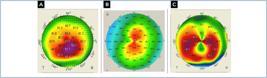
Keratoconus types. (A) Oval; (B) astigmatic; (C) pellucid marginal degeneration or pellucid marginal degeneration like.
Intrastromal ring segment criteria
Patients with PMD and PMD-like were included in the same group due to similar topographic characteristics. Because of the asymmetric shape of the keratoconus, they received a single 210° arc segment implant. Astigmatic keratoconus patients received a pair of 160° arc segments, while patients with asymmetric oval keratoconus received a single 160° segment. Symmetric oval keratoconus patients (50/50% or 60/40% distribution in opposite meridians) received a pair of 160° arc segments.
Surgical technique
We performed standard manual ICRS implantation with the Castaneda suction ring (Ferrara Ophtalmics, Belo Horizonte, Brazil) used for eye fixation.
Inclusion criteria
Patients over 10 years of age, regardless of sex or gender; astigmatic and oval keratoconus, PMD and PMD-like; ICRS implant of 160° and 210° arch rings before 2010, with a post-surgical follow-up of at least 7 years; visual acuity with correction less than +0.1 logMAR, high topographic AST (> 4.00 D); maximum keratometry less than 75D (Kmax); a thinner point in the ring path greater than 300 µm; intolerant to the use of contact lenses.
Exclusion criteria
Keratitis or history of ocular herpes; dystrophies and other corneal degeneration; previous eye surgeries; glaucoma; autoimmune diseases or systemic connective tissue diseases; pregnant patients; individuals who had follow-up time less than 1 year were exclusion criteria.
Statistical data analysis
Initially, the normality and homogeneity of all variables were tested using the Shapiro-Wilk and Levene tests, respectively. As the data did not present normality and homogeneity, the Wilcoxon test was used to compare the pre-and post-intervention of the right eye, left eye and both eyes. A significance level of 5% was considered. The Statistical Package for the Social Sciences version 17.0 (SPSS Inc, Chicago, IL, United States) was used for all analyses.
RESULTS
Sample characterization
Thirty-five individuals aged between 10 and 30 participated in the study. The majority of patients underwent surgery on both eyes, completing 62 eyes.
The types of keratoconus are different concerning the predominant age group, with more individuals aged between 15 to 20 years old with oval-type keratoconus (Table 1).
In the oval type, the following complication was observed: removal of the nasal segment, with an average time of 82.00 (± 55.75); in the PMD type, the following complications were observed: removal of the nasal arc, removal of the temporal arc, removal of a ring, and removal of two segments with an average time of 42.00 (± 34.64); and in the astigmatic type, removal of the nasal segment was observed with an average time of 27.00 (± 29.70).
Overall results despite keratoconus classification
A decrease was found in topographic AST (p = 0.001) (Figure 2) and in specular microscopy cell count (p < 0.001) (Table 2). We also found an improvement in corrected visual acuity (VA) (p < 0.001) (Figure 2).

Pre and post – operative box plots. (A) Topographic astigmatism; (B) BCVA: Best corrected visual acuity; (C) spherical equivalent; (D) keratometry K1; (E) keratometry K2.
On the other hand, no difference was found in pachymetry (p=0.033) (Table 2).
We had statistically significant improvement also in keratometric values and spherical equivalent (SE) (Figure 2).
We had an 83.9% improvement in patients’ lines of vision. The vast majority had 1 or 2 gains in lines of vision, as both represent 25.8% of cases (each). In our group, 1.6% gained nine lines of vision, 12.9% did not show a change in line of sight, and 3.2% lost lines of sight, with half of this amount losing 1 line and the other half losing two lines.
The ICRS (pre versus postoperative) with different strategies (one 160° arc ring, one 210° arc ring, two 160° arc rings) and different classifications (oval, astigmatic, PMD or PMD-like) were compared, as well as the results obtained with ICRS of two segments with different thicknesses in cases of oval cones, the results obtained with ICRS with different arc lengths in oval (asymmetric) keratoconus cases, the effects of ICRS of two segments of 160° in astigmatic and oval keratoconus; and the results of ICRS with different lengths (160° or 210°) in PMD and PMD-like cases.
The behavior of these variables before and after the intervention does not depend on the type of keratoconus evaluated, the type of arc, or the thickness.
Comparison of the results obtained with intrastromal ring segment of two segments of 160° arc with intrastromal ring segment of 210° arc
The ICRS of two 160° arcs showed smaller differences in topographic AST before the intervention (p = 0.002) compared to the values after the intervention. No differences were found in the ICRS of a 210° arc. The other variables did not show differences between groups.
Comparison of the results obtained by the intrastromal ring segment of two segments with different thicknesses in cases of astigmatic cones
The ICRS of two segments with a thickness of 0.20 mm showed lower values after intervention than before (p = 0.043). All other variables were not statistically significant.
As for the following three comparisons, no differences were found between groups: the results obtained with ICRS of two segments with different thicknesses in cases of oval cones, an ICRS with different arches in PMD and PMD-like cases, one ICRS of 210° in cases of PMD and PMD-like and 160° in cases of oval keratoconus (asymmetric); the effects of ICRS of 210° in cases of PMD or PMD-like and 160° in cases of oval keratoconus (asymmetric); the effects of ICRS of 210° in cases of PMD or PMD-like and 160° in cases of oval keratoconus (asymmetric).
Comparison of variables in keratoconus types
We found a statistically significant difference between the Keratoconus groups for K1 Keratometry (Delta: postoperative values minus pre-operative values) and K2 Keratometry (Delta).
Comparing Keratometry K1 (Delta) with a p-value = 0.032 (analyzing the p-values), we conclude that the difference occurs between Astigmatic, with a median of −2.83, and PMD, with a median of −0.07 (p-value = 0.031).
We had statistically significant improvement in all groups in SE and BCVA. (Figure 2)
We had statistically significant improvement in K1 and K2 in astigmatic and oval keratoconus. When we analyzed topographic AST, there was only improvement in oval cones.
Comparison of variables in different arc rings
Finally, we compared the arc rings for the parameters analyzed three times.
We found a statistically significant difference between the types of Rings for K2 and Keratometry (Delta).
We had statistically significant improvement in all groups in the following variables: SE and CDVA. We had statistically significant improvement in K1 with two 160-arc rings, regarding K2 in all variables except for one 210°-arc ring. When we analyzed topographic AST, we only had improvement with two 160°-arc rings.
Changes between 6 months and last follow-up
Our sample did not show statistically significant changes between the sixth month before surgery and the final visit.
DISCUSSION
Studies have already demonstrated decreased quality of life and mental health issues such as depression in keratoconic patients. So, it is imperative to improve vision quality in these patients, as demonstrated by Rodrigues et al. A multidisciplinary treatment is recommended. Still, the role of ophthalmologists is to improve these patients’ vision quality.(9,12-19)
The vast majority of studies show promising results with ICRS, with a reduction in SE, AST, keratometry (K), and improvement in VA. A literature review showed an improvement in SE (in ring implantation of the models: Keraring and Ferrara) from 0.06D to 5.8D, with an average of 3D and with a gain in lines of sight between 48.7% and 90.6%, with an average of 70%.(6) Other studies showed more effective improvement in patients with more advanced keratoconus and a gain in lines of vision by more than 77%. Another review showed visual acuity improvement of 0.23 ± 0.28 logMAR without correction and 0.06 ± 0.21 logMAR with correction at 12 months of follow-up. Regarding refraction, the spherical degree improved by 2.81 ± 1.54 D and the cylinder improved by 1.49 ± 0.83 D in 12 months of follow-up. Preoperative mean keratometry demonstrated mean flattening of 3.41 ± 2.13 D one year after ICRS implantation.(5-8,13)
Agreeing with the results in the literature, we found statistical improvement in all keratometric and visual variables. As expected, we did not find a pachymetry difference, but we had a significant Specular Microscopy cell count reduction compared to the literature results, mainly with two segments implanted. Since the surgeries were performed with manual technique, this could cause more endothelial damage due to the trauma and deeper tunnels. Another hypothesis is that the device did not capture the endothelium adequately after ICRS. This explanation could be suitable for two rings, leading to a more significant decrease than for one ring. Nevertheless, despite the significance, these reduction values were small and should not lead to clinical influence in most cases, except for patients with Fuchs dystrophy.(1-5)
We had improvement in vision lines in most patients, but we had some patients without improvement and even two with loss of two vision lines. We attribute these bad results to the fact that we used a manual technique when we did not have a femtosecond laser and to the implant nomograms that improved over time. We also needed to explant some arc rings due to the difficulty of creating a perfect tunnel using the manual technique, facilitating its superficialization and displacement. Regarding refractive AST, we found a reduction with 210 and 160 rings but did not have a topographic astigmatic reduction with 210 rings. This corroborates the theory that rings greater than 180 do not significantly generate the coupling effect or reduce corneal AST but can reduce coma. We also had only statistical improvement in K1 and K2 with two segments implanted, strengthening the theory that two segments greatly decrease these values and topography AST.(5-10)
Evaluating the keratoconus types, K1 and K2 improved only in astigmatic and oval keratoconus. We only had improvement in topographic AST in oval keratoconus.
Sandes et al. studied 140 arc rings in PMD patients with good results. The actual tendency in PMD cases is using a 140 arc ring or the combination of a 160 inferiorly and a 90 arc superiorly (since this segment does not act in keratometric values and reduces AST). The problem in using 210 arc rings in PMD cases is that these corneas are usually oblated with a central corneal flattening. This arc type normally prolates the cornea and flattens the central region. Some authors try combining a 140 arc implanted in the 5 mm region and one 210 ring in the 6 mm region.(14)
In astigmatic cones, we usually have a more regular cornea, so it is recommended that we use more symmetrical implantation with two arc rings to maintain the cornea shape, only reducing its values.
We usually have an asymmetrical shape in oval cones, sometimes with superior cornea flattening, similar to PMD cases. In cases with a superior cornea, flattening is recommended for only one ring or asymmetrical implantation with the thicker ring in the inferior region and the thinner or smaller one in the opposite site so that we can benefit from the coupling effect.(19-24)
This is the first study comparing long-term progress in different types of keratoconus and results with different arc ring implantation strategies. Since we had improvement in almost all variables in all cases and we did not find statistically significant changes between six months and the last visit, we can assume that we did not have significant disease progression.
The only procedure that demonstrably halts the progression of the disease is corneal CXL, but some professionals believe that ICRS can interfere with corneal biomechanics. There is a focal change in biomechanics in the thinnest and most curved areas (evidenced by Brillouin microscopy), creating a vicious circle of instability and curving of the cornea. ICRS can stop this trend. Furthermore, ICRS can redistribute corneal stress by shortening the lamellae and altering the shape of the cornea without altering its intrinsic properties. This causes the cornea to undergo viscoelastic changes and acquire a more regular shape over time, making the biomechanical injury caused by intraocular pressure and gravity more distributed and smoothed.(11,20,21,25,26)
In a study carried out with OCT elastography, ICRS was shown to reduce general tissue tension under increased IOP and cause subsequent tissue relaxation. This effect is more prominent the longer the arc length and the smaller the ICRS optical zone are. ICRS has not only a geometric but also a mechanical impact on the corneal tissue.(21)
In a recent publication, de Araujo et al. studied disease progression over 5 years after ICRS implementation. Thirty-four patients were divided by age (over or under 21), and most were classified as stage 3 keratoconus. This work showed that the percentage of patients who experienced disease progression was higher in patients under 21 years of age (42%) than in patients over 21 years of age (7%), as well as the average progression of maximum keratometry, which in this group was lower than 2D.(15)
Vega-Estrada et al., in a similar study, also analyzed 18 eyes. The mean age of the patients was 25.75 years ± 3.59 (SD), and the implanted ICRS showed good postoperative results. However, from 6 months postoperatively to 5 years, the mean K value increased by 3.36 D, indicating disease progression.(16)
Moscovici et al. studied the progression of the disease after ICRS. They found that when comparing 3-month postoperative results and data from the final visit, approximately 40% of patients showed a keratometric increase in K1 or K2 by more than one diopter, and 60% showed an increase of less than one diopter. Patients with more than one diopter were younger (26 to 27 years old) than patients with an increase of less than one diopter (28 to 29 years old).(8) This conclusion agrees with the theory that keratoconus has a greater tendency to progress in younger patients. In this study, one patient, aged between 30 and 35 and another, between 20 to 25 years old, showed progression of more than five diopters in 15 months. These cases showed that, despite the implementation of ICRS, progression of keratoconus can occur, and patients must be periodically evaluated to ensure control of the disease.(8)
Our study has some limitations, such as a small sample size and the fact that the study was retrospective, and that we could only access some of the information we wanted.
CONCLUSION
Our study found good results, even in a long-term follow-up, with an improvement of almost all variables, especially spherical equivalent and corrected distance visual acuity. We also found a gain of vision lines in most cases and a reduced endothelial cell count, especially with implanting two arcs. Additionally, we found more significant astigmatism reduction with thicker rings, more keratometric reduction with two-ring segment arcs, and less in the pellucid marginal degeneration keratoconus type. Finally, with 210 arc rings, we did not find a reduction in topographic astigmatism but a decrease in refractional astigmatism.
REFERENCES
- 1 Hashemi H, Heydarian S, Hooshmand E, Saatchi M, Yekta A, Aghamirsalim M, et al. The prevalence and risk factors for keratoconus: a systematic review and meta-analysis. Cornea. 2020;39(2):263-70.
- 2 Nosé W, Neves RA, Burris TE, Schanzlin DJ, Belfort Júnior R. Intrastromal corneal ring: 12-month sighted myopic eyes. J Refract Surg. 1996;12(1):20-8.
- 3 Gomes JA, Rapuano CJ, Belin MW, Ambrósio R Jr; Group of Panelists for the Global Delphi Panel of Keratoconus and Ectatic Diseases. Global Consensus on Keratoconus Diagnosis. Cornea. 2015;34(12):e38-9.
- 4 Belin MW, Duncan JK. Keratoconus: The ABCD grading system. Klin Monbl Augenheilkd. 2016;233(6):701-7.
- 5 Ziaei M, Barsam A, Shamie N, Vroman D, Kim T, Donnenfeld ED, et al.; ASCRS Cornea Clinical Committee. Reshaping procedures for the surgical management of corneal ectasia. J Cataract Refract Surg. 2015;41(4):842-72.
- 6 Zadnik K, Money S, Lindsley K. Intrastromal corneal ring segments for treating keratoconus. Cochrane Database Syst Rev. 2019;5(5):CD011150.
- 7 Ferrara P, Torquetti L. Clinical outcomes after implantation of a new intrastromal corneal ring with a 210-degree arc length. J Cataract Refract Surg. 2009;35(9):1604-8.
- 8 Moscovici BK, Rodrigues PF, Rodrigues RA, Rios LC, Simoncelli R, Freitas MM, et al. Evaluation of keratoconus progression and visual improvement after intrastromal corneal ring segments implantation: A retrospective study. Eur J Ophthalmol. 2021;31(6):3483-9.
- 9 Rodrigues PF, Moscovici BK, Hirai F, Mannis MJ, de Freitas D, Campos M, et al. Vision-related quality of life in patients with keratoconus with enantiomorphic topography after bilateral intrastromal corneal ring implantation. Cornea. 2024;43(2):190-4.
- 10 Piñero DP, Alio JL, Morbelli H, Uceda-Montanes A, El Kady B, Coskunseven E, et al. Refractive and corneal aberrometric changes after intracorneal ring implantation in corneas with pellucid marginal degeneration. Ophthalmology. 2009;116(9):1656-64.
- 11 Rodrigues PF, Moscovici BK, Ferrara G, Lamazales L, Freitas MM, Torquetti L, et al. Corneal densitometry in patients with keratoconus undergoing intrastromal Ferrara ring implantation. Eur J Ophthalmol. 2021;31(6):3505-10.
- 12 Moschos MM, Gouliopoulos NS, Kalogeropoulos C, Androudi S, Kitsos G, Ladas D, et al. Psychological Aspects and Depression in Patients with Symptomatic Keratoconus. J Ophthalmol. 2018;2018:7314308.
- 13 Kang MJ, Byun YS, Yoo YS, Whang WJ, Joo CK. Long-term outcome of intrastromal corneal ring segments in keratoconus: Five-year follow up. Sci Rep. 2019;9(1):315.
- 14 Sandes J, Stival LR, Ávila MP, Ferrara P, Ferrara G, Magacho L, et al. Clinical outcomes after implantation of a new intrastromal corneal ring with 140-degree of arc in patients with corneal ectasia. Int J Ophthalmol. 2018;11(5):802-6.
- 15 Araujo BS, Kubo L, Marinho DR, Kwitko S. Keratoconus progression after intrastromal corneal ring segment implantation according to age: 5-year follow-up cohort study. Int Ophthalmol. 2020;40(11):2847-54
- 16 Vega-Estrada A, Alió JL, Plaza-Puche AB. Keratoconus progression after intrastromal corneal ring segment implantation in young patients: Five-year follow-up. J Cataract Refract Surg. 2015;41(6):1145-52.
- 17 Santos RT, Moscovici BK, Hirai FE, Nosé W. Association between keratoconus, ocular allergy, and sleeping behavior. Arq Bras Oftalmol. 2021;84:17-21.
- 18 Carvalho N, Guerra TF, Moutinho CF, Santos FS, Miyashiro RA, Simoncelli RA, et al. Therapeutic Analysis in Patients with Keratoconus. Int J Kerat Ect Cor Dis 2019;8(2):40-2.
- 19 Ambrósio R Jr, Salomão MQ, Barros L, da Fonseca Filho JB, Guedes J, Neto A, et al. Multimodal diagnostics for keratoconus and ectatic corneal diseases: a paradigm shift. Eye Vis (Lond). 2023;10(1):45.
- 20 Vinciguerra R, Fernández-Vega-Cueto L, Roberts CJ, Poo-Lopez A, Eliasy A, Merayo-Lloves J, et al. Effect of intrastromal corneal ring segments on in vivo corneal biomechanics in keratoconus: 1-year results. J Cataract Refract Surg. 2023;49(12):1258-63.
- 21 Torres-Netto EA, Hafezi F, Kling S. Intracorneal ring segment implantation results in corneal mechanical strengthening visualized with optical coherence elastography. J Refract Surg. 2022;38(7):459-64
- 22 Barugel R, David C, Kallel S, Borderie M, Cuyaubère R, Goemaere I, et al. Comparative study of asymmetric versus non-asymmetric intrastromal corneal ring segments for the management of keratoconus. J Refract Surg. 2021;37(8):552-61.
- 23 Costa JV, Monteiro T, Franqueira N, Faria-Correia F, Alfonso J, Vaz F. Five-year long-term outcomes of intrastromal corneal ring segment implantation using the manual technique for keratoconus management. J Cataract Refract Surg. 2021;47(6):713-21.
- 24 Neves Cruz C, Franqueira N, Mendes JC, Oliveira M, Monteiro T. Intrastromal corneal ring segments: effect of depth of implantation in visual, refractive, and topographic outcomes in patients with keratoconus. J Cataract Refract Surg. 2023;49(9):949-55.
- 25 Rodrigues PF, Moscovici BK, Lamazales L, Freitas MMS, Gomes JÁP, Nosé W, Campos MS. Measurement of the visual axis through two different methods: quantification and differences for measuring chord μ. Arq Bras Oftalmol. 2023;87(4):e2022-0035.
- 26 Rios LC, Silva PG, Canamary Junior AM, Rodrigues P, Regatieri CVS, Campos M, et al. Centration in refractive surgery. Arq Bras Oftalmol. 2020;83(1):76-81.


