Avellino corneal dystrophy: corneal stars
Keywords:
Corneal dystrophy; Corneal dystrophies, hereditary
Descritores:
Distrofias da córnea; Distrofias hereditárias da córnea
Keywords:
Corneal dystrophy; Corneal dystrophies, hereditary
Descritores:
Distrofias da córnea; Distrofias hereditárias da córnea
Corneal dystrophy is a hereditary non-inflammatory disorder in which substances accumulate in the cornea.
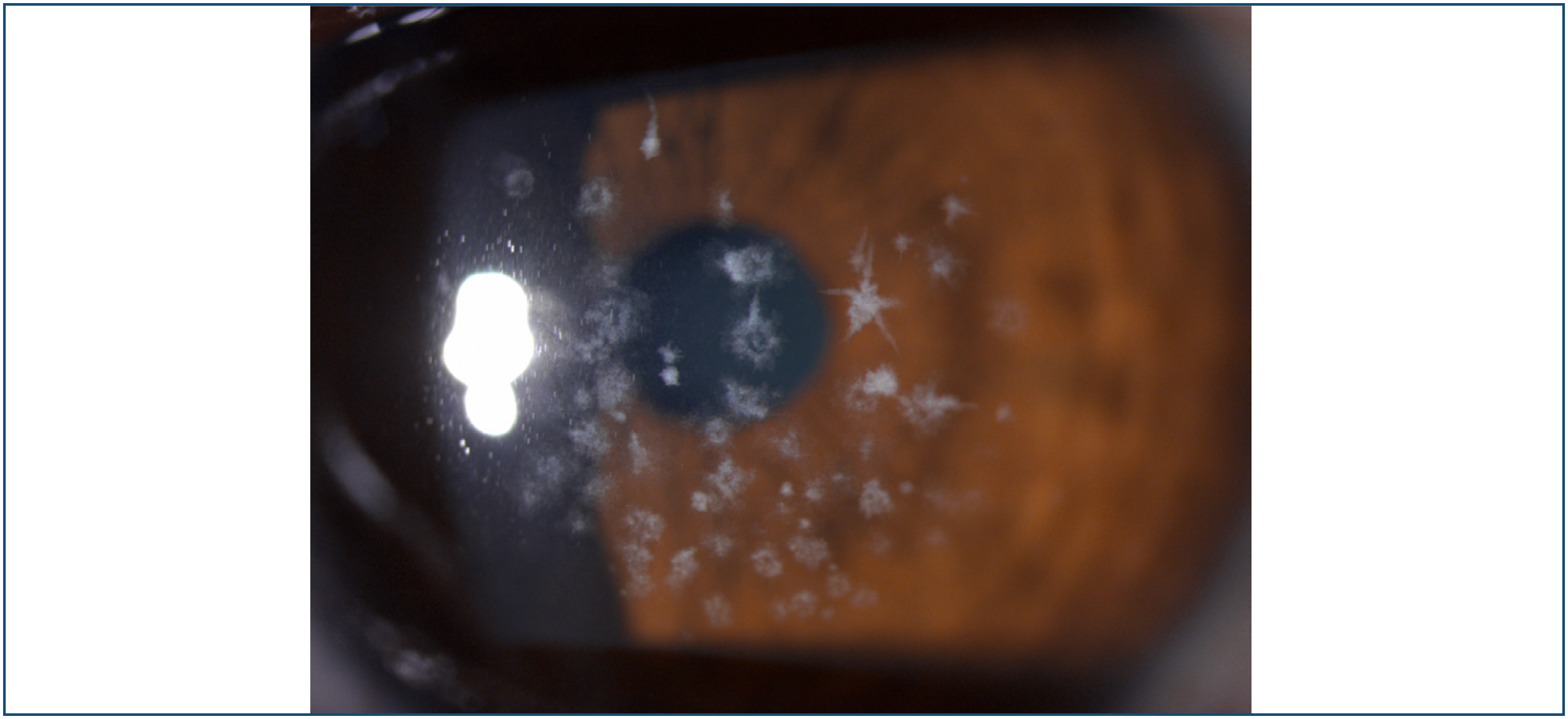
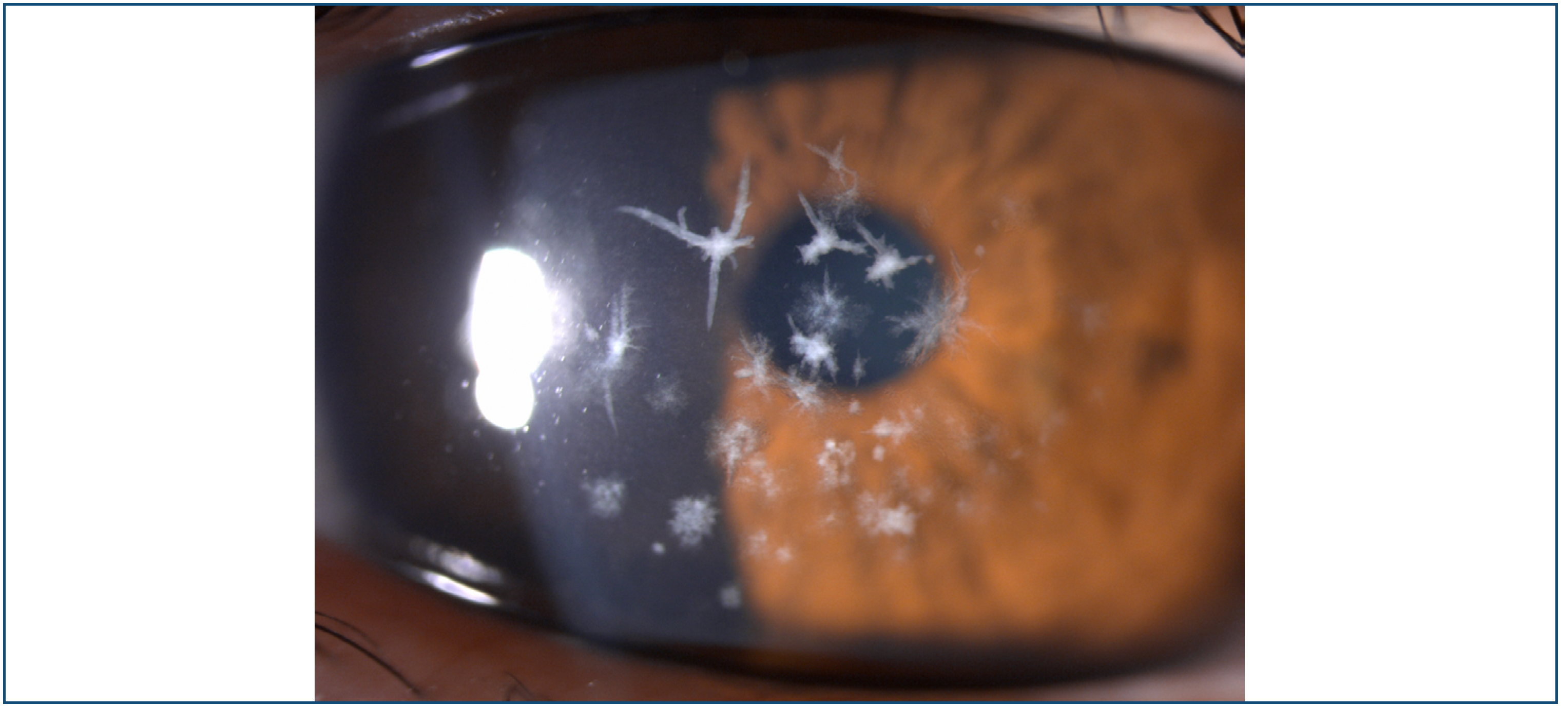
A 32-year-old woman presented for routine ophthalmic examination. The patient has myopia of −6.25 in both eyes with best corrected visual acuity at 8/10 bilaterally. On slit-lamp examination, she was found to have crumb-like and star opacities, grey-white deposits in the anterior corrneal stroma (Figures 1-3). Clear areas were present between the opacities. The peripheral cornea was clear. Her best corrected visual acuity was 8/10 bilaterally. A diagnosis of granular dystrophy type II was made.

Slit-lamp photograph, at high magnification, of the left eye showing the star deposits on the anterior corneal stroma.
Granular corneal dystrophy type II or Avellino corneal dystrophy was first described in individuals from Avellino in 1988.(1) Avellino dystrophy is one of the transforming growth factor-β-induced (TGFBI) associated corneal dystrophies, of which the clinical aspect is the coexistence of granular and amyloid deposits in the cornea.(2) It is characterized clinically by corneal opacities that are shaped like stars, crumb and snowflakes. This corneal dystrophy is an autosomal dominant disorder and appears to progress with age. Cases of exacerbated granular corneal dystrophy Type II after refractive surgery have been reported.(3) The reason for the worsening is that corneal trauma activates and overproduces TGFBI.(4)The refractive surgeries were contraindicated in patients with this corneal dystrophy.(5)
REFERENCES
- 1 Folberg R, Alfonso E, Croxatto JO, Driezen NG, Panjwani N, Laibson PR, et al. Clinically atypical granular corneal dystrophy with pathologic features of lattice-like amyloid deposits. A study of these families. Ophthalmology. 1988;95(1):46-51.
- 2 Klintworth GK. Advances in the molecular genetics of corneal dystropies. Am J Ophthalmol. 1999;128(6):747-54
- 3 Banning CS, Kim WC, Randleman JB, Kim EK, Stulting RD. Exacerbation of Avellino corneal dystrophy after LASIK in North America. Cornea. 2006;25(4):482-4.
- 4 Han KE, Kim TI, Chung WS, Choi SI, Kim BY, Kim EK. Clinical findings and treatments of granular corneal dystrophy type 2 (avellino corneal dystrophy): a review of the literature. Eye Contact Lens. 2010;36(5):296-9.
- 5 Weiss JS, Møller HU, Aldave AJ, Seitz B, Bredrup C, Kivelä T, et al. IC3D classification of corneal dystrophies - edition 2. Cornea. 2015;34(2):117-59