Phacoviscocanalostomy versus phacoemulsification alone for glaucoma associated with cataract: a meta-analysis
ABSTRACT
Objective: This systematic review and meta-analysis compare the safety and efficacy of phacoemulsification alone versus combined phacoviscocanalostomy in glaucoma and cataract patients.
Methods: We followed PRISMA guidelines. PubMed®, Web of Science, Cochrane, and Embase were searched for randomized clinical trial and non-randomized clinical trial studies comparing phaco versus phacoviscocanalostomy in patients with any glaucoma and cataract type. Statistical analysis was conducted using RevMan software.
Results: The study included 889 patients from one randomized clinical trial and three non-randomized clinical trials studies, of whom 499 (56.1%) received phacoviscocanalostomy. Patients treated with phacoviscocanalostomy had significantly lower intraocular pressure at 1 (mean difference: 3.90; p < 0.00001), 3 (mean difference: 2.52; p = 0.03), 6 (mean difference: 1.90; p = 0.04), 18 (mean difference: 3.23; p < 0.00001) and 24 months (mean difference: 2.55; p < 0.00001), except for 12 months (mean difference: 2.26; p = 0.08). A lower medication at six months (mean difference: 0.68; p < 0.00001), no difference in the visual acuity (mean difference: −0.02; p = 0.77), and a higher rate of adverse events (odds ratios: 236.96; p = 0.004) were found.
Conclusions: The combined phacoviscocalanostomy approach showed positive outcomes, without increasing the severe complication rates. Registry PROSPERO - CRD42024501313
Keywords:
Glaucoma; open angle; Cataract; Phacoemulsification
RESUMO
Objetivo: Esta revisão sistemática e meta-análise comparam a segurança e eficácia da facoemulsificação isolada versus a facoviscocanalostomia combinada em pacientes com glaucoma e catarata.
Métodos: Seguimos as diretrizes PRISMA. Foram realizadas buscas nas bases PubMed®, Web of Science, Cochrane e Embase para estudos de ensaios clínicos randomizados e não randomizados que comparassem faco versus facoviscocanalostomia em pacientes com qualquer tipo de glaucoma e catarata. A análise estatística foi conduzida utilizando o software RevMan.
Resultados: O estudo incluiu 889 pacientes provenientes de um ensaio clínico randomizado e três estudos clínicos não randomizados, dos quais 499 (56,1%) receberam facoviscocanalostomia. Os pacientes tratados com facoviscocanalostomia apresentaram pressão intraocular significativamente menor em 1 (diferença média: 3,90; p < 0,00001), 3 (diferença média: 2,52; p = 0,03), 6 (diferença média: 1,90; p = 0,04), 18 (diferença média: 3,23; p < 0,00001) e 24 meses (diferença média: 2,55; p < 0,00001), exceto aos 12 meses (diferença média: 2,26; p = 0,08). Foi observada uma redução no uso de medicação aos seis meses (diferença média: 0,68; p < 0,00001), nenhuma diferença na acuidade visual (diferença média: −0,02; p = 0,77) e uma maior taxa de eventos adversos (odds ratio: 236,96; p = 0,004).
Conclusões: A abordagem combinada de facoviscocanalostomia demonstrou resultados positivos, sem aumento nas taxas de complicações graves. Registro PROSPERO - CRD42024501313
Descritores:
Glaucoma de ângulo aberto; Catarata; Facoemulsificação
INTRODUCTION
Glaucoma is the main cause of irreversible blindness in the world, and at least 70 million people in the world live with this pathology. However, its timely treatment can significantly delay visual loss.(1,2) The main risk factor associated with glaucoma is elevated intraocular pressure.(3) Several treatments aimed at reducing intraocular pressure, including medications, laser therapies and surgeries, have been used to prevent the progression of glaucoma damage.(4-7)
Cataracts are the main cause of reversible blindness in the world(8,9) and around 12 million people worldwide are blind due to cataracts.(10) The coexistence of cataracts and glaucoma may become common in the elderly population.(11) The treatment of these two pathologies is a significant challenge in ophthalmology, as the treatment of each can influence the progression of the other. Cataract removal surgery can only reduce intraocular pressure levels, whereas glaucoma surgery associated with cataract surgery is subject to several complications.(12)
Several surgical techniques for the treatment of glaucoma aim to reduce intraocular pressure, including viscocanalostomy, a procedure in which Schlemm's canal is identified under a scleral flap and then dilated with viscoelastic, promoting the opening of the eye's drainage system.(13,14)
This systematic review and meta-analysis article will compare the different aspects that affect the choice and outcome of surgical treatment between phacoemulsification alone for cataract removal and the combination of phacoemulsification and viscocanalostomy in patients coexisting with glaucoma and cataract.
METHODS
This meta-analysis was performed according to the guidelines of the Preferred Reporting Items for Systematic Reviews and Meta-Analysis (PRISMA) and the recommendations of the Cochrane Collaboration.(15)
Eligibility criteria
Studies that met the following eligibility criteria were included: randomized clinical trial (RCT) or observational studies; comparing phacoviscocanalostomy to phacoemulsification alone; patients aged ≥18 years with any type of glaucoma associated with cataract; follow-up time of at least 1 week; and reporting any of the clinical outcomes of interest. Studies with overlapping populations, case reports, animal studies, and in vitro experiments were excluded.
Information source
Two authors independently searched at PubMed®, Embase, Web of Science and Cochrane Library from inception to January 2024. Furthermore, the references from all studies included were also searched manually for any additional studies. Eventual conflicts were resolved by consensus among the authors.
Search strategy
The following terms were used in this search strategy: "(phaco OR phacoemulsification OR cataract) AND (phacoviscocanalostomy OR viscocanalostomy)". We did not use publication dates or language restrictions in our electronic search for the studies.
Study selection
We imported search results into the Zotero software, and duplicated records were excluded. Two independent authors applied eligibility criteria to screen the titles and abstracts. After that, the full text of potentially eligible studies was appraised. Any disagreements were resolved by contacting the senior author.
Data extraction
Two authors extracted the following data from selected studies: country, study design, number of patients and eyes allocated for each arm, inclusion and exclusion criteria, time to follow-up, and main patient's baseline characteristics. Also, the same authors collected pre-specified baseline characteristics and outcome data and recorded them in an Excel template.
Endpoints and subgroup analysis
The primary outcomes of interest were mean change in intraocular pressure (IOP), mean change in the number of medications, mean change in visual acuity, and ocular adverse events. General adverse events reported IOP spike, Microperfuration of Descemet's membrane, Iris prolapse, hyphema, Filtering bleb, and fibrin formation.
Risk of bias assessment
The risk of bias was analyzed in randomized studies using version 2 of the Cochrane Risk of Bias assessment tool.(16) Non-randomized studies were assessed with the Risk of Bias in Non-randomized Studies - of Interventions (ROBINS-I) tool.(17) Two independent authors completed the risk of bias assessment. Disagreements were resolved through a consensus after discussing reasons for the discrepancy.
Statistical analysis
For each outcome analysis, the study with a higher number of patients from among those with likely overlapping populations was chosen. Treatment effects for binary endpoints were compared using pooled odds ratios (OR) with 95% of confidence interval. Mean differences (MD) were used to analyze continuous outcomes. The Cochrane Q-test and I2 statistics assessed heterogeneity; p-values > 0.10 and I2 values >25% were considered significant for heterogeneity. Statistical significance was defined as p-values < 0.05. The Sidik-Jonkman estimator was used to calculate the tau2 variance between studies. We used a random-effects model for all pooled outcomes. For statistical analysis, we used the Review Manager (version 5.3).(18)
RESULTS
Study selection and baseline characteristics
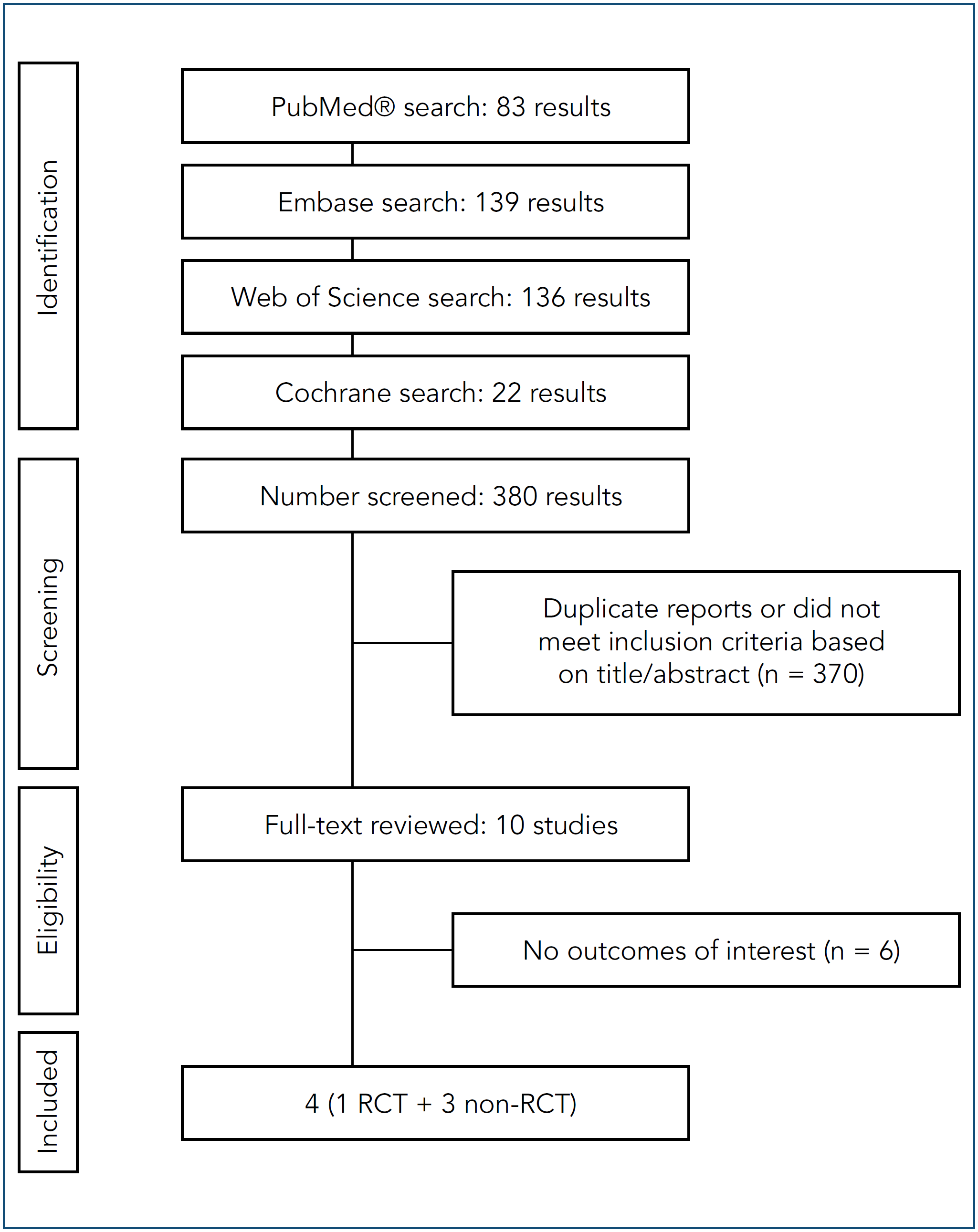
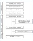
As detailed in Figure 1, we found 380 articles, with 83 in PubMed® (MEDLINE®), 139 in Embase (Elsevier), 136 in Web of Science, and 22 in Cochrane databases. Of these, 166 were removed as duplicates. After removing duplicate records and ineligible studies, ten remained and were thoroughly reviewed based on inclusion criteria. Next, six articles were excluded as per our exclusion criteria. Finally, four studies were included in this review, one RCT(19) and three nonrandomized cohorts.(4,20,21) Study characteristics are reported in table 1.
Pooled analysis of all studies
Intraocular pressure
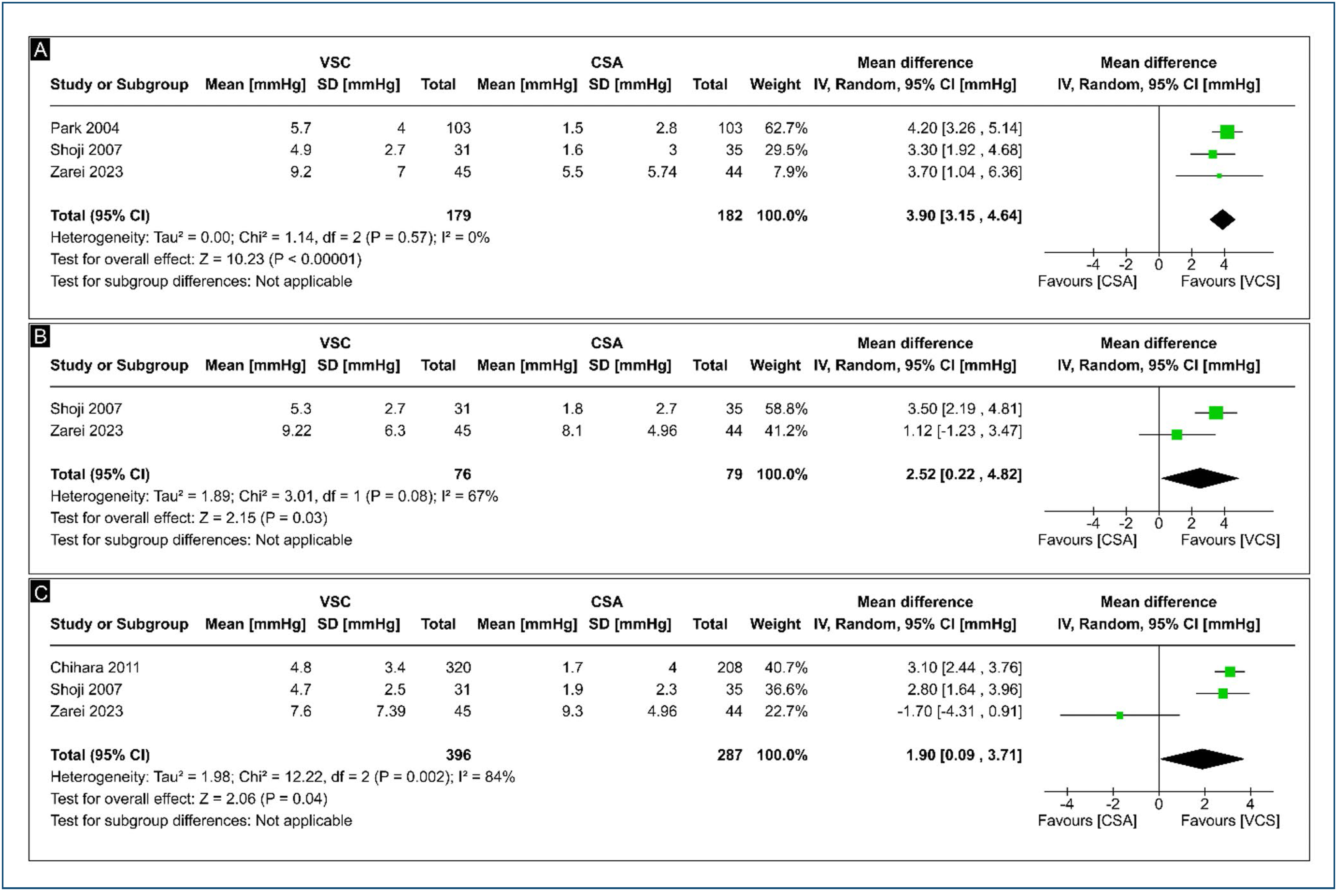
Regarding the mean change in IOP after 1 month, the pooled results revealed a higher decrease in mean IOP in phacoviscocanalostomy when compared with phaco alone group (MD: 3.90; 95%CI 3.15-4.64; p < 0.00001; I2 = 0%; Figure 2A).

(A) Intraocular pressure at 1 month forest plot. (B) Intraocular pressure at 3 months forest plot. (C) Intraocular pressure at 6 months forest plot.
Regarding the mean change in IOP after 3 months, the pooled results revealed a higher decrease in mean IOP in phacoviscocanalostomy when compared with the phaco alone group (MD: 2.52; 95%CI 0.22-4.82; p = 0.03; I2 = 67%; Figure 2B).
Regarding the mean change in IOP after 6 months, the pooled results revealed a higher decrease in mean IOP in phacoviscocanalostomy when compared with the phaco alone group (MD: 1.90; 95%CI 0.09-3.71; p = 0.04; I2 = 84%; Figure 2C).
Regarding the mean change in IOP after 12 months, the pooled results revealed no significant difference between patients in the phacoviscocanalostomy and phaco alone groups (MD: 2.26; 95%CI −0.25-4.77; p = 0.08; I2 = 89%; Figure 3A).
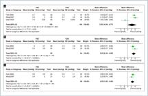
(A) Intraocular pressure at 12 months forest plot. (B) Intraocular pressure at 18 months forest plot. (C) Intraocular pressure at 24 months forest plot.
Regarding the mean change in IOP after 18 months, the pooled results revealed a higher decrease in mean IOP in phacoviscocanalostomy when compared with the phaco alone group (MD: 3.23; 95%CI 2.18-4.29; p < 0.00001; I2 = 7%; Figure 3B).
Regarding the mean change in IOP after 24 months, the pooled results revealed a higher decrease in mean IOP in phacoviscocanalostomy when compared with the phaco alone group (MD: 2.55; 95%CI 1.62-3.49; p < 0.00001; I2 = 0%; Figure 3C).
Medications
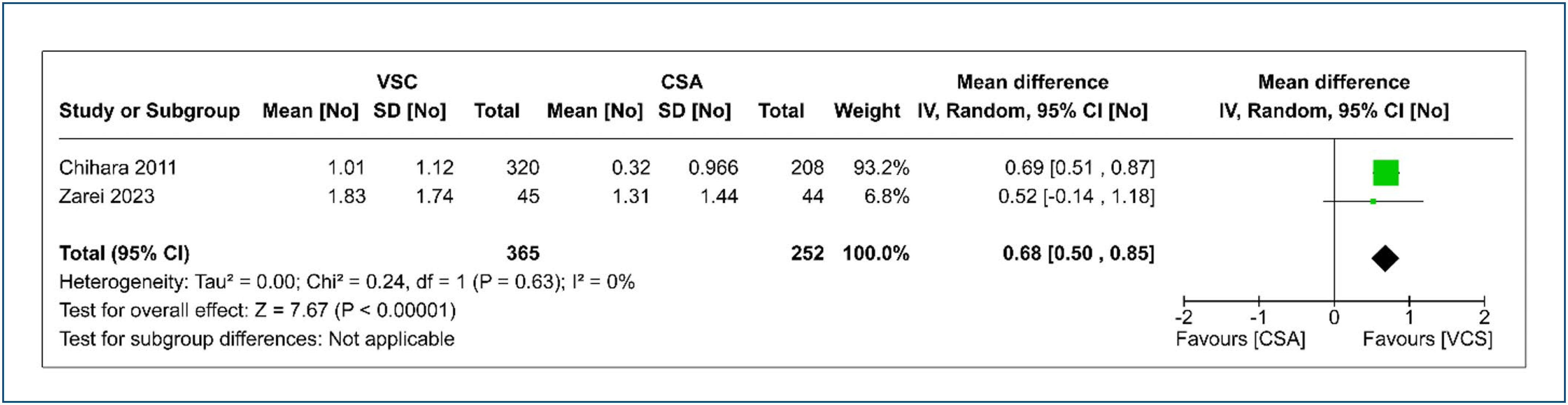
Among the studies included, two reported the mean change medications after 6 months. The pooled results revealed a higher decrease in mean medication in phacoviscocanalostomy when compared with phaco alone group (MD 0.68; 95%CI 0.50-0.85; p < 0.00001; I2 = 0%; Figure 4).

Visual acuity
Among the studies included, two reported the mean change of best-corrected visual acuity in a range of 6-12 months after surgery. For these studies, the pooled results revealed no significant difference between patients in the phacoviscocanalostomy and phaco alone groups (MD −0.02; 95%CI −0.15-0.11; p = 0.77; I2 = 37%; Figure 5).

Adverse events
Two studies reported adverse events. In the phacoviscocanalostomy group, there were 58 instances of hyphema, while no such events were observed in the phaco alone group. Fibrin formation occurred in 19 cases within the phacoviscocanalostomy group, compared to 3 cases in the phaco alone group. A detailed breakdown of the total adverse events is presented in table 2. No serious adverse events were reported.
Quality and evidence assessment
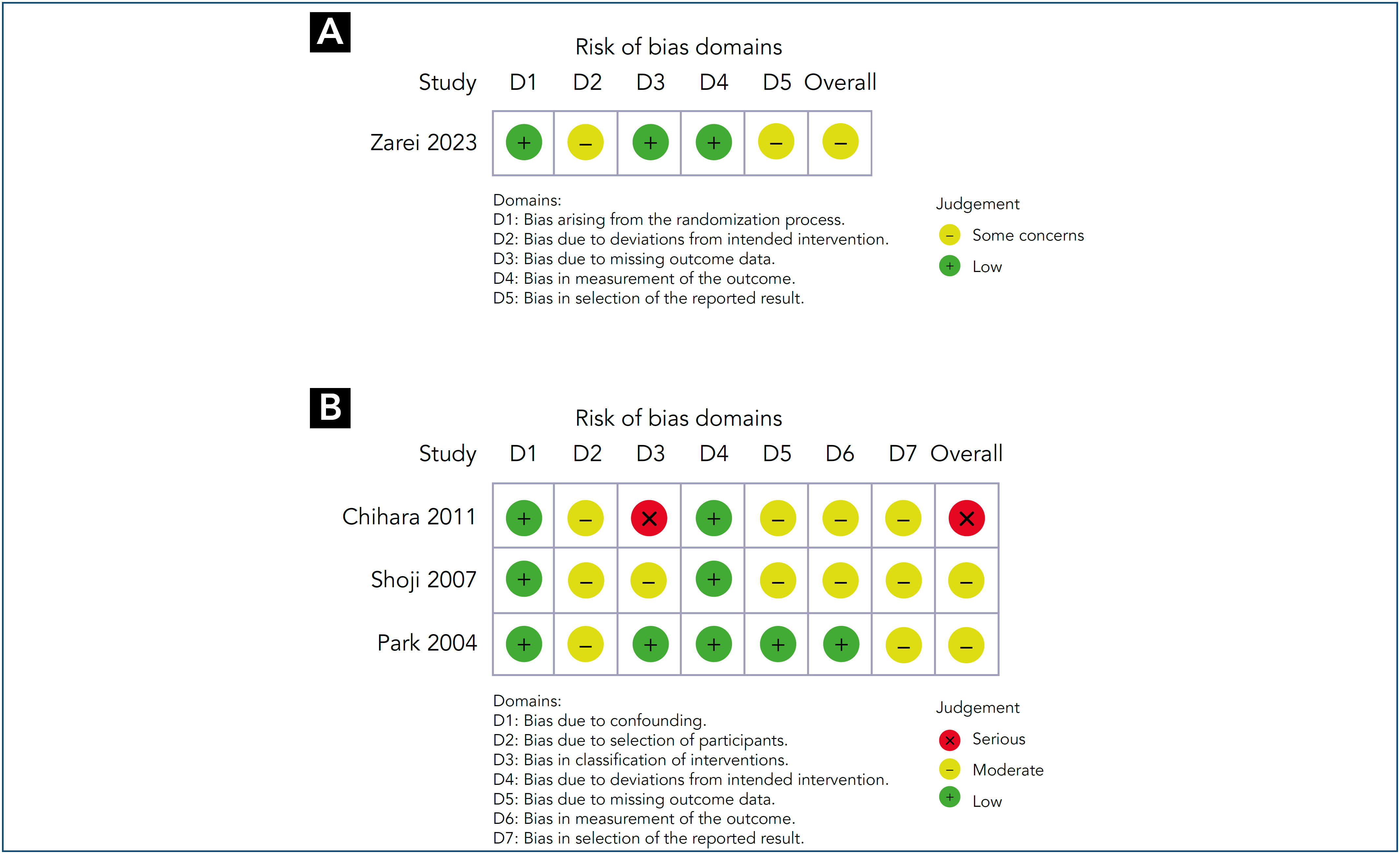
One study presented moderate concerns regarding the risk of bias due to deviations from the intended intervention and selection of the reported result.(19) One study considered a moderate risk of bias due to the selection of participants, classification interventions, missing data, measurement of outcomes, and selection of reported results.(21) One study considered a moderate risk of bias due to the selection of participants and the selection of reported results.(4) One study considered a serious risk of bias for the classification of interventions.(20) The risk of bias appraisal for individual studies is reported in Figure 6.

(A) Risk of bias assessment for RCTs using the ROB-2 tool. (B) Risk of bias assessment for non-RCT studies using the ROBINS-I tool.
DISCUSSION
To our knowledge, this meta-analysis of combining viscocanalostomy with phacoemulsification versus phacoemulsification alone for glaucoma treatment is the first one in the literature. In our meta-analysis, the study selection and characteristics are outlined, and four studies analyzing a total of 842 eyes were included in the review. Compared to the phaco alone group, those receiving phacoviscocanalostomy showed better results in IOP at 1, 3, 6, 18, and 24 months. However, no statistical differences were observed at 12 months. In the same way, a better result in medication change at 6 months was observed. The visual acuity between 6-12 months does not show a statistically significant difference. Complication rates, both general and specific like hyphema and fibrin formation, except for the IOP spike, were significantly higher in the phacovisco group, indicating the potential impact of combined procedures on patient outcomes.
The assessment of IOP consistently demonstrated favorable outcomes across 1, 3, 6, 18 and 24 months, except at 12 months. The impact of phacoemulsification on reducing IOP is well-established in both normal eyes and those with glaucoma, as indicated in prior studies. The extent of IOP reduction varies based on the baseline IOP and the specific type and progression of glaucoma.(22,23) Most of the available data suggest the effectiveness of phacoviscocanalostomy in individuals with ocular hypertension and primary open angle glaucoma. Nevertheless, the procedure tends to result in a final IOP within the mild level.(24-29) The higher decrease in the average number of medications at 6 months in the phacovisco group, compared to the phaco group, could be the contributing factor (MD: 0.68; p < 0.00001). Due to this, the long-term decrease in IOP in the phacoviscocanalostomy group is likely to be more substantial than the recorded reduction, as the postoperative medication count was lower than the preoperative count in the phaco group.(4) The variations in results at 12 months when compared with 18 and 24 months can occur because some studies did not report data for all visits analyzed. Consequently, only three studies were considered for analysis at 12 months, and the RCT study, which failed to report a notable difference in long-term IOP between groups, did not provide data for the 18 and 24-month periods.(19) In the short term, phacoviscocanalostomy effectively lowers IOP and offers improved IOP control compared to phaco alone. However, the long-term difference remains controversial, necessitating further RCTs with extended follow-up periods (>12 months).
The assessment of best-corrected visual acuity at 6 to 12 months post-surgery indicated that the group exclusively undergoing phacoviscocanalostomy demonstrated visual outcomes comparable to the phaco alone group (MD: −0.02; p = 0.77). Consequently, these patients exhibited a satisfactory quality of vision following combined cataract and glaucoma surgery, aligning with findings in previous literature.(12,21) The scarce data on visual acuity post-surgery underscores the need for a more comprehensive analysis to delve deeper into visual outcomes.
Striving for a low IOP goal may lead to an increased occurrence of severe postoperative complications and a diminished quality of life.(30,31) In the analysis, the adverse events (OR = 236.96; p = 0.004), hyphema (OR = 53.35; p = 0.01) and fibrin formation (OR = 7.47; p = 0.002) were significantly higher in the phacoviscocanalostomy when compared with the phaco alone group. This variation might be influenced by factors such as differences in surgical protocols, patient demographics, and variations in definitions of hyphema and fibrin formation, as proposed in a prior study.(4) However, concerning complications, various published case series agree that viscocanalostomy is linked to a lower incidence of complications than trabeculectomy.(25,27,32,33) Furthermore, none of the eyes in the phacoviscocanalostomy group experienced severe complications. Minor complications observed were temporary and did not pose a threat to eyesight. Performing viscocanalostomy is considered one of the most challenging glaucoma surgeries, and several studies have documented surgical outcomes following the learning curve of the procedure.(34,35)
Limitations
Although the results yielded significant insights into the safety and efficacy of phacoviscocalanostomy as a comparison therapy to phacoemulsification for glaucoma and cataracts, it is crucial to consider this study's limitations. Our analysis included a relatively small number of RCTs due to the limited research on phacoemulsification combined with viscocalanostomy. The use of different types of glaucoma varies significantly among studies, leading to a lack of consensus. Despite age and sex matching, the glaucoma severity may be more advanced in one group than another, potentially affecting the baseline IOP values and the number of glaucoma medications. Furthermore, the variability may be influenced by factors such as differences in surgical protocols. For example, one study did not employ any tissue spacer during the operation.(19)
Many studies included in this analysis reported follow-up periods at a maximum of 12 months. However, long-term outcomes (≥ 12 months) beyond the follow-up period were assessed in just a few studies. Evaluating the long-term stability of surgical effects and glaucoma progression is crucial. Furthermore, there was also no set protocol for discontinuation of preoperative glaucoma medications. This could have led to potential discrepancies in the management between different clinicians. The involvement of different surgeons is acknowledged as a possible confounding factor that may affect the generalizability of the results. The meta-analysis relied on aggregated data from published studies, and individual patient data were unavailable for analysis, limiting a more detailed understanding of the treatment effects and predictors of response.
CONCLUSIONS
The combined phacoviscocalanostomy approach demonstrated positive outcomes in terms of sustained intraocular pressure reduction and decreased medication dependence. These insights are valuable for considering therapeutic options in individuals with coexisting glaucoma and cataracts, emphasizing the need for individualized treatment based on patient characteristics and surgeon expertise. Further research, particularly with larger randomized clinical trial, and longer-term follow-up, is warranted to validate these findings and address the identified limitations. The results of this meta-analysis contribute with valuable insights into the potential advantages and challenges associated with combining viscocalanostomy with phacoemulsification for glaucoma and cataract treatment.
REFERENCES
- 1 Jayaram H, Kolko M, Friedman DS, Gazzard G. Glaucoma: now and beyond. Lancet. 2023;402(10414):1788-801.
- 2 Tham YC, Li X, Wong TY, Quigley HA, Aung T, Cheng CY. Global prevalence of glaucoma and projections of glaucoma burden through 2040: a systematic review and meta-analysis. Ophthalmology. 2014;121(11):2081-90.
- 3 Jiang N, Zhao GQ, Lin J, Hu LT, Che CY, Wang Q, et al. Meta-analysis of the efficacy and safety of combined surgery in the management of eyes with coexisting cataract and open angle glaucoma. Int J Ophthalmol. 2018;11(2):279-86.
- 4 Park M, Tanito M, Nishikawa M, Hayashi K, Chichara E. Combined viscocanalostomy and cataract surgery compared with cataract surgery in Japanese patients with glaucoma. J Glaucoma. 2004;13(1):55-61.
- 5 Weinreb RN, Aung T, Medeiros FA. The pathophysiology and treatment of glaucoma: a review. JAMA. 2014;311(18):1901-11.
- 6 Amaral DC, Louzada RN, Moreira PHS, et al. Combined Endoscopic Cyclophotocoagulation and Phacoemulsification Versus Phacoemulsification Alone in the Glaucoma Treatment: A Systematic Review and Meta-Analysis. Cureus. Mar 2024;16(3):e55853.
- 7 Amaral DC, Guedes J, de Oliveira Caneca K, Pereira SF, Alves MR, Manso JE. Manual small incision cataract surgery combined with trabeculectomy versus phacoemulsification combined with trabeculectomy for coexisting glaucoma and cataract: a systematic review and meta-analysis. Expert Rev Ophthalmol. 2024;19(4):281-9.
- 8 Lam D, Rao SK, Ratra V, Liu Y, Mitchell P, King J, et al. Cataract. Nat Rev Dis Primers. 2015;1:15014.
- 9 Hashemi H, Hatef E, Fotouhi A, Feizzadeh A, Mohammad K. The prevalence of lens opacities in Tehran: the Tehran Eye Study. Ophthalmic Epidemiol. 2009;16(3):187-92.
- 10 Flaxman SR, Bourne RR, Resnikoff S, Ackland P, Braithwaite T, Cicinelli MV, et al.; Vision Loss Expert Group of the Global Burden of Disease Study. Global causes of blindness and distance vision impairment 1990-2020: a systematic review and meta-analysis. Lancet Glob Health. 2017;5(12):e1221-e1234.
- 11 Cantor L, Lindfield D, Ghinelli F, Świder AW, Torelli F, Steeds C, et al. Systematic Literature Review of Clinical, Economic, and Humanistic Outcomes Following Minimally Invasive Glaucoma Surgery or Selective Laser Trabeculoplasty for the Treatment of Open-Angle Glaucoma with or Without Cataract Extraction. Clin Ophthalmol. 2023;17:85-101.
- 12 Zhang ML, Hirunyachote P, Jampel H. Combined surgery versus cataract surgery alone for eyes with cataract and glaucoma. Cochrane Database Syst Rev. 2015;2015(7):CD008671.
- 13 Wild GJ, Kent AR, Peng Q. Dilation of Schlemm's canal in viscocanalostomy: comparison of 2 viscoelastic substances. J Cataract Refract Surg. Aug 2001;27(8):1294-7.
- 14 Ghate D, Wang X. Surgical interventions for primary congenital glaucoma. Cochrane Database Syst Rev. 2015;1:CD008213.
- 15 Page MJ, McKenzie JE, Bossuyt PM, Boutron I, Hoffmann TC, Mulrow CD, et al. The PRISMA 2020 statement: an updated guideline for reporting systematic reviews. BMJ. 2021;372:n71.
- 16 Sterne JAC, Savović J, Page MJ, Elbers RG, Blencowe NS, Boutron I, et al. RoB 2: a revised tool for assessing risk of bias in randomised trials. BMJ. 2019;366:l4898.
- 17 Sterne JA, Hernán MA, Reeves BC, Savović J, Berkman ND, Viswanathan M, et al. ROBINS-I: a tool for assessing risk of bias in non-randomised studies of interventions. BMJ. 2016;355:i4919.
- 18 T C. Review Manager (RevMan) 5.3. Copenhagen: The Nordic Cochrane Centre. 2008. p. 373.
- 19 Zarei R, Azimi A, Fakhraei G, Eslami Y, Naderan M, Nouri-Mahdavi K, et al. Combined phacoviscocanalostomy versus phacoemulsification alone in patients with coexisting cataract and mild-to-moderate open-angle glaucoma; a randomized-controlled trial. Eye (Lond). 2023;37(7):1390-96.
- 20 Chihara E, Hayashi K. Different modes of intraocular pressure reduction after three different nonfiltering surgeries and trabeculectomy. Jpn J Ophthalmol. 2011;55(2):107-14.
- 21 Shoji T, Tanito M, Takahashi H, Park M, Hayashi K, Sakurai Y, et al. Phacoviscocanalostomy versus cataract surgery only in patients with coexisting normal-tension glaucoma: midterm outcomes. J Cataract Refract Surg. 2007;33(7):1209-16.
- 22 Masis Solano M, Lin SC. Cataract, phacoemulsification and intraocular pressure: Is the anterior segment anatomy the missing piece of the puzzle? Prog Retin Eye Res. 2018;64:77-83.
- 23 Siegel MJ, Boling WS, Faridi OS, Gupta CK, Kim C, Boling RC, et al. Combined endoscopic cyclophotocoagulation and phacoemulsification versus phacoemulsification alone in the treatment of mild to moderate glaucoma. Clin Exp Ophthalmol. 2015;43(6):531-9.
- 24 Wishart MS, Shergill T, Porooshani H. Viscocanalostomy and phacoviscocanalostomy: long-term results. J Cataract Refract Surg. 2002;28(5):745-51.
- 25 Wishart MS, Dagres E. Seven-year follow-up of combined cataract extraction and viscocanalostomy. J Cataract Refract Surg. 2006;32(12):2043-9.
- 26 Tanito M, Park M, Nishikawa M, Ohira A, Chihara E. Comparison of surgical outcomes of combined viscocanalostomy and cataract surgery with combined trabeculotomy and cataract surgery. Am J Ophthalmol. 2002;134(4):513-20.
- 27 Yalvac IS, Sahin M, Eksioglu U, Midillioglu IK, Aslan BS, Duman S. Primary viscocanalostomy versus trabeculectomy for primary open-angle glaucoma: three-year prospective randomized clinical trial. J Cataract Refract Surg. 2004;30(10):2050-7.
- 28 Lüke C, Dietlein TS, Jacobi PC, Konen W, Krieglstein GK. A prospective randomized trial of viscocanalostomy versus trabeculectomy in open-angle glaucoma: a 1-year follow-up study. J Glaucoma. 2002;11(4):294-9.
- 29 Gimbel HV, Penno EE, Ferensowicz M. Combined cataract surgery, intraocular lens implantation, and viscocanalostomy. J Cataract Refract Surg. 1999;25(10):1370-5.
- 30 Jongsareejit B, Tomidokoro A, Mimura T, Tomita G, Shirato S, Araie M. Efficacy and complications after trabeculectomy with mitomycin C in normal-tension glaucoma. Jpn J Ophthalmol. 2005;49(3):223-7.
- 31 Daugeliene L, Yamamoto T, Kitazawa Y. Effect of trabeculectomy on visual field in progressive normal-tension glaucoma. Jpn J Ophthalmol. 1998;42(4):286-92.
- 32 Carassa RG, Bettin P, Fiori M, Brancato R. Viscocanalostomy versus trabeculectomy in white adults affected by open-angle glaucoma: a 2-year randomized, controlled trial. Ophthalmology. 2003;110(5):882-7.
- 33 O’Brart DP, Shiew M, Edmunds B. A randomised, prospective study comparing trabeculectomy with viscocanalostomy with adjunctive antimetabolite usage for the management of open angle glaucoma uncontrolled by medical therapy. Br J Ophthalmol. 2004;88(8):1012-7.
- 34 Jonescu-Cuypers C, Jacobi P, Konen W, Krieglstein G. Primary viscocanalostomy versus trabeculectomy in white patients with open-angle glaucoma: A randomized clinical trial. Ophthalmology. 2001;108(2):254-8.
- 35 Stegmann R, Pienaar A, Miller D. Viscocanalostomy for open-angle glaucoma in black African patients. J Cataract Refract Surg. 1999;25(3):316-22.