 Revision of taxonomy, immature stages, host associations, and oviposition strategy of the genus Xanthozona Townsend, 1908 (Diptera: Tachinidae), with an identification key to similar genera of Neotropical Tachinini
ABSTRACT
The Neotropical genus Xanthozona Townsend, 1908 (Diptera: Tachinidae) comprises species that serve as important parasitoids of palm tree pests, particularly the larvae of Brassolis Fabricius, 1807 and Opsiphanes Doubleday, [1849]. However, its host range, distribution, taxonomy, and relationships with similar genera have remained unclear. Here, the taxonomy of Xanthozona is revised, with two new junior synonyms proposed: Jurinia scutellaris Robineau-Desvoidy, 1830 = Jurinia indica Robineau-Desvoidy, 1830, syn. nov. = Tachina melanopyga Wiedemann, 1830, syn. nov. As a result, X. scutellaris (Robineau-Desvoidy, 1830) is recognized as the sole representative of the genus. An illustrated key to Tachinini genera that are similar to Xanthozona Townsend, 1908, is provided, highlighting the most useful diagnostic characters. Redescriptions and illustrations of males, females, and the puparium are presented, alongside the first description of the first-instar larva. The classification, distribution and host range of X. scutellaris have been reassessed, and significant misconceptions regarding its oviposition strategy are discussed.
Keywords:
Larval morphology; Lepidoptera; Oviposition strategies; Palm defoliators; Parasitoid flies
Introduction
Xanthozona Townsend, 1908 is a small Neotropical genus within the tribe Tachinini (Diptera: Tachinidae: Tachininae), historically comprising two valid species: X. melanopyga (Wiedemann, 1830) and X. scutellaris (Robineau-Desvoidy, 1830) (O’Hara et al., 2020). The description of Tachina melanopyga Wiedemann was based on a single female from Suriname (Wiedemann, 1830), whereas Jurinia scutellaris Robineau-Desvoidy was originally described from a male collected in Guaratuba, state of Paraná, Brazil (Robineau-Desvoidy, 1830). Townsend (1908a) described the genus for T. melanopyga and later included J. scutellaris (Townsend, 1931).
Crosskey (1976) subsequently added another nominal species to Xanthozona by synonymizing Jurinia indica Robineau-Desvoidy, 1830 with T. melanopyga. Based on the evidence available at the time, Crosskey considered the precedence of the publication of Wiedemann (1830) over that of Robineau-Desvoidy (1830). However, the correct dating of these works was clarified by Evenhuis et al. (2010) and Evenhuis and Pont (2013), establishing that Robineau-Desvoidy’s work, published on 26 June 1830, preceded Wiedemann’s, which was issued on 1 September 1830. As a result, the principle of priority (ICZN 1999, Article 23.1) has not been properly applied in the taxonomy of Xanthozona species until now.
The name Xanthozona melanopyga has been widely used in taxonomic and bionomic studies to refer to specimens recorded as important parasitoids of palm tree defoliators (Lepidoptera: Nymphalidae: Brassolini), namely Brassolis astyra astyraGodart, [1824], B. sophorae sophorae (Linnaeus, 1758), and Opsiphanes invirae (Hübner, [1808]) (Lepesme, 1947; Mariconi and Zamith, 1954). Additional hosts in Erebidae and Notodontidae have also been recorded (Guimarães, 1977). Travassos Filho and Carrera (1941) and Piza and Zamith (1944) provided detailed descriptions of the morphology and bionomy of these specimens under the name X. melanopyga, based mainly on material collected from the state of São Paulo, Brazil.
Due to the unresolved nomenclatural issues mentioned above and the limited focus on local fauna, a comprehensive revision of the taxonomy and distribution of Xanthozona species in the Neotropical Region remains incomplete. Moreover, given its valuable role in the biological control of palm tree pests (Lima, 1950; Mariconi and Zamith, 1954), it is crucial to verify its host range. Finally, the validity of the genus Xanthozona needs to be assessed, as several genera of Neotropical Tachinini share similar body color and traits.
The taxonomy of the genus Xanthozona is revised here, with two new junior synonyms proposed for Jurinia scutellaris Robineau-Desvoidy, 1830: Jurinia indica Robineau-Desvoidy, 1830, syn. nov. and Tachina melanopyga Wiedemann, 1830, syn. nov. Males, females, puparium, and first-instar larva of X. scutellaris are described and illustrated, including notes on the type material and distribution. An identification key to the New World Tachinini genera that are similar in appearance to Xanthozona is also provided. Finally, a thorough revision of host records of X. scutellaris is included in an annotated host catalog, discussing its host range and addressing significant misconceptions regarding its oviposition strategy.
Material and methods
The examined material of Xanthozona and similar genera of Tachinini discussed here is deposited in the following institutions:
AMNH American Museum of Natural History, New York, United States of America
CEAH Coleção Entomológica “Adolph Hempel”, Instituto Biológico, São Paulo, Brazil
CEIOC Coleção Entomológica do Instituto Oswaldo Cruz, Rio de Janeiro, Brazil
CNC Canadian National Collection of Insects, Ottawa, Canada
DZUP Coleção Entomológica Padre Jesus Santiago Moure, Universidade Federal do Paraná, Curitiba, Brazil
MACN Museo Argentino de Ciencias Naturales “Bernardino Rivadavia”, Buenos Aires, Argentina
MNHN Muséum National d’Histoire Naturelle, Paris, France
MNRJ Museu Nacional da Universidade Federal do Rio de Janeiro, Rio de Janeiro, Brazil
MZSP Museu de Zoologia da Universidade de São Paulo, São Paulo, Brazil
NHMUK Natural History Museum, London, United Kingdom
SFMF Senckenberg Forschungsinstitut und Naturmuseum Frankfurt, Frankfurt, Germany
UEFS Universidade Federal de Feira de Santana, Feira de Santana, Brazil
UFMG Coleção Entomológica do Centro de Coleções Taxonômicas da Universidade Federal de Minas Gerais, Belo Horizonte, Brazil
USNM National Museum of Natural History, Washington, DC, United States of America
The morphological terminology of adults and immature stages follows Cumming and Wood (2017) and Courtney et al. (2000), respectively. For details of larval body sensilla the reader is referred to Thompson (1963).
For the study of terminalia and first-instar larvae, the abdomens were detached from the pinned specimens, placed in 10% KOH solution, and heated to 60 °C for 10 min to soften the membranes and clear the tissues. The terminalia were then dissected and heated for 10 min to lighten the structures. Both the terminalia and abdomens were neutralized in 5% acetic acid solution and rinsed with water. Samples of first-instar larvae were slide-mounted in Hoyer’s medium, resulting in a total of ten slides. The abdomens were dried and glued back to the specimens, and the terminalia and first-instar larvae were placed in glycerin in plastic microvials and pinned to the respective specimens. Slides with first-instar larvae were deposited in MZSP.
Photographs and measurements of adults and puparia were taken with a Leica MC170 HD digital camera attached to a Leica MZ16 stereomicroscope using the software Leica Application Suite version 4.12.0, stacked with Helicon Focus 6.7.1, and edited in Gimp 2.10. First-instar larvae were examined and photographed with a Leica DFC295 digital camera attached to a Leica DMLB light microscope. Illustrations of terminalia were made using a Leica MZ16 stereomicroscope with a camera lucida and edited using Inkscape 1.1.
The label data of type material are quoted within double quotation marks, with different lines separated by a forward slash. Specimens in the “Additional material examined” sections are listed alphabetically according to country, with localities organized from the most inclusive administrative level to the most precise locality when available. Species distribution was based on the examined material, literature, iNaturalist, and Global Biodiversity Information Facility (GBIF, 2024) records. All specimens dissected and photographed in the present study are identified in the examined material section, with notes provided in square brackets. Distributional coordinates were obtained directly from labels, estimates from Global Gazetteer Version 2.3, iNaturalist, or GBIF records, and plotted on a map using SimpleMappr (Shorthouse, 2010).
In the host catalog of X. scutellaris below, published records of parasitism and new data obtained from the examined material are given with the name of the lepidopteran host, followed by the author of the record, year of publication, page number, locality, and notes on the record. Comments and emendations regarding the taxonomy, nomenclature or reliability of the record are included in square brackets or remarks. Secondary references citing only previous records were not included. The classification of lepidopteran hosts follows Barbut (2008), Cock (2017), Laguerre (2021), and Piovesan et al. (2022).
Results
Taxonomy
Genus XanthozonaTownsend, 1908a
XanthozonaTownsend, 1908a, p. 116. Type-species: Tachina melanopyga Wiedemann, 1830, by original designation.
References. Townsend (1908a, p. 115, comparison with Euepalpus Townsend, 1908, and type designation); Townsend (1916c, p. 314, comparison with XanthozonopsisTownsend, 1916c); Townsend (1927, p. 254, key to Neotropical Muscoidea genera [sensu Townsend]); Townsend (1931, p. 165, combination of Jurinia scutellaris); Curran (1934, p. 447, key to Tachinidae genera); Townsend (1936, p. 183, notes on hosts, 186, key to Juriniini genera [sensu Townsend]); Townsend (1939, p. 163, redescription); Guimarães (1971, p.74, catalog); Crosskey (1976, p. 256, combination of Jurinia indicaRobineau-Desvoidy, 1830); Guimarães (1977, p. 83, host catalog); Wood and Zumbado (2010, p. 1359, key to Central American genera of Tachinidae, 1413, synopsis of the genus and record in Costa Rica); Evenhuis et al. (2015, p. 282, catalog of Townsend’s genera); Nihei (2016, p. 926, catalog of Tachinidae of Colombia); O’Hara et al. (2020, p. 887, checklist of Tachinidae); Gudin (2024, p. 7, comparison with Euepalpus).
Diversity and distribution. Xanthozona now includes only one valid species, X. scutellaris, and is known from Argentina (province of Misiones), Bolivia (department of Santa Cruz), Brazil (states of Amazonas, Bahia, Espírito Santo, Goiás, Mato Grosso, Minas Gerais, Pará, Paraná, Pernambuco, Rio de Janeiro, Rio Grande do Sul, Santa Catarina, and São Paulo), Colombia (departments of Nariño and Santander), Costa Rica (provinces of Guanacaste, Heredia, and Puntarenas), Ecuador (provinces of Morona Santiago, Napo, Santo Domingo de los Tsáchilas, Tungurahua, and Zamora-Chinchipe), French Guiana (commune of Cayenne), Mexico (state of Chiapas) Panama (provinces of Coclé and Panama Oeste), Paraguay (departments of Canindeyú, Concepción, Cordillera, Guairá, Itapúa, and Paraguarí), Peru (regions of Junín, San Martín, and Ucayali), and Suriname (see Fig. 1 and Supplementary Material for details).

Distribution map of Xanthozona scutellaris (Robineau-Desvoidy, 1830) based on museum collections, literature, iNaturalist, and GBIF data.
Included species. Xanthozona scutellaris (Robineau-Desvoidy, 1830).
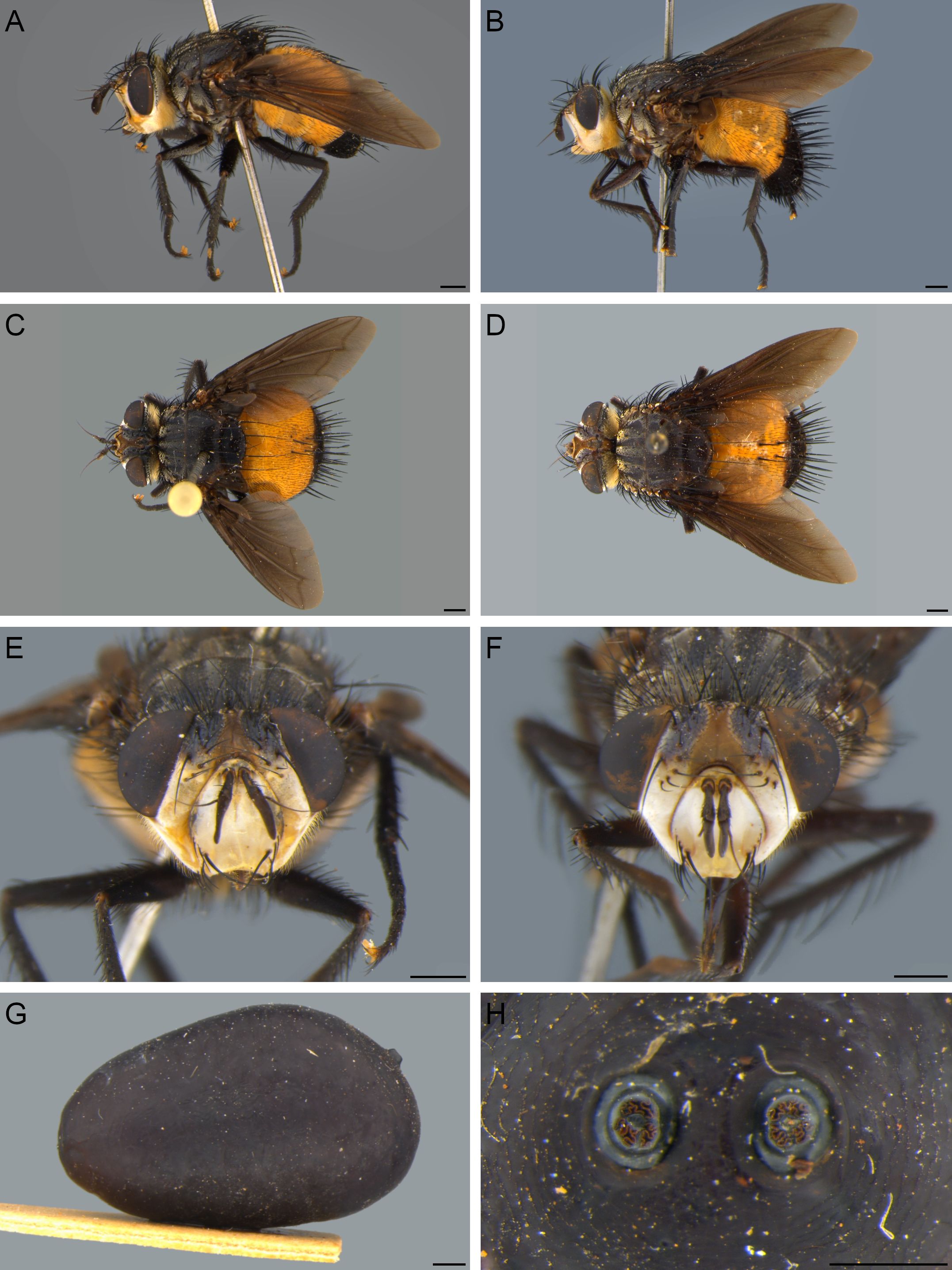
Diagnosis. Xanthozona can be distinguished from the remaining Tachinini genera by having two pairs of proclinate orbital setae in males (Fig. 2E); ocellar setae absent; palpi absent (Fig. 2B); prementum short, about half height of head; wings heavily infuscate, with posterior margin slightly hyaline (Figs. 2A-D); abdomen yellow with black or dark brown tip, with one pair of median marginal setae on tergite 3 and one pair of median discal setae on tergite 4; and male terminalia with postgonite almost vestigial, as a short sclerite adjacent to basiphallus (Fig. 4C).

Adults and puparium of Xanthozona scutellaris (Robineau-Desvoidy, 1830). A, C, E. Lateral habitus, dorsal habitus, and head in frontal view of male, respectively. B, D, F. Lateral habitus, dorsal habitus, and head in frontal view of female, respectively. G. Puparium in lateral view. H. Posterior spiracle of puparium. Scale bars: 1 mm.
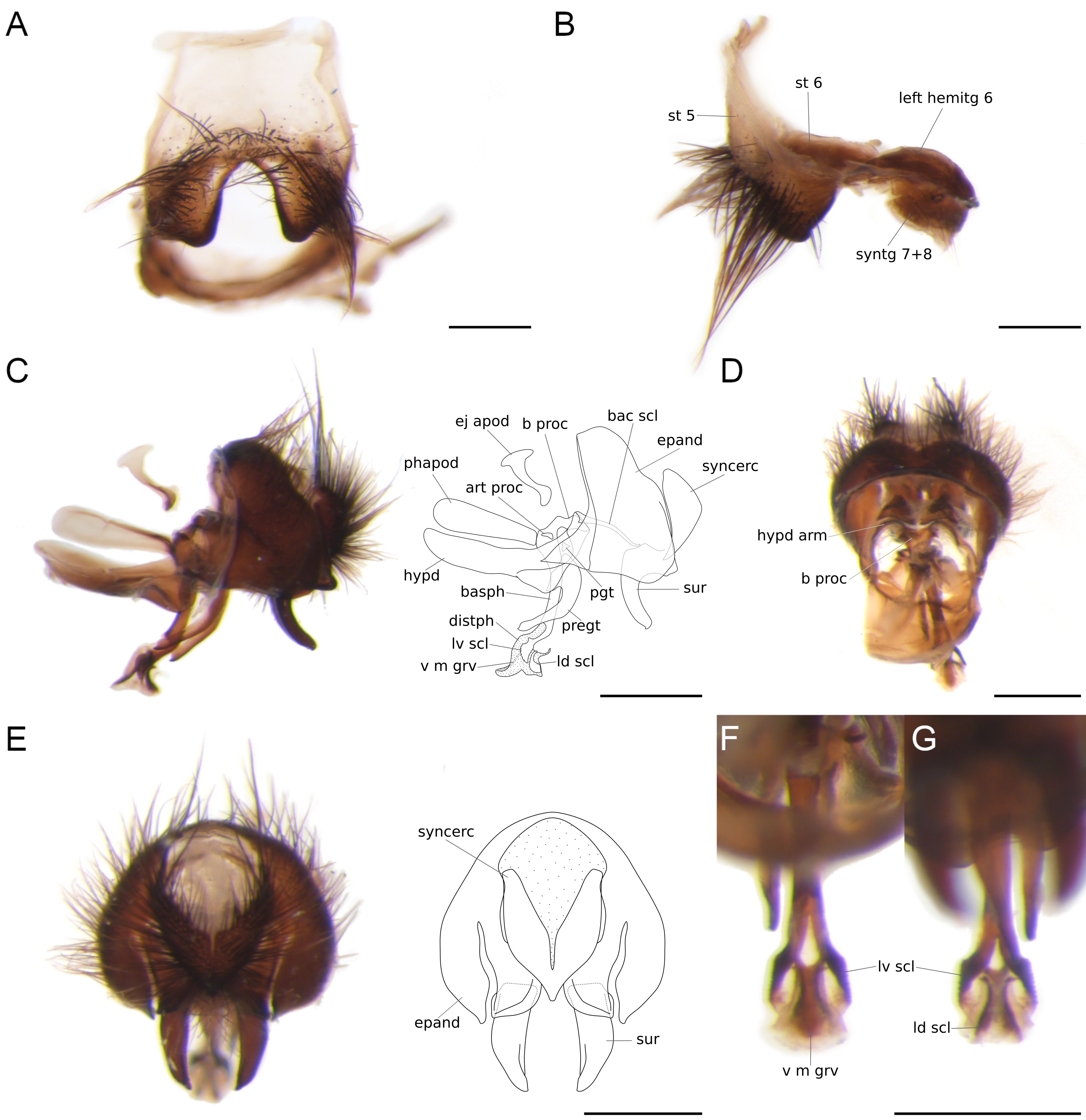
Male terminalia of Xanthozona scutellaris (Robineau-Desvoidy, 1830). A. Sternite 5, ventral view. B. Segments 5–8, lateral view. C. Genitalia and line drawing highlighting outer and inner structures (gray dashed lines) and membranous surfaces (dotted areas), lateral view. D. Hypandrial arms and basal process, internal view. E. Genitalia and line drawing, posterior view. F. Distiphallus, ventral view. G. Distiphallus, dorsal view. Scale bars: 0.5 mm. Abbreviations: art proc, articular process; b proc, basal process; bac scl, bacilliform sclerite; basph, basiphallus; distph, distiphallus; epand, ej apod, ejaculatory apodeme; epandrium; hemitg, hemitergite; hypd, hypandrium; hypd arm, hypandrial arm; ld scl, laterodorsal sclerite; lv scl, lateroventral sclerite; pgt, postgonite; phapod, phallapodeme; pregt, pregonite; st, sternite; sur, surstylus; syncerc, syncercus; syntg, syntergite.; v m grv, ventral median groove.
Remarks. Five genera of New World Tachinini are very similar to Xanthozona, as all of them lack palpi and have a yellow abdomen with a black or dark brown tip, namely: DiaphanomyiaTownsend, 1917 (two species), Euepalpus Townsend, 1908a (two species), Oxyepalpus Townsend, 1927 (one species), XanthoepalpusTownsend, 1914 (four species), and Zonoepalpus Townsend, 1927 (two species) (O’Hara et al., 2020; Gudin, 2024). Although there are other Neotropical Tachinini genera with similar color pattern, such as AdejeaniaTownsend, 1913, ParadejeaniaBrauer and Bergenstamm, 1893, and ProtodejeaniaTownsend, 1915, they have well-developed palpi and can be clearly identified using the keys of Wood (1987) and Wood and Zumbado (2010).
The following key will identify males and females of the small genera of Tachinini discussed above, some of them not included in Wood and Zumbado (2010):
1. Male with two pairs of proclinate orbital setae (Fig. 2E); abdomen with one pair of median marginal setae on tergite 3 and one pair of median discal setae on tergite 4 (Figs. 2C, D); male terminalia with postgonite almost vestigial, as a short sclerite adjacent to basiphallus (Fig. 4C) (Mexico to Argentina) … Xanthozona (Townsend, 1908)
Male without proclinate orbital setae; abdomen either without discal setae or with more than one pair on tergites 3 and 4; male terminalia with postgonite well-developed, bacilliform … 2
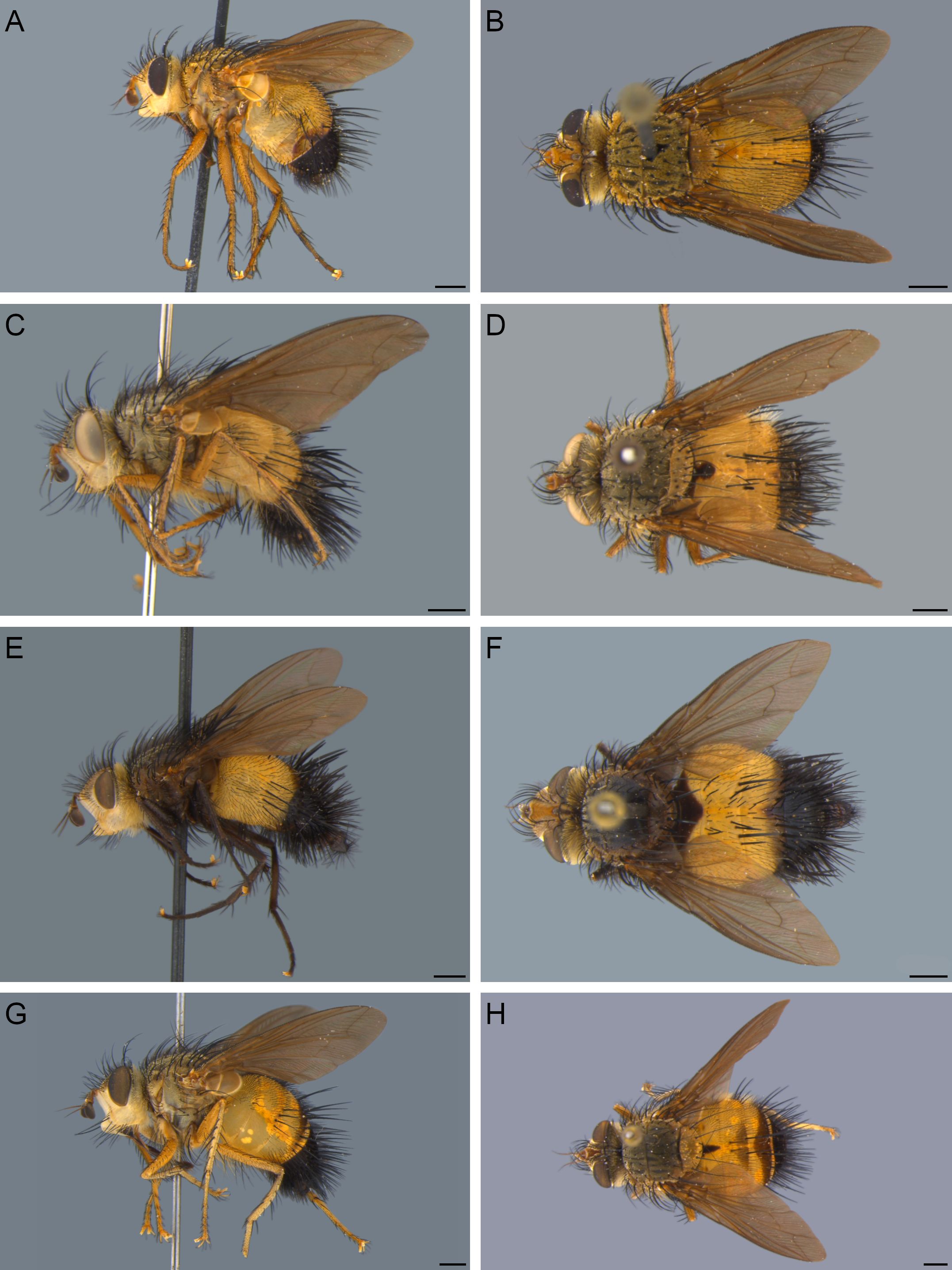
2. Parafacial covered with light setulae, with one strong proclinate seta (Fig. 7A); abdomen with one pair of median discal setae on tergites 3 and 4 (Fig. 4B) (Trinidad and Tobago to southern Brazil) … Diaphanomyia Townsend, 1917

Representatives of New World Tachinini genera that are similar to Xanthozona Townsend, 1908, lateral and dorsal habitus, respectively. A, B. Diaphanomyia aurifacies (Robineau-Desvoidy, 1830), male. C, D. Oxyepalpus flavoscutellatus (Bischof, 1904), male. E, F. Xanthoepalpus bicolor (Williston, 1886), male. G, H. Zonoepalpus testaceus (Robineau-Desvoidy, 1830), male. Scale bars: 1 mm.
Parafacial covered with black or light setulae, without proclinate setae or, at most, with one weak seta (see Zonoepalpus); abdomen without discal setae or with more than one pair on tergites 3 and 4 … 3
3. Abdomen without discal setae; labella with basal sclerites heavily sclerotized and apex of the lateral process of furca wide (see Figs. 3b, 4b in Gudin, 2024); male with tergite 5 covered with dense fine appressed setae (sexual patch), with narrow pale area above terminalia (see Figs. 5A, B in Gudin, 2024); female with abdomen dark brown with a triangular yellow spot on posterior margin of tergite 5 (see Figs. 4G, H in Gudin, 2024) (Costa Rica to southern Brazil) … Euepalpus Townsend, 1908

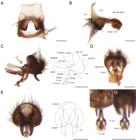
Holotype female of Jurinia scutellaris Robineau-Desvoidy, 1830. A. Lateral habitus. B. Dorsal habitus. C. Head in frontolateral view. D. Labels. Scale bars: 1 mm. Image C originally lacking scale. Muséum national d’Histoire naturelle, Paris (France). Collection: Insects - Diptera (ED). Specimen MNHN-ED-ED8301: http://coldb.mnhn.fr/catalognumber/mnhn/ed/ed8301.

Female terminalia of Xanthozona scutellaris (Robineau-Desvoidy, 1830), with line drawings of lateral and ventral views highlighting outer and inner structures (gray dashed lines) and membranous surfaces (dotted areas). A. Lateral view. B. Ventral view. Scale bars: 0.5 mm. Abbreviations: cerc, cercus; epiprct, epiproct; hemitg, hemitergite; hyprct, hypoproct; l arm, lateral arm; spr, spiracle; st, sternite.
Abdomen with pairs or rows of discal setae; labella with basal sclerites not heavily sclerotized and apex of the lateral process of furca narrow (except for some Xanthoepalpus species that have labella characters similar to those of Euepalpus); male without sexual patches, with tergite 5 covered with discal setae; male and female without sexual dimorphism in body color … 4
4. Tergites 3 and 4 with dark brown bands on posterior margin (Fig. 7H); abdominal setae regular, not spine-like (Argentina, southern and southeastern Brazil) … Zonoepalpus Townsend, 1927
Tergites 3 and 4 without dark brown bands on posterior margin or at most with narrow band on tergite 4 alone; abdominal setae spine-like (Fig. 7F) … 5
5. Parafacial and abdomen densely covered with light setulae (Fig. 7C); abdominal tergite 3 with median marginal setae in a straight row (Fig. 7D); male terminalia with surstyli arched and curved towards each other and syncercus with apex divided into two points (southeastern Brazil) … Oxyepalpus Townsend, 1927
Parafacial and abdomen densely covered with black setulae (Fig. 7E); abdominal tergite 3 with median marginal setae in a sagittate row, with median ones arising slightly farther forward than lateral ones (Fig. 7F); male terminalia with surstyli long and parallel to each other and syncercus with apex subquadrate (Canada to southeastern Brazil) … Xanthoepalpus Townsend, 1914
Xanthozona scutellaris (Robineau-Desvoidy, 1830)

First-instar larvae of Xanthozona scutellaris (Robineau-Desvoidy, 1830). A. Lateral habitus. B. Pseudocephalon and first thoracic segment, dorsal view. C. Ventral habitus. D. Pseudocephalon and first thoracic segment, lateral view. E. Cephaloskeleton, dorsal view. F. Cephaloskeleton, lateral view. G. Cephaloskeleton, lateroventral view. H. Thoracic segments highlighting each Keilin’s organ, lateral view. I. Thoracic segments and first abdominal segment, ventral view. J. Abdominal segments 1–3, lateral view. K. Anal division, dorsal view. L. Abdominal segment 7 and anal division, ventral view. Scale bars: 100 μm (A, C), 50 μm (I–L), and 20 μm (B, D–H). Abbreviations: a, abdominal segment; ad, anal division; ant, antenna; cs, circular sensillum; dc, dorsal cornu; dp, dorsal papilla; ds, dorsal surface; is, intermediate sclerite; ko, Keilin’s organ; lb, labrum; ldp, laterodorsal papilla; lvp, lateroventral papilla; ls, lateral surface; mh, mouth-hook; mx plp, maxillary palpus; p, papilla; pb, parastomal bar; pp, posterior papilla; ps, posterior spiracle; s, spiracle; sp, spine; t, thoracic segment; vc, ventral cornu; vp, ventral papilla; vpl, vertical plate.
Jurinia scutellaris Robineau-Desvoidy, 1830, p. 36. Holotype female (MNHN), examined (Fig. 3). Type locality: Guaratuba, [Paraná, see Remarks section below], Brazil.
Jurinia indica Robineau-Desvoidy, 1830, p. 36. Holotype missing [sex unknown]. Type locality: “Indes orientales” [erroneous locality, as discussed by Crosskey (1976)]. Syn. nov.
Tachina melanopyga Wiedemann, 1830, p. 292. Holotype female (SFMF), missing [see Remarks section below]. Type locality: Suriname. Syn. nov.
Jurinia indica: Macquart (1844, p. 195, distribution of species of JuriniaRobineau-Desvoidy, 1830); Crosskey (1976, p. 256, synonymy with T. melanopyga); Evenhuis et al. (2010, p. 185, list of Robineau-Desvoidy’s species).
Tachina melanopyga: Walker (1849, p. 702, list of accessions of NHMUK, diagnostic characters, locality not given); Townsend (1892, p. 14, as T. melanoppyga [misspelling], catalog of South American species of Calyptratae).
Echinomya melanopyga: Robineau-Desvoidy (1863, p. 662, unpublished combination of Macquart in MNHN’s catalog); Public Library, Museums, and National Gallery (Vic.) (Australia, 1889, p. 55, list of accessions of NHMUK, record in Pará, [Brazil]).
Jurinia melanopyga: Robineau-Desvoidy (1863, p. 662, redescription of female, record in Cayenne [French Guyana]).
Jurinia scutellaris: Robineau-Desvoidy (1863, p. 663, redescription); Townsend (1892, p. 8, catalog of South American species of Calyptratae); Crosskey (1976, p. 256, comparison with J. indica); Evenhuis et al. (2010, p. 196, list of Robineau-Desvoidy’s species).
Belvosia melanopyga: Jaennicke (1867, p. 87, combination).
Xanthozona melanopyga:Townsend (1908a, p. 116, redescription of female, records in Campinas and São Paulo, [São Paulo, Brazil]); Townsend (1916a, p. 20, description of male, record in Chapada [dos Guimarães, Mato Grosso, Brazil]); United States National Museum (1920, p. 195, list of accessions of USNM, record in Ancón, Canal Zone, [Panama]); Townsend (1931, p. 165, comparison with X. scutellaris); Travassos Filho and Carrera (1941, p. 46, description of third instar larva and puparium, 50, redescription of male, 54 redescription of female, 61, bionomy); Mariconi and Zamith (1954, p. 186, notes on taxonomy, distribution, and hosts, 187, records in Mauá [city of state of São Paulo], and Goiás, [Brazil]); Guimarães (1971, p. 74, catalog); Crosskey (1976, p. 256, synonymy with J. indica); Ziegler (1998, p. 163, discussion on Tachinini puparia); O’Hara et al. (2020, p. 887, checklist of Tachinidae); Santis and Nihei (2022, p. 5, phylogeny of Dufouriini, included as root, illustrations of first-instar larva, male terminalia, and spermathecae); Gudin (2024, p. 11, misidentification of the holotype of Euepalpus vestitus (Townsend, 1916c), as Parepalpus melanopygus).
Xanthozona scutellaris: Townsend (1931, p. 165, combination in Xanthozona); Guimarães (1971, p. 74, catalog); O’Hara et al. (2020, p. 887, checklist of Tachinidae).
Males (Figs. 2A, C, E, and 4). Body length: 8.57–12.81 mm (mean = 10.39 mm). Wing length: 8.38–10.82 mm (mean = 9.55 mm) (n = 10).
Coloration. Head with silver pruinosity; frontal vitta brown; ocellar triangle and fronto-orbital plate dark brown; parafacial, gena, and face light yellow. Antenna dark brown. Clypeus and labella brown; prementum dark brown. Occiput covered with silver pruinosity. Thorax with silver pruinosity; scutum dark brown with four vittae, fainter on postsutural scutum. Lateral thoracic sclerites brown to dark brown. Scutellum dark brown with brownish pruinosity. Wing heavily infuscate, slightly hyaline at posterior margin; tegula, basicosta, and veins dark brown. Halter brown. Calypteres dark brown. Legs dark brown, with silver pruinosity. Tarsal claws yellow with black tip. Pulvilli light yellow. Abdomen without pruinosity; yellow from syntergite 1 + 2 to tergite 4; tergite 5 black or dark brown.
Head. Ratio of head height/head width in frontal view 0.70–0.74 (mean = 0.71). Ratio of vertex width/head width in dorsal view 0.35–0.41 (mean = 0.38). Ratio of lower frontal vitta width/upper frontal vitta width 1.36–2.44 (mean = 1.95). Ratio of upper frontal vitta width/fronto-orbital plate width in dorsal view 0.55–1.03 (mean = 0.74). Ratio of parafacial width/fronto-orbital plate width 0.85–1.12 (mean = 0.99). Ratio of first flagellomere length/pedicel length 1.21–1.83 (mean = 1.49). Ratio of genal height/head height in lateral view 0.27–0.34 (mean = 0.30). Dichoptic. Eye bare. Antenna inserted at middle level of eye, about two-thirds height of face. Scape short, about one-third length of pedicel. Pedicel setose dorsally, with one apical erect seta. First flagellomere narrower at apex with anterior margin convex, kidney-shaped; outer surface with one minute sensory pit closer to posterior margin. Arista long, micropubescent, thickened at basal half, and arising at base of anterior margin of first flagellomere; first aristomere short, about half length of second aristomere. Frontal vitta wider at lunule, narrowing abruptly right before ocellar triangle. Six or seven frontal setae from level of pedicel to level of upper quarter of eye, with anterior setae inclinate and longer than posterior setae, posterior setae reclinate. Two pairs of proclinate orbital setae, with anterior seta arising at level of last inclinate frontal seta and posterior seta arising at level of last reclinate frontal seta. Two reclinate orbital setae, with anterior seta about two-thirds length of posterior seta. Ocellar triangle setulose with all ocelli of similar size; ocellar setae absent. Two postocellar setae. Inner vertical setae crossed. Outer vertical setae lateroclinate, subequal to length of inner vertical setae. Fronto-orbital plate setulose. Parafacial large, subequal to width of fronto-orbital plate, covered with light setulae and sometimes with black setulae in lower half. Facial ridge bare, except on lowest quarter with one to three supravibrissal setae. Vibrissa strong and crossed, arising above lower facial margin, with three or four subvibrissal setae. Facial margin well projected forward, clearly visible in profile. Gena with four to six genal setae on ventral margin. Occiput densely covered with long, white setulae. Clypeus long and flat. Palpi absent, not represented as a tubercle or short setae. Prementum bare and short, about half height of head; shaped as a sheath, completely covering labrum. Labellum with ventral margin forming a short lobe emerging from central process of furca; basal sclerite membranous; apex of lateral process of furca narrow.
Thorax. Prosternum bare. Proepisternum without setulae, with two upcurved proepisternal setae. Two or three upcurved proepimeral setae, with setulae at base. Anterior spiracle with both lappets well developed, covering almost entire opening. Posterior spiracle with posterior lappet shaped as an operculum. Six postpronotal setae. Scutum with 3–4 + 2 acrostichal setae; 3 + 4 dorsocentral setae; 2 + 3 intra-alar setae; 1 + 3 supra-alar setae, strong and subequal; postsutural setae stronger than presutural setae. Two notopleural setae. Four postalar setae. Anepisternum anterodorsal corner with patch of upcurved setae; posterior margin with row of five to seven setae. Three katepisternal setae, with lower seta closer to anterior seta. Two strong anepimeral setae, with patch of setulae at base. Katepimeron bare. Six or seven meral setae. Katatergite and anatergite bare. Scutellum with one pair of basal setae; two pairs of lateral setae, with outer pair stronger than inner pair, subequal to subapical setae; one pair of subapical setae; one pair of apical setae, crossed; one pair of discal setae. Wing. Vein C ending right after vein R4+5, just before wing apex; costal spine absent. Vein R4+5 setose only at base. Bend of vein M at angle of 90º, vein beyond bend straight and reaching wing margin well before wing apex. Cell r4+5 open, with length subequal to crossvein r-m. Legs. Fore coxa with two longitudinal rows of setae on anterior surface. Fore femur with dorsal, posterodorsal, and posteroventral rows of setae from base to apex. Fore tibia with complete dorsal row of setae, one strong apical seta on posterodorsal surface, two strong median setae, and one apical seta on posterior surface. Mid coxa with row of setae on anterior surface. Mid femur with patch of seven to nine strong median setae on anterior surface, two or three basal setae on ventral surface. Mid tibia with complete row of strong setae on anterodorsal surface, one median seta on anterior surface, two median setae on posterior surface, three or four apical setae on ventral surface. Hind coxa with row of setae on apical and outer lateral margin, posterior margin setulose. Hind femur with complete rows of setae on anterodorsal and anteroventral surfaces, and basal patch of setae on posteroventral surface. Hind tibia with sparse anterodorsal and posterodorsal rows of setae, one strong median setae, and one apical seta on anteroventral surface. Basal tarsomeres long, subequal to combined length of remaining tarsomeres. Tarsal claws long, almost as long as last tarsomere.
Abdomen. Rounded and wider than thorax, with spine-like setae. Syntergite 1 + 2 with mid-dorsal depression reaching hind margin; one pair of lateral marginal setae. Tergite 3 with one pair of median marginal setae and one pair of lateral marginal setae. Tergite 4 with row of marginal setae and one pair of median discal setae. Tergite 5 with row of marginal setae and discal setae evenly distributed throughout tergite. Sternites partially overlapped by tergites, visible from base to apex bearing row of marginal setae. Sternite 5 concave and subquadrate, with base subequal to width of apex (Fig. 4A); anterior margin with pair of well-developed lateral apodemes; posterior lobes densely covered with setae; inner margin of posterior lobes covered with spicules.
Terminalia (Fig. 4). Tergite 6 with median region narrow, giving general appearance of two lateral hemitergites (Fig. 4B); right hemitergite more developed than left hemitergite; pair of spiracles on membrane. Sternite 6 asymmetrical, connected to left hemitergite of tergite 6 by wide area, with anterior margin widening towards connection to sternite 5, forming two lobes, ending right after connection to sternite 5. Syntergite 7 + 8 narrow and setulose, with anterior half bearing pair of spiracles. Anterior epandrial process at same level of ventral epandrial margin (Fig. 4C). Epandrium with dorsal surface about half length of ventral surface in lateral view, covered with long setae; posterior margin forming rounded lower lobe in lateral view. Surstylus fused with epandrium, with inner part densely covered with long setae; median part bent forward, forming a pronounced median posterior lobe (Fig. 4C); distal part subparallel in posterior view, with inner surface concave and with pointed tip (Fig. 4E). Bacilliform sclerites not fused with each other, wider towards connection to hypandrial arms. Hypandrium with hypandrial apodeme concave, with wide rounded apex in ventral view, almost as long as central plate of hypandrium; posterior margin of central plate of hypandrium not fused with pregonites; hypandrial arms fused with lateral part of basal process of basiphallus (Fig. 4D). Phallapodeme with longitudinal keel on anterior surface, subequal to length of hypandrium, without phallic guide. Ejaculatory apodeme about two-thirds length of phallapodeme, T-shaped in lateral view. Phallus with epiphallus absent; basiphallus long and slightly sinuous, with well-developed basal process; distiphallus wide and sclerotized, connected to basiphallus and slightly bent towards anterior region; pair of sclerotized lateroventral sclerites, serrated, fused with median sclerite on ventral median groove of distiphallus (Fig. 4F); apical dorsal surface with a pair of laterodorsal sclerites surrounding a thin and membranous lobe (Figs. 4C, E). Pregonite bare, long and bent towards anterior region, with anterior base wide and apex pointed. Postgonite almost vestigial, as a short sclerite adjacent to basiphallus, with well-developed articular process. Syncercus entirely setose, convex and globose, heart-shaped (Fig. 4E).
Females (Figs. 2B, D, F, and 5). Differ from males as follows: Body length: 9.55–12.35 mm (mean = 11.04 mm). Wing length: 9.11–11.62 mm (mean = 10.47) (n = 10).
Head. Ratio of head height/head width in frontal view 0.65–0.71 (mean = 0.69). Ratio of vertex width/head width in dorsal view 0.34–0.41 (mean = 0.38). Ratio of lower frontal vitta width/upper frontal vitta width 1.65–2.14 (mean = 1.90). Ratio of upper frontal vitta width/fronto-orbital plate width in dorsal view 0.49–0.90 (mean = 0.70). Ratio of parafacial width/fronto-orbital plate width 0.88–1.19 (mean = 1.00). Ratio of first flagellomere length/pedicel length 1.13–1.51 (mean = 1.33). Ratio of genal height/head height in lateral view 0.25–0.33 (mean = 0.29).
Thorax. Foreleg with tarsomeres 2–5 enlarged and flattened, wider than basitarsus, bearing patches of sensilla on ventral surface.
Terminalia (Fig. 5). Ovipositor very short and flat. Three spermathecae, spherical with rough surface. Tergites 6 and 7 divided into two hemitergites, setose; hemitergite of tergite 6 stout, bearing spiracles 6 and 7; hemitergite of tergite 7 about two-fifths length of hemitergite of tergite 6. Sternite 6 wide and subtrapezoidal, setose. Sternite 7 subtrapezoidal, about two-thirds width of sternite 6, setulose. Tergite 8 divided into two hemitergites; hemitergite of tergite 8 reduced to a narrow strip, bare. Sternite 8 subtriangular, setulose on medial surface. Epiproct very reduced, with four pairs of setae. Hypoproct subtriangular, setulose; medioventral surface concave, covered with short spiniform setae; basal margin with a pair of well-developed lateral arms extending about one-third length of hypoproct. Cerci not fused, setose on posterior half.
Puparia (Figs. 2G, H). Length: 12.25–12.92 mm (mean = 12.57) (n = 4).
Amphipneustic, oval shaped; posterior region wider than anterior region in lateral view. Anterior spiracle small, adjacent to mouth scar, with four slits arranged in a longitudinal row. Posterior spiracles slightly displaced dorsally, with spiracle plates flat; spiracle slits sinuate and arranged in a circle.
First-instar larva (Fig. 6). Small, about 600 μm; cylindrical, with segments of similar length (Fig. 6A).
Head. Pseudocephalon membranous; with large patch of spines on ventral surface (Figs. 6B, D); antenna short, with distal joint semioval, and one adjacent inner and outer circular sensilla and one outer papilla; maxillary palpus flat and rounded; one median patch of short spines below antennae. Cephaloskeleton with labrum long, slender and subequal to width of parastomal bar, with apex slightly emarginate (Fig. 6F); vertical plate forming an acute angle with ventral cornu; dorsal cornu large, with pointed apex; ventral cornu thin, about one-fourth width of dorsal cornu; mouth-hooks not fused at tip (Fig. 6E) with anterior region broad in lateral view (Fig. 6F), subequal to width of labrum and located right before its apex, and posterior region as a narrow strip, located below apex of labrum; intermediate sclerite H-shaped with posterior arms short in ventral view (Fig. 6G), narrow and curved in lateral view (Fig. 6F).
Thorax. Ventral surface with Keilin’s organ and one adjacent papilla, four marginal and one lateral circular sensilla (Figs. 6I, J). Segment 1 membranous, with lateral band of spines (Figs. 6B, D); dorsal surface with one discal circular sensillum and four marginal circular sensilla; lateral surface with three circular sensilla and one upper papilla. Segments 2 and 3 covered with sclerotized, dotted plates on dorsal and lateral surfaces, separated by narrow laterodorsal plates (Figs. 6A, J); dorsal surface with six discal and two lateral circular sensilla (Fig. 6J); lateral surface with two lateral circular sensilla and one lower papilla between them; ventral surface with band of spines on basal third.
Abdomen. Similar to thoracic segments 2 and 3, except the following: lateral surface with small spiracle on upper margin (Fig. 6J); ventral surface of segments 1–7 with six marginal circular sensilla, one lateral circular sensillum, and without papillae (Fig. 6I); ventral surface of anal division without band of spines (Fig. 6L), with anterior half bearing four marginal circular sensilla and one papilla inserted at middle of outer circular sensilla, and posterior half bearing a triangular patch of short spines, with two marginal circular sensilla and one lateroventral circular sensilla. Anterior half of anal division with dorsal surface bearing two basal rows of nodule-like spines, anterior row with six strong spines and posterior row with ten weaker spines (Fig. 6K). Posterior half of anal division with one pair of dorsal, laterodorsal, posterior, lateroventral, and ventral papillae around spiracular field (Figs. 6K, L); posterior spiracles and tracheal trunks well-developed, with similar width.
Type material examined. Holotype ♀ of Jurinia scutellaris Robineau-Desvoidy, 1830: “Guaratuba/ ouest, mars/ 1820”/ “T. scutellaris”/ “Jurinia/ scutellaris/ R.D.”/ “Holotype”/ “Holotype ♀/ Jurinia/ scutellaris/ Robineau-Desvoidy/ det. P.H. Arnaud 1972”/ “MNHN, Paris/ ED8301” (MNHN).
Additional material examined. Argentina: 1♀ (MACN), Misiones, iii.1941, R. Kohler col., ex Brassolis, colección Blanchard; 1♂, 2♀♀ (MACN), Misiones, 15.xi.1941, [no collection data], S/ Brassolis astyra, colección Blanchard. Bolivia: 1♂, 1♀ (MACN), [Santa Cruz], Provincia Sara, [north of] Santa Cruz de la Sierra, [no date], José Steinbach col. Brazil: 1 specimen (AMNH), Amazonas, Benjamin Constant, Rio Javary, 25.i–15.ii.1942; 1♀ (UEFS), Bahia, Muritiba, Morro de Aporá, 3.i.1991, J. Becker col.; 1♀ (UEFS), Bahia, Salvador, Rio Vermelho, i.1960, J. Becker col.; 1 specimen (USNM), Bahia, Salvador, 20.xii.1966; 1♀ (CEIOC) [dissected, first-instar larvae photographed], Bahia, Salvador, Canela, 18.viii.1951, Afonso Garcia col.; 1♀ (CEIOC) Bahia, Engenho Velho de Brotas [as E. Velho], 28.viii.1951, D. Albuquerque col.; 1♂, 1♀ (MNRJ) [male dissected], Espírito Santo, Linhares, x.1972, P.C. Elias col.; 2♀♀ (DZUP), same as preceding, except DZUP 250909/250910; 1♂ (MZSP) [dissected], Minas Gerais, Belo Horizonte, Campus UFMG, 6.xi.1996, M.S. Silva col.; 1♀ (UFMG), same as preceding, except 10.iv.1997, D. Yanega col., UFMG IDI 1300067; 1♂ (MZSP), Minas Gerais, Presidente Olegário, Fazenda Gigante, 1000 m, 2–5.iv.2007, 18°31'S 46°18'W, Amorim, Ribeiro, Capellari and Berbert col.; 3♀♀ (DZUP), Minas Gerais, Sabará, 13–18.iv.1981, C. Elias col., DZUP 459777/459778/459779; 1♂ (AMNH), [Mato Grosso], Chapada [dos Guimarães], xii [no year], H.H. Smith col.; 1♂ (DZUP) [dissected], Mato Grosso, Chapada dos Guimarães, 28.iii–iv.1983, Exc. Dep. Zool. UFPR (Polonoroeste), DZUP 250194; 1♂ (MNHN), Pará, [no date], de Mathan col., 1♀ (CEIOC), Pará, Óbidos, Colônia Rio Branco, v.1953, José Brazilino col.; 2♀♀ (DZUP), Paraná, Curitiba, xii.1993, F. Giacomel col., DZUP 252998/252999; 1♂ (DZUP), same as preceding, except C. Politec., 11.xi.1997; 3♂♂, 2♀♀ (MZSP) [one male dissected], Pernambuco, Recife, x.1974, E.M.C. col.; 1 specimen (USNM), Pernambuco, Tapera, 9.v.1935; 1♀ (CEIOC), Rio de Janeiro, Rio de Janeiro, 1.ii.1934, H. Souza Lopes col., ex Brassolis astyra; 1♀ (CEIOC), same as preceding, except Jacarepaguá, 1931, Berla col., 12121; 1♂ (MNRJ) [dissected], same as preceding, except Jardim Botânico, Embrapa, viii.1933, P. Dehioni col.; 1♀ (MZSP), Rio Grande do Sul, Pelotas, 14.i.1962, C.M. Biezanko col., 1♀ (CEIOC), [Santa Catarina], Joinville, [no date], Brückner col., 12122; 1 specimen (AMNH), Santa Catarina, Nova Teutônia, 3.vi.1939, Fritz Plaumann col., 27°11'S, 52°23'W, AMNH_IZC_00337561; 1 specimen (CNC), same as preceding, i.1966, CNC1808277; 1♀ (MZSP), São Paulo, Barueri, 28.xii.1965, K. Lenko col.; 2♀♀ (MZSP, USNM), São Paulo, Caieiras, i.1953, M. Carrera col; 1♀ (MZSP) [dissected], São Paulo, Cássia dos Coqueiros, ii.1947, M.P. Barreto col.; 4 specimens (USNM) [one male dissected], São Paulo, Campinas, 7.xi–5.xii.1937, USNMENT_01518086; 1♀ (MZSP) same as preceding, except Instituto Agronômico, 20.iv.1948, R.A. Rodrigues col., ex brassolídeo; 1♀ (MZSP), São Paulo, Mauá, xi.1940, H. Hardinan col.; 2♀♀ (AMNH, MZSP), São Paulo, Piracicaba, i.1953, Famm col.; 1♂ (MZSP), São Paulo, Porto Cabral [former village of the city of Presidente Epitácio, see more details in (Travassos, 1945)], Rio Paraná, 1-25.iv.1944, Travassos Filho [as Trav. Fo.], Carrera and E. Dente col.; 1♂ (MZSP), São Paulo, São Carlos, iv.1954, C.L. Pereira col., ex larva Brassolis sophorae; 2♂♂, 3♀♀ (MZSP) [one male dissected], same as preceding, except 8.xii.1974, [no collection data]; 1♂ (MZSP), São Paulo, São Paulo, Cidade Universitária-USP, 25.iv.1985, [no collection data]; 1♂ (MZSP), same as preceding, except iv.1991, Reginaldo col.; 1♂ (MZSP) [dissected], same as preceding, except 12.iv.1991, S.A. Vanin col.; 1♀ (MZSP), same as preceding, except 17.iii.2011, 23°33'57”S 46°43'46”W, J. Audino col., T2345; 1♂ (MZSP), São Paulo, São Paulo, 14.xii.1925, R. Spitz col.; 2♀♀ (MZSP) [one female dissected], same as preceding, except 1930, [no collection data]; 1♂, 2♀♀ (CEAH, MNRJ), same as preceding, except xii.1930, D. Braz col.; 1 specimen (USNM), same as preceding, except xii.1931, [no collection data]; 5♂♂, 5♀♀ (AMNH, CEAH, MZSP), same as preceding, except xii.1943, R.L. Araújo col.; 1 specimen (CNC), same as preceding, except Ipiranga [as Ypiranga], i.1941; 1♀ (MZSP) [dissected and photographed], same as preceding, except Pinheiros, iv.1980, D.S. Amorim col.; 1♀ (MZSP), São Paulo, Sorocaba, iv.2012, Almeida N. col. Colombia: 1 specimen (AMNH), [Santander], Río Opón, 20.xii.1947–7.i.1948. Ecuador: 1 specimen (NHMUK), Napo, Muyuna, 6km W of Tena, 30.xi.1978, Martin Cooper col., NHMUK013741345; 1 specimen (NHMUK), Napo, Tena, 14.iv.1976, Martin Cooper col., NHMUK013741346; 2♂♂ (MNHN), Napo, viii.1930, R. Benoist; 1♂ (CEIOC), [Tungurahua], Jauregui [as Jarugui], v.1937, O. Staudinger and Bans-Haas col.; 1♂ (MNHN) [dissected and photographed], [Santo Domingo de los Tsáchilas], Santo Domingo [as Santo Domingo de los Colorados], viii–ix.1930, R. Benoist col. French Guiana: 1 specimen (CNC), Cayenne, iv.1977; 2♂♂ (MNHN), Cayenne, Montravel, 23.vi.1975; 6♂♂ (MNHN) [one dissected], [no locality]; 1♂ (MNHN), [no locality], iii.1985, ex Brassolis sophorae. Panama: 1 specimen (AMNH), Barro Colorado, Canal Zone, 3.xii.1930, AMNH_IZC_00337574. Paraguay: 1♂ (MZSP) [dissected], [Canindeyú], Salto del Guairá, 1–3.xii.1971; 1 specimen (USNM), [Concepción], Concepción, 27.i.1933; 2 specimens (USNM), [Cordillera], San Bernardino, [no date]; 1♂ (MNHN), [Guairá], Carlos Pfannl [as Carlos Pfanni], 29.ii.1946, Duret col.; 1 specimen (USNM), [Guairá], Villarrica, v.1928, F.H. Schade col., USNMENT_01345564; 13 specimens (2 in CNC, 11 in USNM), [Guairá], Villarrica, xii.1936–v.1937, F.H. Schade col., USNMENT_01634216, USNMENT_01635141, USNMENT_01635141.1, USNMENT_01635141.2; 2♂♂ (MNHN), [Guairá], Villarrica, xi.1945, F.H. Schade col.; 1 specimen (USNM), [Itapúa], Trinidad [as Santísima Trinidad], viii.1913; 1♀ (MZSP), [Paraguarí], Sapucai, 23–24.xi.1971. Peru: 2 specimens (USNM), [Junín], Satipo, xii.1948 and i.1949; 1 specimen (USNM), [Ucayali], Pucallpa, 26.xi.1947.
Remarks. Xanthozona scutellaris is the sole representative of this genus and is widely distributed in the Neotropical Region. Given the challenging classification of Neotropical Tachinidae, particularly Tachinini, with numerous restricted genera, it is possible that X. scutellaris could belong to a more inclusive Tachinini genus when a comparative study with additional species is conducted. In fact, the male terminalia of X. scutellaris shares several traits with species of the Neotropical genus TrichophoraMacquart, 1847, and related genera, such as Diaphanomyia and Zonoepalpus, having a heart-shaped syncercus and distiphallus with apical dorsal surface bearing a pair of laterodorsal sclerites surrounding a thin and membranous lobe (Figs. 4C, G). However, X. scutellaris does not have the characteristic strong parafacial setae observed in these groups (Fig. 7A). Phylogenetic studies are needed to clarify how X. scutellaris is related to the species of these genera, which will provide evidence and support to propose a more stable classification for Neotropical Tachinini.
On examining the holotype female of J. scutellaris, we conclude that this species shares the same diagnostic characters listed in the original descriptions of T. melanopyga and J. indica and observed in the examined material. As noted by Crosskey (1976, p. 256), Robineau-Desvoidy (1830, p. 36) acknowledged the similarity between J. scutellaris and J. indica, distinguishing them primarily by the color of the scutellum, which was also used as a diagnostic character by Townsend (1931, p. 165), and the shape of the pruinose vittae on the scutum. Although the scutellum of the J. scutellaris holotype (Fig. 3B) is lighter than that of other examined specimens (Figs. 2C, D), this variation is found within the range observed in the examined material, with no apparent differences in the male terminalia and other diagnostic characters. Specimens from South and Southeast Brazil, localities closer to the type locality of J. scutellaris, show no morphological evidence supporting the existence of a separate species. According to the principle of priority (ICZN, 1999, Article 23.1), the valid name for this species of Xanthozona must be one of the two Robineau-Desvoidy names. The name J. scutellaris is the more appropriate choice given that its name-bearing type is still extant, unlike the type of J. indica. Acting as the First Reviser (ICZN, Article 24.2.2), we hereby select J. scutellaris as the senior synonym over J. indica.
Regarding the type locality of J. scutellaris, there is another place named Guaratuba, a beach in the city of Bertioga, state of São Paulo, Brazil. The type specimen was collected by the French naturalist Augustin Saint-Hillaire (Robineau-Desvoidy, 1830). Papavero (1971) reviewed Saint-Hilaire’s field trips and verified that he did not visit the coast of São Paulo; instead, he passed through Guaratuba in the state of Paraná (see Papavero, 1971, p. 120 and Map 10).
Concerning the holotype of T. melanopyga, the SFMF curatorial staff was contacted to verify the type material of Tachinidae; however, no specimen matching the type locality of Suriname indicated by Wiedemann (1830) was found. There is a single specimen identified as X. melanopyga in the SFMF collection, collected by the German naturalist Georg Wilhelm Freyreiss, which is mistakenly labeled as the type. This specimen is severely damaged, with only parts of the thorax and wings preserved, and it was collected in Brazil without a recorded date. As the locality does not match the type locality of T. melanopyga, this specimen cannot be considered the original type specimen. Therefore, the holotype of T. melanopyga may be considered as lost.
The known distribution of X. scutellaris covers most of the Neotropical Region, except for Chile and some countries in Central America (Fig. 1). Despite its wide distribution, the specimens examined in this study show very little variance. Some specimens from northern areas of South America and Central America have parafacial covered mostly with black setae, as noted by Wood and Zumbado (2010) when studying the Central American fauna. However, the male terminalia of these specimens do not show sufficient differences to recognize different species.
The first-instar larvae of X. scutellaris are described here for the first time. They share many similarities with other Tachinini species, such as the shape of antennae, arrangement of body sensilla, and the presence of rows of nodule-like spines at the base of the dorsal surface of anal division (Thompson, 1923, 1963; Cantrell, 1988). These rows of nodule-like spines are also found in other Tachininae groups, all of which exhibit indirect oviposition strategies, laying incubated membranous eggs with well-developed first-instar larvae on the host path (Nakamura et al., 2013). These larvae lie on the substrate sheathed in the remnants of the membranous chorion of the egg, waiting for the host (Mellini, 1990). As soon as the host approaches, the larvae stand up and begin to sway to reach the host (Townsend, 1908b). Such rows of spines in the anal division are absent in other groups with well-developed first-instar larvae that actively seek out the hosts, such as Dexiini (Thompson, 1963; Santis and Couri, 2024; Santis et al., 2024). This indicates that these rows of nodule-like spines may be correlated with the movement these larvae do to reach their hosts. Further studies are necessary to confirm this hypothesis.
Thompson (1963) and Cantrell (1988) made significant contributions to understanding the larval morphology of Tachinidae. However, their terminology for the cephaloskeleton and other characters of first-instar larvae requires clarification in light of more recent studies on dipteran larval morphology (Courtney et al., 2000). The terms hook or mouth-hook actually refer to the labrum, the intermediate region to the parastomal bars, the lateral plates or accessory sclerites to the mouth-hooks, and the sclerite of the salivary gland to the intermediate sclerite, which is clearly visible in ventral view (Fig. 6G).
Regarding the body segmentation of the first-instar larva, Courtney et al. (2000) recognize only seven abdominal segments in brachyceran larvae, with the posteriormost division termed the anal division due to its composite nature. The number, position, and shape of papillae (rod-shaped sensoria as defined by Thompson, 1963) around the spiracular field seem to have significant taxonomic and systematic value within Oestroidea. For instance, species of Calliphoridae (Szpila et al., 2014), Polleniidae (Szpila 2003), and Sarcophagidae (Szpila et al., 2024) typically exhibit seven pairs of papillae, with varied degrees of development. However, Xanthozona scutellaris and other Tachinini (Thompson, 1963) present only five pairs (Figs. 6K, L). A comprehensive comparative study of papillae positioning and morphology is needed to clarify their homologies and evolutionary transformations.
Host catalog of Xanthozona scutellaris
Numerous articles have published information regarding the hosts of X. scutellaris (mentioned as X. melanopyga), especially in Brazil, although not all have included new host records. Readers are referred to the following articles dealing with science outreach, discussions on the potential of X. scutellaris as a biological control agent of palm tree pests, or comparisons with other parasitoids of palm tree pests: Silva Prado Júnior (1942), Blanchard (1941b), Lordello (1949, 1952), Lima (1950), and Marcicano et al. (2009).
Xanthozona scutellaris has been recorded in at least six species of Lepidoptera of the families Erebidae, Nymphalidae, and Notodontidae (Table 1). In all records, adult flies emerged from the host’s pupa, with one adult per pupa. Ochraceous fluid is usually exuded from parasitized pupae (Travassos Filho and Carrera, 1941; Piza and Zamith, 1944; Clark and Silva, 1977). Adult flies typically emerge near the thoracocephalic suture of the host’s pupa, with scars of the posterior spiracles of the fly larvae visible on the host’s abdominal segments 3, 4, or 5 (Travassos Filho and Carrera, 1941). However, the reliability of some host associations is pending revision, as discussed in the following remarks.
List of lepidopteran hosts of Xanthozona scutellaris (Robineau-Desvoidy, 1830) (Diptera: Tachinidae), with localities of recorded parasitism. Host species with a question mark (?) indicate unreliable records that require further verification. Additional details regarding these records, localities, and respective references can be found in the host catalog in the main text.
Family Nymphalidae
Brassolis astyra astyra Godart, [1824]
Recorded by Townsend (1908a, p. 116) in Campinas and São Paulo, São Paulo, Brazil; Andrade (1927, p. 72, as Xanthozona sp.) in Rio Claro, São Paulo, Brazil; by Hugo Souza Lopes, pinned specimen collected in February 1934 [see Additional material examined] in Rio de Janeiro, Rio de Janeiro, Brazil; by Blanchard (1941a, p. 16) in Posadas, Misiones, Argentina; by Travassos Filho and Carrera (1941, p. 44) in São Paulo, São Paulo, Brazil; by R. Kohler, pinned specimen collected in March and November 1941 [see Additional material examined] in Misiones, Argentina; by Sauer (1946, p. 22, as Xanthosoma [misspelling] melanopyga) in Campinas, São Paulo, Brazil; by Mariconi and Zamith (1954, p. 187) in São Paulo, São Paulo, Brazil; by Blanchard and Santis (1975, p. 23), repetition of record of Blanchard (1941a); and by Salgado-Neto (2011, p. 414) in Santa Maria and Tupanciretã, Rio Grande do Sul, Brazil. Remarks: Salgado-Neto (2011, p. 414) mentioned the species Rachiplusia nu (Guenée, 1852b) (Noctuidae) as a host of X. scutellaris based on the catalog of Lima (1948, p. 26). Although X. scutellaris is really associated with R. nu in Lima (1948), this is a mistake due to a mismatch in the format of the parasite and host columns in the original catalog. Xanthozona scutellaris is preceded by Winthemia rufopicta (Bigot, 1889), followed by Protypophaemyia haywardi (Blanchard, 1942) [as YpophaemyiaTownsend, 1916b]. Rachiplusia nu is a host of W. rufopicta (Guimarães, 1977), followed by B. astyra, a host of X. scutellaris, and Spodoptera ornithogalli (Guenée, 1852a) [as ProdeniaGuenée, 1852a], a host of P. haywardi. Therefore, the association between X. scutellaris and R. nu is misguided.
Brassolis sophorae sophorae (Linnaeus, 1758)
Recorded by Piza and Zamith (1944, p. 205) in Piracicaba, São Paulo, Brazil; by C. L. Pereira, pinned specimen collected in April 1954 [see Additional material examined] in São Carlos, São Paulo, Brazil; by Santos and Rocha (1981, p. 156) in Quatro Barras, Paraná, Brazil; pinned specimen [no collection data] collected in March 1985 [see Additional material examined] in French Guiana [locality not given]; and by Ruszczyk (1996, p. 357) in Campinas, São Paulo, Brazil.
Unidentified BrassolisFabricius, 1807 [1938]
Recorded by Clark and Silva (1977, p. 176) in Viçosa, Minas Gerais, Brazil.
Caligo illioneus oberon Butler, 1870
Recorded by Bustillo Pardey (2011:[unpaginated]) in Colombia [locality not given]. Remarks: Salgado-Neto (2011) listed C. illioneus as a host of X. scutellaris referencing Gómez Laverde and Lastra Borja (1998) and Bustillo Pardey (2011). Gómez Laverde and Lastra Borja (1998) recorded a dipteran parasitoid in this species, but they did not specify the species. Later, Bustillo Pardey (2011) revised the natural enemies of C. i. oberon, a pest on sugarcane plantations in Valle del Cauca, and included X. scutellaris based on identifications made by researchers of Centro de Investigación de la Caña de Azúcar de Colombia (Bustillo Pardey, personal communication). However, this record is pending revision because the original record could not be traced.
Opsiphanes invirae (Hübner, [1808])
Recorded by Mariconi and Zamith (1954, p. 187) in Campinas, São Paulo, Brazil; and by Mendes (1959, p. 582) in Campinas, São Paulo, Brazil [based on material examined by Mariconi and Zamith (1954)].
Family Erebidae
Robinsonia praphoea Dognin, 1906
Recorded by Biezanko (1960, p. 7, as R. prophaea [misspelling]) in Pelotas, Rio Grande do Sul, Brazil. Remarks: The host species is probably a misidentification, as R. praphoea has only been recorded in the Amazon rainforest of Peru and Brazil (Travassos, 1964; Vincent and Laguerre, 2014; Teston and Ferro, 2016; Laguerre, 2021). According to Travassos (1964) and Vincent and Laguerre (2014), Robinsonia longimaculaSchaus, 1915, is the species with the southernmost distribution in Brazil, occurring in Joinville, state of Santa Catarina. Therefore, the identification of this host is pending revision.
Family Notodontidae
Hemiceras pallidula Guenée, 1852b
Recorded by Biezanko (1963, p. 8) in Pelotas, Rio Grande do Sul, Brazil.
Discussion
Although several aspects of X. scutellaris biology have been clarified (Travassos Filho and Carrera, 1941), information regarding its oviposition behavior and the cues that females use to find their hosts remains scarce. Moreover, some significant misconceptions regarding its oviposition strategy have persisted in the literature without proper discussion. Piza and Zamith (1944) observed numerous white, elongated eggs on the cuticle of late instars of B. sophorae sophorae caterpillars and assumed they belonged to X. scutellaris, providing some notes on the eggs and first-instar larvae. Considering the types of oviposition strategies in Tachinidae (Nakamura et al., 2013), this assumption implies that X. scutellaris exhibits a direct oviposition strategy by laying unincubated macrotype eggs directly on the cuticle of the host. The work of Piza and Zamith (1944) has influenced many authors studying the biology of X. scutellaris (Lordello, 1949; Mariconi, 1952; Mariconi and Zamith, 1954; Santos and Rocha, 1981; Ruszczyk and Ribeiro, 1998; Salgado-Neto, 2011). However, Piza and Zamith (1944) actually observed eggs that are very similar to those of species of Winthemia Robineau-Desvoidy, 1830, which lay macrotype eggs directly on the cuticle of the host and also have been recorded in Brassolis species (Lordello, 1952; Mariconi and Zamith, 1954; Gonçalves and Gonçalves, 1973).
Xanthozona scutellaris, like all Tachinini species, actually exhibits an indirect oviposition strategy, laying numerous tiny membranous eggs with well-developed first-instar larvae (Fig. 6) on the host path (Mellini, 1990; Nakamura et al., 2013). Although a single caterpillar might be parasitized by several larvae, only one larva of X. scutellaris can complete its development. In the records listed above, X. scutellaris adults were reared from their host’s pupa, which indicates that the first-instar larvae probably parasitize the late instars of caterpillars.
How X. scutellaris females locate their hosts is unclear, but evidence from other Tachinini species may provide a clearer picture. In Tachinidae, females can locate their hosts using cues derived from the host plant, such as herbivore-induced volatile compounds, or from the host itself, such as pheromones (Nakamura et al., 2013). Immatures of Brassolis, CaligoHübner, [1819], OpsiphanesDoubleday, [1849], and RobinsoniaGrote, 1866, feed at dusk and hide in nests, under leaves, or in crevices of the host plant during the day (Malo and Willis, 1961; Harrison, 1963; Travassos, 1964; Ambrogi et al., 2016). Little is known about the feeding behavior of larvae of HemicerasGuenée, 1852b. Larvae of these lepidopteran groups attack a wide variety of host plant species in the families Arecaceae, Cannaceae, Euphorbiaceae, Fabaceae, Heliconiaceae, Marantaceae, Monimiaceae, Musaceae, Myrtaceae, Poaceae, Rubiaceae, Sapotaceae, and Zingiberaceae (Robinson et al., 2023). Despite the diversity of host plant species, similar sets of herbivore-induced volatile compounds are emitted by most plant species regardless of their taxonomic affinities, which might be detected by natural enemies (McCormick et al., 2012).
Regarding cues derived from the host itself, studies on the oviposition of Tachinini species demonstrate that females prefer to lay their eggs on stems and leaves, preceded by tarsal contact with the host (Allen, 1926; Hughes, 1975). Townsend (1908b) observed that females of Tachinini hover and lay their eggs preferably in places where a silken thread was made by caterpillars. Ambrogi et al. (2016) verified that the trail-following behavior of B. sophorae sophorae larvae is mediated by pheromonal compounds found in the silk. In fact, Ruszczyk and Ribeiro (1998) observed a female of X. scutellaris landing at the entrance of a nest of B. sophorae sophorae, closer to the last caterpillars of the group. Although there are no observations on the oviposition of X. scutellaris, it is highly probable that females lay their eggs on or near the silken threads left by their hosts, near where the caterpillars rest during the day. Further studies are required to confirm this hypothesis.
Conclusions
Xanthozona scutellaris is the sole representative of this genus and is distributed from southern Mexico to northern Argentina. This species is a parasitoid of late instars of B. astyra astyra, B. sophorae sophorae, O. invirae, and H. pallidula, emerging in the pupal stage. Records in the immatures of C. illioneus oberon and Robinsonia species are pending revisions. Instead of laying unincubated macrotype eggs directly on the cuticle of the host, X. scutellaris females deposit incubated membranous eggs with well-developed first-instar larvae on the host path, probably on or near silken threads left by their hosts. Further studies are necessary to clarify how X. scutellaris is related to other Tachinini species and to confirm the details of its oviposition strategy.
Supplementary material
The following online material is available for this article:
Supplementary
Acknowledgments
We want to thank the following museum curators and associated researchers for providing access, loans, and assistance during visits or contacts to the Diptera collections: David Grimaldi and Courtney Richenbacher (AMNH), Sergio Ide (CEAH), Jane Costa and Márcio Felix (CEIOC), James O’Hara (CNC), Claudio de Carvalho (DZUP), Pablo Mulieri (MACN), Christophe Daugeron and Emmanuel Delfosse (MNHN), Marcia Couri (MNRJ), Carlos Lamas and Camila Conti (MZSP), Nigel Wyatt (NHMUK), Peter Haase and Nathalie Kaffenberger (SFMF), Freddy Bravo (UEFS), Lica Haseyama (UFMG), and Torsten Dikow (USNM). We are also grateful to Pablo Mulieri and the staff of the library of the Instituto de Biociências da Universidade de São Paulo, Brazil (IBUSP) for kindly helping with access to references, to Rodrigo Dios for valuable help when searching for the type material in SFMF, to José Eduardo Marian and Jorge Audino (IBUSP) for access to the light microscope, and to James O’Hara and one anonymous reviewer for valuable comments and suggestions on an earlier version of the manuscript.
-
Funding
FMG received financial support from Coordenação de Aperfeiçoamento de Pessoal de Nível Superior (CAPES, proc. 88881.189228/2018-01), Conselho Nacional de Desenvolvimento Científico e Tecnológico (CNPq, proc. 101992/2011-4 and 140113/2016-9), and Fundação de Amparo à Pesquisa do Estado de São Paulo (FAPESP, proc. BEPE 2014/01952-8). SSN received financial support from CNPq (proc. 310630/2021-5 and 409847/2021-6).
References
- Allen, H. W., 1926. Life history of the variegated cutworm tachina fly, Archytas analis. J. Agric. Res. 32, 417-435.
-
Ambrogi, B. G., Pinto‐Zevallos, D. M., Leite, J. C., Sousa‐Souto, L., Queiroz, A. F. O., Moreira, S. M., 2016. Trail‐following behaviour and biological aspects of the gregarious caterpillar Brassolis sophorae (Lepidoptera: nymphalidae). Austral Entomol. 55 (4), 366-370. http://doi.org/10.1111/aen.12197
» http://doi.org/10.1111/aen.12197 - Andrade, E. N., 1927. Contribuição para o estudo da Entomologia Florestal Paulista. Bol. Biol. 6, 66-72.
- Australia. Public Library, Museums, and National Gallery (Vic.), 1889. Report of the Trustees of the Public Library, Museums, and National Gallery of Victoria, for 1888, with a Statement of Income and Expenditure for the Financial Year 1887-88. Government Printer, Melbourne, 55 pp.
-
Barbut, J., 2008. Révision du genre Rachiplusia Hampson, 1913 (Lepidoptera, Noctuidae, Plusiinae). Bull. Soc. Entomol. Fr. 113 (4), 445-452. http://doi.org/10.3406/bsef.2008.3039
» http://doi.org/10.3406/bsef.2008.3039 - Biezanko, C.M., 1960. IV. Satyridae, Morphidae et Brassolidae da Zona Sueste do Rio Grande do Sul. Arq. Entomol. Ser. A, 1-12 + 1 pl.
- Biezanko, C.M., 1963. VIII. Notodontidae et Dioptidae da Zona Sueste do Rio Grande do Sul. Arq. Entomol. Ser. A, 1-14 + 1 pl.
- Bigot, J.-M.-F., 1889. Diptères nouveaux ou peu connus. 34e partie. XLII. Diagnoses de nouvelles espèces. Ann. Soc. Entomol. Fr. 6, 253-270.
- Bischof, J., 1904. Beitrag zur Kenntnis der Muscaria schizometopa. Verh. K.-K. Zool. Bot. Ges. Wien 54, 79-101.
- Blanchard, E. E., 1941a. Insectos útiles. Bol. Inf. Dir. Sanid. Veg. Buenos Aires 4, 15-16.
- Blanchard, E. E., 1941b. Insectos útiles. Bol. Inf. Dir. Sanid. Veg. Buenos Aires 5, 16-17.
- Blanchard, E. E., 1942. Nuevos dípteros e himenópteros parásitos, de la República Argentina. Rev. Soc. Entomol. Argent. 11, 340-379.
- Blanchard, E. E., Santis, L., 1975. Primera lista anotada de Oestromuscarios entomófagos argentinos. Rev. Invest. Agropec. 12, 7-76.
-
Brauer, F., von Bergenstamm, J. E., 1893. Die Zweiflügler des Kaiserlichen Museums zu Wien. VI. Vorarbeiten zu einer Monographie der Muscaria schizometopa (exclusive Anthomyidae). Pars III. Wien. http://doi.org/10.5962/bhl.title.9289
» http://doi.org/10.5962/bhl.title.9289 - Bustillo Pardey, A. E., 2011. Parasitoides, predadores y entomopatógenos que afectan las plagas de la caña de azúcar en Colombia. Cent. Investig. Caña Azúcar Colomb. Doc. Trab. 719, 1-24.
-
Butler, A. G., 1870. Descriptions of some new diurnal Lepidoptera, chiefly Hesperiidae. Trans. R. Entomol. Soc. Lond. 1870 (4), 485-520. http://doi.org/10.1111/j.1365-2311.1870.tb01881.x
» http://doi.org/10.1111/j.1365-2311.1870.tb01881.x -
Cantrell, B. K., 1988. The comparative morphology of the male and female postabdomen of the Australian Tachinidae (Diptera), with descriptions of some first-instar larvae and pupae. Invertebr. Syst. 2 (1), 81-221. http://doi.org/10.1071/IT9880081
» http://doi.org/10.1071/IT9880081 - Clark, E. W., Silva, N. A., 1977. A ocorrência, em Minas Gerais, de Xanthozona melanopyga Wied, 1830 (Diptera: Tachinidae) um parasito de lagarta-das-palmeiras Brassolis spp. (Lepidoptera: Brassolidae). Rev. Arvore 1, 176-177.
- Cock, M., 2017. A preliminary catalogue of the moths (Lepidoptera except Papilionoidea) of Tobago, West Indies. Insecta Mundi 585, 1-58.
- Courtney, G. W., Sinclair, B. J., Meier, R., 2000. Morphology and terminology of Diptera larvae. In: Papp, L., Darvas, B. (Eds.), Contributions to a Manual of Palaeartic Diptera (with Special Reference to Flies of Economic Importance). Science Herald Press, Budapest, pp. 85-161.
-
Crosskey, R. W., 1976. A taxonomic conspectus of the Tachinidae (Diptera) of the Oriental Region. Bull. Br. Mus. Nat. Hist. Entomol. 26, 1-357. http://doi.org/10.5962/p.78194
» http://doi.org/10.5962/p.78194 - Cumming, J. M., Wood, D. M., 2017. 3. Adult morphology and terminology. In: Kirk-Spriggs, A.H., Sinclair, B.J. (Eds.), Manual of Afrotropical Diptera: Introductory Chapters and Keys to Diptera families. Vol. 1. South African National Biodiversity Institute, Pretoria, pp. 89-133.
-
Curran, C. H., 1934. The Families and Genera of North American Diptera. Ballou Press, New York. http://doi.org/10.5962/bhl.title.6825
» http://doi.org/10.5962/bhl.title.6825 -
Dognin, P., 1906. Hétérocères nouveaux de l’Amérique du Sud. Ann. Soc. Entomol. Bel. 50, 178-186. http://doi.org/10.5962/bhl.title.10597
» http://doi.org/10.5962/bhl.title.10597 - Doubleday, E., 1849. s.n. In: Doubleday, E., Westwood, J.O. (Eds.), The Genera of diurnal Lepidoptera: Comprising Their Generic Characters, a Notice of Their Habits and Transformations, and a Catalogue of the Species of Each Genus. Vol. 2. Longman, Brown, Green, and Longmans, London, pp. pls. 45-52 + 56-58 + 60-62 + 64.
-
Evenhuis, N. L., Hara, J. E. O., Pape, T., Pont, A. C., 2010. Nomenclatural studies toward a world list of Diptera genus-group names. Part I: André-Jean-Baptiste Robineau-Desvoidy. Zootaxa 2373 (1), 1-265. http://doi.org/10.11646/zootaxa.2373.1.1
» http://doi.org/10.11646/zootaxa.2373.1.1 -
Evenhuis, N. L., Pont, A. C., 2013. Nomenclatural studies toward a world catalog of Diptera genus-group names. III. Christian Rudolph Wilhelm Wiedemann. Zootaxa 3638 (1), 1-75. http://doi.org/10.11646/zootaxa.3638.1.1
» http://doi.org/10.11646/zootaxa.3638.1.1 -
Evenhuis, N. L., Pont, A. C., Whitmore, D., 2015. Nomenclatural studies toward a world list of Diptera genus-group names. Part IV: charles Henry Tyler Townsend. Zootaxa 3978 (1), 1-362. http://doi.org/10.11646/zootaxa.3978.1.1
» http://doi.org/10.11646/zootaxa.3978.1.1 -
Fabricius, J. C., 1938. Systema glossatorum secundum ordines, genera, species, adiectis, synonymis, locis, observationibus, descriptionibus. In: Bryk, F.J. (Ed.), J. Chr. Fabricius Systema Glossatorum (Brunovici 1807). Gustav Feller, Neubrandenburg, pp. [i]–xii, 13-112 [facsimile of the incomplete proof of the Systema Glossatorum, printed in 1807 in Brunovici by Karl Reichard]. http://doi.org/10.5962/bhl.title.137098
» http://doi.org/10.5962/bhl.title.137098 -
GBIF, 2024. GBIF Occurrence Download for Xanthozona melanopyga (Wiedemann, 1830). GBIF. http://doi.org/10.15468/dl.9ksgdm
» http://doi.org/10.15468/dl.9ksgdm - Godart, J. B., 1824. Papillon. In: Latreille, P.A., Godart, J.B. (Eds.), Encyclopédie Méthodique. Histoire Naturelle. Entomologie, ou histoire naturelle des crustacés, des arachnides et des insectes. Tome Neuvième. Agasse, Paris, pp. 1-828.
- Gómez Laverde, L. A., Lastra Borja, L. A., 1998. Caligo illioneus: el gusano cabrito. Cent. Investig. Caña Azúcar Colomb. Carta Trimest. 1, 8-14.
- Gonçalves, C. R., Gonçalves, A. J. L., 1973. Novas observações sobre insetos hospedeiros de moscas da família Tachinidae (Diptera). Esc. Nac. Agron. Rio J. 31, 9-15.
- Grote, A. R., 1866. Notes on the Bombycidae of Cuba. Proc. Entomol. Soc. Phila. 5, 227-260.
-
Gudin, F. M., 2024. Taxonomic revision of the sexually dimorphic flies of the Neotropical genus Euepalpus Townsend, 1908 (Diptera: Tachinidae), with notes on sexual patches in Tachinini. Can. J. Zool. 102 (1), 1-13. http://doi.org/10.1139/cjz-2023-0048
» http://doi.org/10.1139/cjz-2023-0048 - Guenée, A., 1852a. Noctuélites. Tome I. In: Boisduval, J.A., Guenée, A. (Eds.), Histoire naturelle des insectes: species général des lépidoptères. Tome V. Librairie Encyclopédique de Roret, Paris, pp. i-xcvi, 1–407.
- Guenée, A., 1852b. Noctuélites. Tome II. In: Boisduval, J.A., Guenée, A. (Eds.), Histoire naturelle des insectes: species général des lépidoptères. Tome VI. Librairie Encyclopédique de Roret, Paris, pp. 1-444.
- Guimarães, J. H., 1971. Family Tachinidae (Larvaevoridae). In: Papavero, N. (Ed.), A Catalogue of the Diptera of the Americas South of the United States. Vol. 104. Departamento de Zoologia, Secretaria de Agricultura, São Paulo, pp. 1-333.
-
Guimarães, J. H., 1977. Host-parasite and parasite-host catalogue of South American Tachinidae (Diptera). Arq. Zool. 28 (3), 1-131. http://doi.org/10.11606/issn.2176-7793.v28i3p1-131
» http://doi.org/10.11606/issn.2176-7793.v28i3p1-131 -
Harrison, J. O., 1963. On the biology of three banana pests in Costa Rica (Lepidoptera: Limacodidae, Nymphalidae). Ann. Entomol. Soc. Am. 56 (1), 87-94. http://doi.org/10.1093/aesa/56.1.87
» http://doi.org/10.1093/aesa/56.1.87 - Hübner, J., 1808. Sammlung exotischer Schmetterlinge. Jacob Hübner, Augsburg.
- Hübner, J., 1819. s.n. In: Hübner, J. (Ed.), Verzeichniss bekannter Schmettlinge. Vol. 2-8. Jacob Hübner, Augsburg, pp. 17-128.
-
Hughes, P. S., 1975. The Biology of Archytas marmoratus (Townsend). Ann. Entomol. Soc. Am. 68 (4), 759-767. http://doi.org/10.1093/aesa/68.4.759
» http://doi.org/10.1093/aesa/68.4.759 - International Commission on Zoological Nomenclature – ICZN, 1999. International Code of Zoological Nomenclature, 4th edition adopted by the International Union of Biological Sciences. International Trust for Zoological Nomenclature, London. xxix + 306 pp.
- Jaennicke, F., 1867. Neue exotische Dipteren. Abh. Senckenb. Naturf. Ges. 6, 311-408 + pls. 43-44.
-
Laguerre, M., 2021. Partial revision of the genus Robinsonia Grote 1866: description of five new species for the Neotropical fauna (Lepidoptera, Erebidae, Arctiinae, Phaegopterina). Zootaxa 4990 (1), 65-80. http://doi.org/10.11646/zootaxa.4990.1.4
» http://doi.org/10.11646/zootaxa.4990.1.4 - Lepesme, P., 1947. Les insectes des palmiers. Paul Lechevalier, Paris.
- Lima, A. M. C., 1948. Entomofagos sul americanos (parasitos e predadores) de insetos nocivos a agricultura. Bol. Soc. Bras. Agron. 11, 1-32.
- Lima, A. M. C., 1950. Insetos do Brasil, 6o Tomo (Lepidópteros), 2a Parte. Esc. Nac. Agron. Rio J. 8, 1-420 + pls. 8.
- Linnaeus, C., 1758. Systema naturae per regna tria naturae: secundum classes, ordines, genera, species, cum characteribus, differentiis, synonymis, locis. 10 editio reformata. Laurentii Salvii, Holmiae [= Stockholm].
- Lordello, L. G. E., 1949. Brassolis sophorae lurida Stich, séria praga de palmáceas. Sitios Fazendas Sao Paulo 14, 9-11.
- Lordello, L. G. E. 1952. Sobre os inimigos naturais da lagarta das palmáceas Brassolis sophorae sophorae (L.). (Lep. Brassolidae). An. Esc. Super. Agric. “Luiz de Queiroz” 9, 23-30. http://doi.org/10.1590/S0071-12761952000100002.
- Macquart, P.-J.-M., 1844. Diptères exotiques nouveaux ou peu connus. Tome deuxième.—3e partie. “1843.” N.E. Roret, Paris.
- Macquart, P.-J.-M., 1847. Diptères exotiques nouveaux ou peu connus. 2.e supplément. N.E. Roret, Paris.
-
Malo, F., Willis, E. R., 1961. Life history and biological control of Caligo eurilochus, a pest of banana. J. Econ. Entomol. 54 (3), 530-536. http://doi.org/10.1093/jee/54.3.530
» http://doi.org/10.1093/jee/54.3.530 -
Marcicano, M.-D. L., Nihei, S. S., Lima, I. M. M., 2009. First host record for Winthemia analis (Macquart) (Diptera: Tachinidae: Exoristinae) in Brazil: Brassolis sophorae laurentii Stichel (Lepidoptera: Nymphalidae: Brassolinae). Neotrop. Entomol. 38 (4), 550-552. http://doi.org/10.1590/S1519-566X2009000400020
» http://doi.org/10.1590/S1519-566X2009000400020 - Mariconi, F. A. M., 1952. As lagartas das palmeiras. Biologico 18, 103-107.
- Mariconi, F. A. M., Zamith, A. P. L., 1954. Contribuição para o conhecimento da Brassolis sophorae (Linnaeus, 1758), B. astyra Godart, 1821 (Lepid.- Brassolidae) e de seus inimigos naturais. An. Esc. Super. Agric. “Luiz de Queiroz” 11, 159-214. http://doi.org/10.1590/S0071-12761954000100009.
-
McCormick, A. C., Unsicker, S. B., Gershenzon, J., 2012. The specificity of herbivore-induced plant volatiles in attracting herbivore enemies. Trends Plant Sci. 17 (5), 303-310. http://doi.org/10.1016/j.tplants.2012.03.012
» http://doi.org/10.1016/j.tplants.2012.03.012 - Mellini, E., 1990. Sinossi di biologia dei Ditteri larvevoridi. Boll. Ist. Entomol. “G. Grandi” Univ. Bologna 45, 1-38.
- Mendes, L. O. T., 1959. Sobre a ocorrência de alguns inimigos naturais de insetos. An. Acad. Bras. Cienc. 31, 577-585.
-
Nakamura, S., Ichiki, R. T., Kainoh, Y., 2013. Chemical ecology of tachinid parasitoids. In: Wajnberg, E., Colazza, S. (Eds.), Chemical Ecology of Insect Parasitoids. Wiley-Blackwell, Oxford, pp. 145-167. http://doi.org/10.1002/9781118409589.ch7
» http://doi.org/10.1002/9781118409589.ch7 -
Nihei, S. S. 2016. Family Tachinidae. Zootaxa, 4122 (1), 904-949. http://doi.org/10.11646/zootaxa.4122.1.76
» http://doi.org/10.11646/zootaxa.4122.1.76 -
O’Hara, J. E., Henderson, S. J., Wood, D. M., 2020. Preliminary Checklist of the Tachinidae of the World. Version 2.1. 1039 pp. Available in: http://www.nadsdiptera.org/Tach/WorldTachs/Checklist/Worldchecklist.html (accessed 25 July 2024).
» http://www.nadsdiptera.org/Tach/WorldTachs/Checklist/Worldchecklist.html -
Papavero, N., 1971. Essays on the History of Neotropical Dipterology, with Special Reference to Collectors (1750-1905). Vol. I. Museu de Zoologia, Universidade de São Paulo, São Paulo. http://doi.org/10.5962/bhl.title.101715
» http://doi.org/10.5962/bhl.title.101715 -
Piovesan, M., Casagrande, M. M., Mielke, O. H. H., 2022. Systematics of Opsiphanes Doubleday, [1849] (Lepidoptera: Nymphalidae, Satyrinae, Brassolini): an integrative approach. Zootaxa 5216 (1), 1-278. http://doi.org/10.11646/zootaxa.5216.1.1
» http://doi.org/10.11646/zootaxa.5216.1.1 - Piza, S., Zamith, A., 1944. Contribuição para o conhecimento da organização e da biologia de Brassolis sophorae (Lep. Brassolidae) e de seu parasita Xanthozona melanopyga (Dip. Tachinidae). Rev. Agric. 19, 204-220.
- Robineau-Desvoidy, J.-B., 1830. Essai sur les myodaires. Imprimerie Royal, Paris, 813 pp. Mémoires présentés par divers savans à l’Académie Royale des Sciences de l’Institut de France (Sciences Mathématiques et Physiques) 2.
-
Robineau-Desvoidy, J.-B., 1863. Histoire naturelle des diptères des environs de Paris. Oeuvre posthume du Dr Robineau-Desvoidy publiée par les soins de sa famille, sous la direction de M. H. Monceaux. Tome premier. Victor Masson et fils, Paris; Franz Wagner, Leipzig; Williams & Norgate, London. http://doi.org/10.5962/bhl.title.8550
» http://doi.org/10.5962/bhl.title.8550 -
Robinson, G. S., Ackery, P. R., Kitching, I., Beccaloni, G. W., Hernández, L. M., 2023. HOSTS: a Database of the World’s Lepidopteran Hostplants. Available in: https://data.nhm.ac.uk/dataset/hosts (accessed 25 July 2024).
» https://data.nhm.ac.uk/dataset/hosts -
Ruszczyk, A., 1996. Spatial patterns in pupal mortality in urban palm caterpillars. Oecologia 107 (3), 356-363. http://doi.org/10.1007/BF00328452
» http://doi.org/10.1007/BF00328452 -
Ruszczyk, A., Ribeiro, J. C., 1998. Mortalidade dos parasitóides Spilochalcis morleyi (Hymenoptera, Chalcididae) e Xanthozona melanopyga (Diptera, Tachinidae) em pupas femininas e masculinas de Brassolis sophorae (Lepidoptera, Nymphalidae). Rev. Bras. Biol. 58 (4), 633-637. http://doi.org/10.1590/S0034-71081998000400010
» http://doi.org/10.1590/S0034-71081998000400010 -
Salgado-Neto, G., 2011. First record of Xanthozona melanopyga (Diptera: Tachinidae) on Brassolis astyra (Lepidoptera: Nymphalidae) for Rio Grande do Sul, Brazil. Biota Neotrop. 11 (3), 413-415. http://doi.org/10.1590/S1676-06032011000300034
» http://doi.org/10.1590/S1676-06032011000300034 -
Santis, M. D., Nihei, S. S., 2022. Phylogenetic analysis of the tribe Dufouriini (Diptera: Tachinidae) using a total evidence approach based on adult and immature stages. Arthropod Syst. Phylogeny 80, 1-38. http://doi.org/10.3897/asp.80.e69618
» http://doi.org/10.3897/asp.80.e69618 -
Santis, M. D., Couri, M. S., 2024. Revision of the Brazilian species of Ptilodexia Brauer and Bergenstamm, 1889 (Diptera: Tachinidae: Dexiinae), with the description of a new species. An. Acad. Bras. Cienc. 96 (2), e20230542. http://doi.org/10.1590/0001-3765202420230542
» http://doi.org/10.1590/0001-3765202420230542 -
Santis, M. D., O’Hara, J. E., Couri, M. S., 2024. Partial revision of the exclusively Neotropical species of Zelia Robineau-Desvoidy, 1830 (Tachinidae: Dexiinae: Dexiini), with the description of a new species. Zootaxa 5419 (1), 1-52. http://doi.org/10.11646/zootaxa.5419.1.1
» http://doi.org/10.11646/zootaxa.5419.1.1 - Santos, H. R., Rocha, M. A. L., 1981. Controle natural da lagarta das palmeiras (Brassolis sophorae L., 1759) Lep. Brassolidae, por doencas virulentas e parasitismo de Xanthozona melanopyga (Wied., 1830) Dip. Tachinidae. Rev. Set. Ciênc. Agrar. 3, 155-157.
- Sauer, H. F. G., 1946. Constatação de himenópteros e dípteros entomófagos no estado de São Paulo. Bol. Fitossanit. 3, 7-23.
- Schaus, W., 1915. New species of Heterocera from Tropical America. Trans. Am. Entomol. Soc. 41, 1-9.
-
Shorthouse, D. P. 2010. SimpleMappr, an Online Tool to Produce Publication-Quality Point Maps. Available in: https://www.simplemappr.net (accessed 25 July 2024).
» https://www.simplemappr.net - Silva Prado Júnior, L., 1942. Combate biológico a uma praga das palmeiras. Chác. Quintais 65, 308-309.
-
Szpila, K., 2003. First instar larvae of nine West-Palaearctic species of Pollenia Robineau-Desvoidy, 1830 (Diptera: calliphoridae). Entomol. Fenn. 14 (4), 193-210. http://doi.org/10.33338/ef.84188
» http://doi.org/10.33338/ef.84188 -
Szpila, K., Hall, M. J. R., Wardhana, A. H., Pape, T., 2014. Morphology of the first instar larva of obligatory traumatic myiasis agents (Diptera: Calliphoridae, Sarcophagidae). Parasitol. Res. 113 (5), 1629-1640. http://doi.org/10.1007/s00436-014-3808-x
» http://doi.org/10.1007/s00436-014-3808-x -
Szpila, K., Walczak, K., Soszyńska, A., Pape, T., 2024. First data on early instar Afrotropical Miltogramminae (Diptera: Sarcophagidae) reveal unexpected morphological diversity. Zool. J. Linn. Soc. 203 (1), zlae030. http://doi.org/10.1093/zoolinnean/zlae030
» http://doi.org/10.1093/zoolinnean/zlae030 -
Teston, J. A., Ferro, V. G., 2016. Arctiini Leach, [1815] (Lepidoptera, Erebidae, Arctiinae) of the Brazilian Amazon. I: Subtribe Phaegopterina Kirby, 1892. Check List 12 (2), 1852. http://doi.org/10.15560/12.2.1852
» http://doi.org/10.15560/12.2.1852 - Thompson, W. R., 1923. Recherches sur les Diptères parasites. Les larves primaires des Tachinidae du groupe des Echinomyiinae. Ann. Epiphyties 9, 137-201.
-
Thompson, W. R., 1963. The tachinids of Trinidad. II. Echinomyiines, dexiinaes, and allies. Can. J. Zool. 41 (3), 335-576. http://doi.org/10.1139/z63-029
» http://doi.org/10.1139/z63-029 -
Townsend, C. H. T., 1892. Catalogue of the described South American species of calyptrate Muscidae. Ann. N. Y. Acad. Sci. 7 (1), 1-44. http://doi.org/10.1111/j.1749-6632.1893.tb55409.x
» http://doi.org/10.1111/j.1749-6632.1893.tb55409.x - Townsend, C. H. T., 1908a. The taxonomy of the muscoidean flies, including descriptions of new genera and species. Smithson. Misc. Collect. 51, 1-138.
- Townsend, C.H.T., 1908b. A record of results from rearings and dissections of Tachinidae. U. S. Dep. Agric. Bur. Entomol. Tech. Ser. 12, 95-118.
-
Townsend, C. H. T., 1913. On the tribe Dejeaniini of the muscoid family Hystriciidae, with five new genera. Psyche 20 (3), 102-106. http://doi.org/10.1155/1913/40259
» http://doi.org/10.1155/1913/40259 -
Townsend, C. H. T., 1914. New muscoid flies, mainly Hystriciidae and Pyrrhosiinae from the Andean Montanya [part.]. Insecutor Inscitiae Menstruus 2, 153-160. http://doi.org/10.5962/bhl.part.9593
» http://doi.org/10.5962/bhl.part.9593 - Townsend, C. H. T., 1915. Proposal of new muscoid genera for old species. Proc. Biol. Soc. Wash. 28, 19-23.
- Townsend, C. H. T., 1916a. New and noteworthy Brazilian Muscoidea collected by Herbert H. Smith. Bull. Am. Mus. Nat. Hist. 35, 15-22.
- Townsend, C. H. T., 1916b. Some new North American muscoid forms. Insect. Insc. Menstr. 4, 73-78.
-
Townsend, C. H. T., 1916c. New genera and species of muscoid flies. Proc. U. S. Natl. Mus. 51 (2152), 299-323. http://doi.org/10.5479/si.00963801.2152.299
» http://doi.org/10.5479/si.00963801.2152.299 - Townsend, C. H. T., 1917. Second paper on Brazilian Muscoidea collected by Herbert H. Smith. Bull. Am. Mus. Nat. Hist. 35, 221-233.
- Townsend, C. H. T., 1927. Synopse dos generos muscideos da região humida tropical da America, com generos e especies novas. Rev. Mus. Paul. 15, 203-385.
- Townsend, C. H. T., 1931. Notes on American oestromuscoid types. Rev. Entomol. 1, 157-183.
- Townsend, C. H. T., 1936. Manual of Myiology in Twelve Parts. Part III. Oestroid Classification and Habits. Gymnosomatidae to Tachinidae. Charles Townsend & Filhos, Itaquaquecetuba.
- Townsend, C. H. T., 1939. Manual of Myiology in Twelve Parts. Part VIII. Oestroid Generic Diagnoses and Data. Microtropesini to Voriini. Charles Townsend & Filhos, Itaquaquecetuba.
-
Travassos Filho, L., Carrera, M., 1941. Xanthozona melanopyga (Wiedmann, 1830) (Diptera: Tachinidae) predadora de Brassolis astyra Godart, 1824 (Lepidoptera: Brassolidae), praga das palmeiras: dados bionômicos dos dois insetos e morfológicos do taquinídeo. Arq. Zool. 3, 43-74. http://doi.org/10.11606/issn.2176-7793.1942343-74
» http://doi.org/10.11606/issn.2176-7793.1942343-74 - Travassos, L., 1945. Relatório da excursão do Instituto Oswaldo Cruz ao Rio Paraná (Porto Cabral), em março e abril de 1944. Mem. I. Oswaldo Cruz 42, 151-165. http://doi.org/10.1590/S0074-02761945000100010.
- Travassos, L., 1964. Contribuição ao conhecimento dos Arctiidae. Gênero Robinsonia Grote, 1865 (Lepidoptera, Heterocera). An. Acad. Bras. Cienc. 36, 515-537.
- United States National Museum, 1920. Report on the Progress and Condition of the United States National Museum for the Year Ending June 30, 1920. Government Printing Office, Washington.
-
Vincent, B., Laguerre, M., 2014. Catalogue of the Neotropical Arctiini Leach, [1815] (except Ctenuchina Kirby, 1837 and Euchromiina Butler, 1876) (Insecta, Lepidoptera, Erebidae, Arctiinae). Zoosystema 36 (2), 137-533. http://doi.org/10.5252/z2014n2a1
» http://doi.org/10.5252/z2014n2a1 -
Walker, F., 1849. List of the Specimens of Dipterous Insects in the Collection of the British Museum. Part IV. London. Available in: https://www.biodiversitylibrary.org/bibliography/57902 (accessed 25 July 2024).
» https://www.biodiversitylibrary.org/bibliography/57902 - Wiedemann, C. R. W., 1830. Aussereuropäische zweiflügelige Insekten. Als Fortsetzung des Meigenschen Werkes. Zweiter Theil. Schulz, Hamm.
-
Williston, S. W., 1886. Dipterological notes and descriptions. Trans. Am. Entomol. Soc. Proc. Entomol. Sect. Acad. Nat. Sci. Phila. 13 (3-4), 287-307. http://doi.org/10.2307/25076477
» http://doi.org/10.2307/25076477 - Wood, D. M., 1987. Tachinidae. In: McAlpine, J.F., Peterson, B.V., Shewell, G.E., Teskey, H.J., Vockeroth, J.R., Wood, D.M. (Eds.), Manual of Nearctic Diptera. Vol. 2. Research Branch Agriculture Canada, Ottawa, pp. 1193-1269.
- Wood, D. M., Zumbado, M. A., 2010. Tachinidae (tachinid flies, parasitic flies). In: Brown, B.V., Borkent, A., Cumming, J.M., Wood, D.M., Woodley, N.E., Zumbado, M.A. (Eds.), Manual of Central American Diptera. Vol. 2. National Research Council of Canada, Ottawa, pp. 1343-1417.
- Ziegler, J., 1998. Die Morphologie der Puparien und der larvalen Cephalopharyngealskelette der Raupenfliegen (Diptera, Tachinidae) und ihre phylogenetische Bewertung. Stud. Dipterol. 3, 1-244.
Edited by
-
Associate Editor:
Marcia Couri