
 Research trends of 3D printing in neurosurgical training and simulation: a bibliometric perspective
ABSTRACT
Introduction: The transition from theoretical knowledge to practical surgical skills in neurosurgery presented significant challenges, particularly due to the restricted field of view and unique spatial orientations encountered under the surgical microscope. While traditional training methods remained essential, the authors recognized their limitations due to resource constraints, ethical concerns, and potential risks.
Objective: This bibliometric review aimed to comprehensively analyze the use of 3D printing in neurosurgical training and simulation, identifying research trends, key contributors, and the impact of technological advancements on the field.
Method: The authors conducted a systematic search in the Scopus database using specific keywords related to 3D printing and neurosurgical simulation. They limited the search to articles published between 2014 and 2024 and written in English. They analyzed bibliometric data using VOSviewer to assess publication trends, country contributions, keyword co-occurrence, and journal impact.
Results: The analysis of 83 articles selected from Scopus (2014-2024) showed a sharp increase in publications over the last two years, with the United States and China leading geographical contributions. Keyword analysis identified “three-dimensional printing,” “neurosurgery,” and “surgical simulation” as central themes, with an increasing trend toward the integration of augmented reality (AR) in recent studies. Notable studies demonstrated the value of patient-specific and hybrid biomodels for complex procedures such as aneurysm clipping. However, high production costs and technical limitations in replicating soft tissues and vascular dynamics remained significant challenges.
Conclusions: While 3D biomodels and hybrid manufacturing techniques offer realistic and risk-free training environments, persistent challenges, such as cost and material limitations, underscore the need for ongoing innovation. The integration of AR and 3D printing represented a promising direction for future research, aiming to further enhance the quality and effectiveness of neurosurgical training. This review highlighted the growing impact of 3D printing in neurosurgical education, driven by technological advancements and increased accessibility.
Keywords:
Printing, Three-Dimensional; Education, Medical; Neurosurgery
RESUMO
Introdução: A transição do conhecimento teórico para as habilidades cirúrgicas práticas em neurocirurgia apresentou desafios significativos, especialmente devido ao campo visual restrito e às orientações espaciais únicas encontradas sob o microscópio cirúrgico. Embora os métodos tradicionais de treinamento permaneçam essenciais, os autores reconheceram suas limitações relacionadas a restrições de recursos, questões éticas e riscos potenciais.
Objetivo: Esta revisão bibliométrica teve como objetivo analisar de forma abrangente o uso da impressão 3D no treinamento e simulação neurocirúrgica, identificando tendências de pesquisa, principais colaboradores e o impacto dos avanços tecnológicos na área.
Método: Foi realizada uma busca sistemática na base de dados Scopus utilizando palavras-chave específicas relacionadas à impressão 3D e simulação neurocirúrgica. A busca foi delimitada a artigos publicados entre 2014 e 2024 e escritos em inglês. Dados bibliométricos foram analisados com o software VOSviewer para avaliar tendências de publicação, contribuições por país, coocorrência de palavras-chave e impacto dos periódicos.
Resultados: A análise dos 83 artigos selecionados do Scopus (2014-2024) revelou crescimento no número de publicações nos últimos dois anos, com os Estados Unidos e a China como principais contribuintes. A análise de palavras-chave identificou “impressão tridimensional”, “neurocirurgia” e “simulação cirúrgica” como temas centrais, destacando uma tendência crescente na integração da realidade aumentada (RA) em estudos recentes. Investigações notáveis demonstraram o valor de biomodelos híbridos e específicos para o paciente em procedimentos complexos, como o clipagem de aneurisma. No entanto, altos custos de produção e limitações técnicas para replicar tecidos moles e a dinâmica vascular permaneceram desafios significativos.
Conclusões: Embora os biomodelos 3D e as técnicas híbridas de manufatura ofereçam ambientes de treinamento realistas e isentos de riscos, desafios persistentes, como custo e limitações dos materiais, destacam a necessidade de inovação contínua. A integração da RA com a impressão 3D representa uma direção promissora para pesquisas futuras, visando aprimorar a qualidade e a efetividade do treinamento neurocirúrgico. Esta revisão evidenciou o impacto crescente da impressão 3D na educação neurocirúrgica, impulsionado pelos avanços tecnológicos e pela maior acessibilidade.
Palavras-chave:
Impressão Tridimensional; Educação Médica; Neurocirurgia
INTRODUCTION
One of the primary challenges in teaching neurosurgical techniques to trainees is the transition from theoretical knowledge to practical application in a surgical environment1. The neuroanatomical perspectives observed through the surgical microscope provide a more restricted field of view compared to traditional textbook illustrations, coupled with their unique three-dimensional spatial trajectories and orientations2.
Despite advancements in educational resources, replicating the delicacy and complexity of neurovascular structures involved in surgeries remains a significant challenge1. Furthermore, traditional methods of training neurosurgeons, while essential and still widely practiced, are not without drawbacks, including potential risks to patients3.
In this context, 3D printing (3DP) has revolutionized the development of educational tools in neurosurgery4. 3D biomodels enable residents to practice both basic and advanced neurosurgical techniques in a controlled setting. These models can be produced from imaging data using various materials that replicate the textures and properties of human tissues5.
We aimed to conduct a bibliometric review of the use of 3D printing in neurosurgical training and simulation. Bibliometric reviews provide valuable insights into research trends over time, frequently used keywords, highly cited authors and sources, as well as the journals with the highest publication output on the topic6.
METHODOLOGY
We searched the Scopus database on December 23, 2024, using the following search strategy: (TITLE-ABS-KEY ((simulation AND training) OR (training AND model) OR (surgical AND simulation))) AND (TITLE-ABS-KEY ((printing AND three-dimensional) OR (3D AND print) OR (3D AND printing) OR (3D AND printed) OR (additive AND manufacturing))) AND (TITLE-ABS-KEY ((neurosurgery) OR (craniocerebral) OR (cerebral))). The search was limited to articles published between 2014 and 2024 and restricted to those written in English.
This search yielded 168 articles. We conducted the selection process manually by analyzing the titles and abstracts. Our team excluded reviews, letters to the editor, studies that did not utilize 3D printing in their methodology, and those unrelated to surgical simulation. The selected articles were then exported to VOSviewer, a tool designed for mapping and analyzing bibliometric data extracted from source databases. In this study, the analysis focused on specific dimensions to identify patterns and relationships within the dataset, including the co-occurrence of keywords, the co-occurrence of countries and institutions, and cited references and sources.
RESULTS
Number of Publications Per Year (2014-2024)
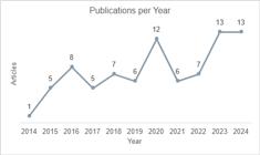
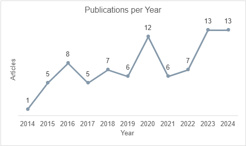
Following the search and selection process based on the predefined criteria, a total of 83 articles (n=83, 100%) were included in the analysis. The distribution of publications per year is illustrated in Figure 1.
Citation Analysis by Country
Research publications from 26 different countries were identified. Among these, the United States accounted for the highest number of publications, contributing a total of 26 articles (31%), followed by China with 13 (16%) articles. The top 10 countries with the highest number of publications, along with their corresponding citation counts, are summarized in Table 1.
In VOSviewer, “link strength” refers to the numerical value that quantifies the strength of the connection between two items in a network visualization. The “total link strength” represents the sum of the strengths of all links that an item has with other items.
Although the United States and China lead in terms of publication volume and citation counts, the analysis of total link strength reveals additional insights into collaborative and thematic connectivity among countries. Notably, Japan and Australia present the same number of published articles; however, Japan exhibits a substantially higher total link strength. This discrepancy suggests that Japanese publications are more strongly integrated within international collaboration networks and thematic linkages, whereas Australian contributions appear more fragmented.
A similar pattern is observed when comparing Australia and Italy. Despite Italy having fewer published articles and a comparable number of citations, its higher total link strength indicates a more central role within the bibliometric network. These findings highlight that total link strength reflects the degree of relational connectivity between countries rather than productivity alone, emphasizing the importance of collaborative structures and shared research themes in shaping scientific influence.
Relevant Sources
A total of 35 journals were identified from the analyzed articles. World Neurosurgery emerged as the most prolific journal in this field, contributing 25 articles (35%), followed by Operative Neurosurgery with 12 articles (12%). The top 20 list of journals and their respective publication counts are provided in Table 1. Additionally, the journals were evaluated based on their SCImago Journal Rank (SJR). The quartile classification organizes journals into four groups, from Q1 to Q4, with Q1 representing the top 25% most prestigious and influential in their field, while Q4 covers the bottom 25% least prominent.
Author citation Analysis
In the author citation analysis, the five authors with the highest number of citations for their articles were identified and examined. The most cited author was Peter Nakaji, with a total of 219 citations. The results are summarized in Table 1. The column “Number of Articles” represents the total number of articles authored by each individual within the dataset. In contrast, the column “Number of Citations” indicates the cumulative number of citations for these articles.
The author citation analysis further demonstrates that total link strength does not necessarily correlate with either the number of publications or total citation counts. While some highly cited authors exhibit low or null total link strength, others with fewer publications show stronger network connectivity. This indicates that authors with higher total link strength may function as key connectors within the research network, linking different research groups or thematic areas. Conversely, highly cited authors with low total link strength may contribute to influential but more isolated works. These findings reinforce the value of network-based metrics in capturing structural roles within the scientific community that are not reflected by traditional citation indicators alone.
Keywords co-occurrence analysis
The dataset contained a total of 933 unique keywords. By applying a minimum occurrence threshold of 5, 78 keywords were selected for analysis. Figure 2 presents an overlay visualization of the identified keywords, enabling an examination of keyword trends over time. In this visualization, the size of the node represents the frequency of the corresponding keyword, while the node colors indicate the year of highest occurrence, thereby highlighting temporal patterns. The lines connecting nodes illustrate the network relationships among the keywords.

Overlay network visualization of keywords, illustrating their frequency, temporal trends, and interconnections within the dataset.
To identify the top 20 keywords, a higher minimum occurrence threshold of 14 was applied. The most frequent keywords include “three-dimensional printing”, “neurosurgery”, and “surgical simulation”. Table 2 provides a detailed list of the top 20 keywords ranked by frequency. This analysis underscores the central terms in this research field and their interconnections within the bibliometric network.
Cited References Analysis
The final analysis performed was the cited references. Unlike the previous analysis, which examined the total number of citations attributed to multiple articles by an author, this analysis focuses on the number of citations received by individual articles. The five most frequently cited articles are detailed in Table 3.
The cited reference analysis reveals that articles with higher total link strength are not necessarily those with the highest citation counts. Instead, total link strength reflects the frequency with which a reference is co-cited alongside other studies, indicating its role as a shared conceptual foundation within the literature. Articles with higher total link strength tend to act as intellectual anchors, supporting multiple lines of research, even when their individual citation numbers are moderate. This distinction highlights the importance of co-citation analysis in identifying foundational works that shape the structure and evolution of the field beyond citation-based impact alone.
DISCUSSION
In recent decades, 3D printing has expanded its applications across various fields of medicine. One prominent area is the use of 3DP to transform imaging studies into manipulable 3D biomodels7. This technology has revolutionized the development of educational tools in neurosurgery and is now widely implemented in hospitals and clinical centers4.
One of the key challenges in teaching neurosurgical techniques to new professionals lies in the restricted neuroanatomical perspectives provided by the surgical microscope. These perspectives offer a limited field of view compared to traditional textbook illustrations and involve unique trajectories and spatial orientations. Additionally, mastering the use of the surgical microscope itself adds another layer of complexity2. While essential, traditional neurosurgical training methods have limitations, particularly concerning the potential risks to patients3.
Neurosurgical simulation can be performed using non-living animals, cadaveric models, or 3D biomodels8. Non-living animal models allow residents to refine their surgical skills without endangering patients; however, these models fail to replicate specific human anatomies and raise ethical concerns as well as challenges related to biological material handling4. Cadaver-based training provides the most realistic experience and is essential for learning neuroanatomy and skull base techniques. Nevertheless, conventional dissection techniques require significant resources, experienced instructors, appropriate facilities, and ethically prepared specimens1. These constraints limit the accessibility of cadaveric models.
3D biomodels have emerged as a viable alternative to address these challenges. They enable residents to practice both basic and complex neurosurgical techniques in a controlled environment while providing realistic representations of anatomical structures and surgical instrument handling9. These biomodels are created from DICOM imaging data, obtained from either healthy patients, individuals with pathologies, or even cadaveric specimens. They can be replicated indefinitely using various types of 3D printers and materials.
Our analysis included 83 articles published between 2014 and 2024 and indexed in the Scopus database on the topic of 3D printing for neurosurgical simulation. We observed a growth in publications over the past two years, reflecting advancements in 3D printing technologies, increased accessibility, and reduced costs. The United States leads in the number of publications, followed by China and Japan. Contributions were identified from a total of 26 countries; however, most countries had three or fewer publications.
Beyond publication volume, the analysis of total link strength provided additional insights into collaborative and thematic connectivity between countries. For instance, Japan and Australia presented the same number of published articles; however, Japan demonstrated substantially higher total link strength, suggesting stronger integration within international collaboration networks and shared research themes. Similarly, Italy exhibited higher total link strength despite having fewer publications and comparable citation counts to Australia. These findings indicate that total link strength reflects relational connectivity and network centrality rather than productivity alone, emphasizing the importance of collaborative structures in shaping scientific influence within this field.
In keyword citation analysis, frequently used terms included Printing, Three-dimensional, Neurosurgery, Surgical Simulation, and Surgical Training. Overlay network analysis revealed that these keywords were predominantly used between mid-2020 and 2021. More recently, there has been an increasing trend in the use of Augmented Reality (AR). Several studies10)-(12 have explored the combination of AR with 3D printing for neurosurgical training. For instance, Lee et al. (2022) demonstrated that combining 3D-printed skull models for trepanation and craniotomies with AR for skull base approaches provided a valuable complement to neurosurgeon training10.
The integration of AR and 3DP has led to significant advancements in medical applications, particularly in surgical planning and training. AR enhances the real-world environment by overlaying interactive virtual information, while 3D printing enables the creation of precise anatomical models tailored to individual patients. Together, these technologies offer detailed and immersive insights into patient anatomy, improving preparation for complex surgeries such as those in neurosurgery. This combination has demonstrated its ability to enhance surgical precision and safety, as well as optimize the training process for both basic and advanced procedures.
In contrast to the analyses of countries, authors, and cited references, keyword analysis demonstrated a more consistent relationship between frequency of occurrence and total link strength. This pattern reflects the intrinsic thematic cohesion of the field, as frequently used keywords tend to co-occur across a wide range of studies. The strong alignment between occurrence and network connectivity suggests that research topics related to 3D printing in neurosurgical training are highly interconnected, even when publication output and citation impact vary among contributors. This distinction further emphasizes the complementary role of keyword-based network analysis in revealing the conceptual structure of literature.
Beyond bibliometric indicators, the analyzed studies reveal clear technological and clinical trends. Most investigations focus on patient-specific anatomical models derived from computed tomography or magnetic resonance imaging data, particularly for vascular pathologies such as intracranial aneurysms and skull base lesions. Hybrid manufacturing approaches, combining additive manufacturing with silicone casting or manual assembly, are frequently employed to overcome material limitations and improve tactile realism. These models are primarily applied in surgical simulation, resident training, and preoperative planning, providing risk-free environments for practicing complex neurosurgical procedures. More recent studies indicate a growing interest in integrating three-dimensional printing with augmented reality systems, aiming to enhance spatial understanding and procedural guidance.
Sufianov et al. (2024) highlighted that low-cost 3D models provide practical and realistic experiences that are essential for mastering complex neurosurgical techniques. Their study evaluated cervical vertebrae models printed with silicone simulating spinal cord tissues and tumors. The results demonstrated high satisfaction regarding tactile sensation and pathology simulation, while also boosting participants’ confidence13.
Author-level analysis further demonstrated that citation impact does not necessarily correspond to network centrality. While some highly cited authors exhibited low or null total link strength, others with fewer publications showed stronger network connectivity, acting as key nodes within collaborative and thematic networks. This finding highlights that total link strength captures the structural role of authors within the scientific network, rather than their individual citation performance alone.
Peter Nakaji was identified as the most-cited author among the analyzed works, with a total of 219 citations across four collaborative studies. One remarkable study focused on fabricating anatomically accurate skull base models derived from CT scans of cadaveric temporal bones for simulating lateral skull base approaches. These models proved to be highly accurate during evaluations15.
At the article level, a similar pattern was observed, as co-citation analysis revealed that papers with higher total link strength were not always the most cited. Instead, these studies often served as shared conceptual references underpinning multiple research directions. In this context, total link strength reflects the integrative role of a publication within literature, rather than its isolated citation count. Foundational studies on aneurysm biomodelling and patient-specific simulation frequently functioned as intellectual anchors across the field.
The most-cited article was authored by Ryan et al. (2016), describing the development of a patient-specific neurosurgical simulator for aneurysm clipping training. This pilot study combined 3D printing and silicone to create a biomodel that replicates the tactile properties of human brain tissue16. Other highly cited studies also focused on creating aneurysm biomodels17. Subsequent authors, such as Mashiko et al. (2017) and Leal et al. (2019), employed similar methodologies to fabricate aneurysm biomodels18),(19, which are essential tools for training in one of the most challenging neurosurgical procedures: aneurysmal clipping20.
Another highly cited article by Coelho et al. (2020) described a hybrid model combining additive manufacturing techniques with manual intervention for preoperative planning of transsphenoidal encephalocele surgery. The fabrication of this model required approximately three weeks, utilizing both additive manufacturing and manual work11. As noted by Grillo et al. (2020), combining different techniques-such as various forms of additive manufacturing or conventional methods like silicone injection into printed molds-can help overcome the material limitations inherent to commonly used methods like Fused Deposition Modeling (FDM)21.
The journal World Neurosurgery, ranked Q2 in the SCImago Journal Rank (SJR) for 2023, had the highest number of publications on this topic, with 25 articles. While 35 journals were identified overall, only ten had more than one publication each. Notably, two of the five most-cited articles were published by World Neurosurgery. Among all the journals analyzed, fifteen were ranked Q1, the highest classification, and eight were ranked Q2 according to SJR metrics. This highlights the significant impact of research on this topic14.
However, despite their advantages, Mery et al. (2020) emphasized that high production costs remain a significant drawback of using 3D biomodels. The necessity of replacing biomodels after each training session further increases these costs. Factors influencing expenses include the type of printer, material requirements, and time-consuming design processes needed to create anatomically accurate models. Additionally, technical challenges persist in replicating soft tissues and vascular structures using current technologies. Specifically, replicating blood flow dynamics is particularly challenging, yet it is critical for procedures such as aneurysm clipping and vascular bypasses. These limitations emphasize the need for ongoing advancements in materials science and biofabrication to improve anatomical realism, functional accuracy, and reproducibility. Overcoming these challenges will be crucial for consolidating three-dimensional printing as a scalable, cost-effective, and broadly applicable educational approach in neurosurgical practice and training.
CONCLUSIONS
Our bibliometric review highlights the increasing interest and advancements in three-dimensional printing for neurosurgical training. In recent years, a notable rise in publications has been observed, driven by technological progress and improved accessibility. Key studies identified in this review focused on patient-specific biomodels and hybrid manufacturing techniques, directly addressing limitations associated with traditional training methods. However, ongoing challenges remain, including high production costs and technical constraints, emphasizing the need for further research and technological development. The analysis also revealed leading contributors and emerging trends, such as the integration of augmented reality, underscoring the concentrated yet impactful nature of research in this field. Overall, this work provides a solid foundation for understanding current developments and for guiding future research aimed at enhancing neurosurgical education and simulation.
ACKNOWLEDGEMENTS
The authors would like to thank the National Council for Scientific and Technological Development (CNPq) for the financial support during the doctoral studies, of which this article is a part.
References
- 1 Graffeo CS, Perry A, Carlstrom LP, Peris-Celda M, Alexander A, Dickens HJ, et al. 3D Printing for Complex Cranial Surgery Education: Technical Overview and Preliminary Validation Study. J Neurol Surg Part B Skull Base. 2022 Jun;83(S 02):e105-12.
- 2 Doron O, Paldor I, Moscovisci S, Spektor S, Chalif DJ, Langer DJ, et al. Acquisition of Basic Microsurgical Skills Using Low-Cost, Readily Available Models: The Orange Model. World Neurosurg. 2021 Feb;146:189-96.
-
3 Thiong’o GM, Bernstein M, Drake JM. 3D printing in neurosurgery education: a review. 3D Print Med [Internet]. 2021;7(1). Available from: https://www.embase.com/search/results?subaction=viewrecord&id=L2010883877&from=export
» https://www.embase.com/search/results?subaction=viewrecord&id=L2010883877&from=export - 4 Maclachlan LR, Alexander H, Forrestal D, Novak JI, Redmond M. Properties and Characteristics of Three-Dimensional Printed Head Models Used in Simulation of Neurosurgical Procedures: A Scoping Review. World Neurosurg . 2021;156((Maclachlan L.R., liam.maclachlan@health.qld.gov.au; Alexander H.; Redmond M.) Kenneth G Jamieson Department of Neurosurgery, Royal Brisbane and Women’s Hospital, Brisbane, Queensland, Australia):133-146.e6.
- 5 You Y, Niu Y, Sun F, Huang S, Ding P, Wang X, et al. Three-dimensional printing and 3D slicer powerful tools in understanding and treating neurosurgical diseases. Front Surg. 2022 Oct 14;9:1030081.
-
6 Hallinger P, Kovačević J. Applying bibliometric review methods in education: rationale, definitions, analytical techniques, and illustrations. In: Tierney RJ, Rizvi F, Ercikan K, editors. International Encyclopedia of Education (Fourth Edition) [Internet]. Oxford: Elsevier; 2023. p. 546-56. Available from: https://www.sciencedirect.com/science/article/pii/B9780128186305050703
» https://www.sciencedirect.com/science/article/pii/B9780128186305050703 - 7 Meyer-Szary J, Luis MS, Mikulski S, Patel A, Schulz F, Tretiakow D, et al. The Role of 3D Printing in Planning Complex Medical Procedures and Training of Medical Professionals-Cross-Sectional Multispecialty Review. Int J Environ Res Public Health. 2022 Mar 11;19(6):3331.
- 8 Iop A, El-Hajj VG, Gharios M, De Giorgio A, Monetti FM, Edström E, et al. Extended Reality in Neurosurgical Education: A Systematic Review. Sensors. 2022 Aug 14;22(16):6067.
- 9 Mery F, Aranda F, Méndez-Orellana C, Caro I, Pesenti J, Torres J, et al. Reusable Low-Cost 3D Training Model for Aneurysm Clipping. World Neurosurg . 2021;147:29-36.
-
10 Lee MH, Lee TK. Cadaver-Free Neurosurgical Simulation Using A 3D Printer and Augmented Reality. J Neurol Surg Part B Skull Base [Internet]. 2022;83(SUPPL 1). Available from: https://www.embase.com/search/results?subaction=viewrecord&id=L637829742&from=export
» https://www.embase.com/search/results?subaction=viewrecord&id=L637829742&from=export - 11 Coelho G, Rabelo NN, Adani LB, Cecilio-Fernandes D, Souza Carvalho FR, Pinto FG, et al. The Craniosynostosis Puzzle: New Simulation Model for Neurosurgical Training. World Neurosurg . 2020;138:e299-304.
- 12 Amini A, Allgaier M, Saalfeld S, Stein KP, Rashidi A, Swiatek VM, et al. Virtual Reality vs Phantom Model: Benefits and Drawbacks of Simulation Training in Neurosurgery. Oper Neurosurg Hagerstown Md. 2024 Nov 1;27(5):618-31.
-
13 Sufianov A, Ovalle CS, Cruz O, Contreras J, Begagić E, Kannan S, et al. Low-Cost 3D Models for Cervical Spine Tumor Removal Training for Neurosurgery Residents. Brain Sci [Internet]. 2024;14(6). Available from: https://www.scopus.com/inward/record.uri?eid=2-s2.0-85196899192&doi=10.3390%2fbrainsci14060547&partnerID=40&md5=3f6b45f9048e0cd131c5b5a896b5726d
» https://www.scopus.com/inward/record.uri?eid=2-s2.0-85196899192&doi=10.3390%2fbrainsci14060547&partnerID=40&md5=3f6b45f9048e0cd131c5b5a896b5726d -
14 SCImago (n.d.). SJR - SCImago Journal & Country Rank [Portal] [Internet]. [cited 2025 Jan 15]. Available from: Available from: http://www.scimagojr.com
» http://www.scimagojr.com - 15 Mooney MA, Cavallo C, Zhou JJ, Bohl MA, Belykh E, Gandhi S, et al. Three-dimensional printed models for lateral skull base surgical training: Anatomy and simulation of the transtemporal approaches. Oper Neurosurg. 2020;18(2):193-201.
- 16 Ryan JR, Almefty KK, Nakaji P, Frakes DH. Cerebral Aneurysm Clipping Surgery Simulation Using Patient-Specific 3D Printing and Silicone Casting. World Neurosurg . 2016;88((Ryan J.R., jrryan@asu.edu; Frakes D.H.) School of Biological and Health Systems Engineering, Arizona State University, Tempe, AZ, United States):175-81.
- 17 Wurm G, Lehner M, Tomancok B, Kleiser R, Nussbaumer K. Cerebrovascular biomodeling for aneurysm surgery: Simulation-based training by means of rapid prototyping technologies. Surg Innov. 2011;18(3):294-306.
- 18 Mashiko T, Kaneko N, Konno T, Otani K, Nagayama R, Watanabe E. Training in Cerebral Aneurysm Clipping Using Self-Made 3-Dimensional Models. J Surg Educ. 2017;74(4):681-9.
-
19 Leal AG, Mori YT, Nohama P, de Souza MA. Three-Dimensional Hollow Elastic Models for Intracranial Aneurysm Clipping Election - A Case Study. In: 2019 41st Annual International Conference of the IEEE Engineering in Medicine and Biology Society (EMBC) [Internet]. Berlin, Germany: IEEE; 2019 [cited 2020 Oct 20]. p. 4137-40. Available from: Available from: https://ieeexplore.ieee.org/document/8856514/
» https://ieeexplore.ieee.org/document/8856514/ - 20 Błaszczyk M, Jabbar R, Szmyd B, Radek M. 3D Printing of Rapid, Low-Cost and Patient-Specific Models of Brain Vasculature for Use in Preoperative Planning in Clipping of Intracranial Aneurysms. J Clin Med. 2021 Mar 13;10(6):1201.
- 21 Grillo FW. Simuladores de Paciente-Específico para Médicos. [São Paulo]: Universidade de São Paulo; 2020.
Research data is only available upon request.

 Source: Elaborated by the authors using VOSviewer, 2025.
 Source: Elaborated by the author using VOSviewer, 2025.