 Facial No-Touch Zones in the Freelift Technique
Abstract
Introduction The Freelift technique for facial and neck rejuvenation is a biplanar approach, which involves extensive sub-superficial musculoaponeurotic system (SMAS)-platysma release and, thus, requires advanced facial anatomy knowledge and surgical skills.
Objective To describe the topographic anatomy of the facial no-touch zones in Freelift surgery.
Materials and Methods This is a prospective study of facial anatomy through standardized cadaveric dissections. The dermoadipose and SMAS-platysma flaps were initially created mimicking the Freelift technique. Then, they were extended for a wide hemiface exposure. The facial no-touch zones topographic characteristics were analyzed, and landmarks were described. Histological analyses of SMAS were performed.
Results Seventy-four families were approached, and 40 consented to take part in the current study (54% consent rate). The hemifaces of 37 cadavers were dissected. The zygomatic no-touch zone starts at 4.5 ± 0.3 cm from the supratragal notch, and contains the zygomaticus major muscle, the zygomatic branch of the facial nerve, and the parotid gland duct. The superior and inferior loops of the buccal branch, the buccal fat pad, and the risorius muscle lie within the buccal no-touch zones. The cervical no-touch zone holds the superficializing superior cervical branches at 3.6 ± 0.3 cm from the gonial angle. The mobile SMAS is inexistent on histology.
Conclusion The current fresh-specimen study allowed the description of the facial no-touch zones in the Freelift technique. These zones contain important structures and must be undisturbed while creating the composite segment of the Freelift flap. Finally, surgical standardization is crucial for safely performing the Freelift approach.
Keywords
anatomy; dissection; facial nerve; plastic; regional; rhytidoplasty; superficial musculoaponeurotic system; surgery
Resumo
Introdução A técnica Freelift para rejuvenescimento cervicofacial é uma abordagem biplanar, com ampla liberação sub-sistema musculoaponeurótico superficial (SMAS)-platisma; e, portanto, requer conhecimento avançado de anatomia facial e destreza cirúrgica.
Objetivo Descrever a anatomia topográfica das zonas faciais no-touch na cirurgia de Freelift.
Materiais e Métodos Estudo prospectivo da anatomia facial por meio de dissecções padronizadas de cadáveres. Os retalhos dermogordurosos e de SMAS-platisma foram inicialmente confeccionados mimetizando o Freelift. Em seguida, foram ampliados para exposição de toda a hemiface. As características topográficas das zonas faciais no-touch foram analisadas. Os pontos de referência para as zonas faciais no-touch foram descritos. Análises histológicas de SMAS foram realizadas.
Resultados Setenta e quatro famílias foram abordadas, 40 consentiram (54% de taxa de consentimento) em tomar parte do estudo. As hemifaces de 37 cadáveres foram dissecadas. A zona zigomática no-touch inicia-se a 4,5 ± 0,3 cm da incisura supratragal e contém o músculo zigomático maior, o ramo zigomático do nervo facial e o ducto da glândula parótida. As alças superior e inferior do ramo bucal, a gordura bucal e o músculo risório situam-se dentro das zonas bucais no-touch. A zona cervical no-touch mantém os ramos cervicais superiores superficiais a 3,6 ± 0,3 cm do ângulo goníaco. O SMAS móvel é inexistente na histologia.
Conclusão Este estudo com espécimes frescos permitiu a descrição das zonas faciais no-touch na técnica Freelift. Essas zonas contêm estruturas importantes e devem permanecer intactas durante a criação do segmento composto do retalho Freelift. Por fim, a padronização cirúrgica é crucial para realizar a abordagem Freelift com segurança.
Palavras-chave
anatomia; cirurgia; dissecção; nervo facial; plástica; regional; ritidoplastia; sistema musculoaponeurótico superficial
Introduction
Face and neck cosmetic surgeries are among the most performed esthetic procedures across the globe.1 In 2023, it has been estimated that over 1 million facial and neck rejuvenation surgeries were executed worldwide, representing an increase of 16.2% compared to 2022.1,2 In the United States, facelift surgery was the most performed procedure for people aged 65 or more in 2023.3
The improved understanding about facial anatomy, particularly with respect to the facial layers and the topographic characteristics of the facial nerve branches, and the push for improved long-lasting results have contributed to the increasing popularity of multiplane surgical techniques.4–7 These multilayer approaches separate the superficial musculoaponeurotic system (SMAS) and mobilize it individually from the dermoadipose flap (DAF). These sub-SMAS-platysma surgical interventions demand advanced facial anatomy knowledge and surgical skills to prevent injuries, especially to the facial nerve. In addition, the previous utilization of non-invasive surgical esthetic procedures, including energy-based collagen stimulants, is invariably common among patients seeking facial rejuvenation surgery.8,9 This is being responsible for the formation of more fibrotic tissues that require wider and deeper undermining to achieve adequate release and pulling of facial flaps during surgery. In keeping with and to address these issues, the senior author has recently described the Freelift technique for facial rejuvenation.10
The detailed surgical technique for the Freelift has been published elsewhere.10 Briefly, it is a biplanar surgical procedure in which the DAF is limited to the ligamentous boundaries of the face while the SMAS-platysma flap (SPF) extends even further towards the midline. It also involves the creation of sub-SMAS-platysma standardized tunnels (dead-end caves), which contain important anatomical structures on their floors and between them. A final composite flap is then formed by the zygomatic and superior and inferior buccal tunnels when connecting their roofs' sidewalls. Furthermore, the SMAS-platysma undermining leaves an untouched area between the subplatysmal and the inferior buccal tunnels. The areas of cautious preservation during sub-SMAS-platysma dissection were named facial no-touch zones. The surgical anatomy applied to the main sub-SMAS platysma facelift procedures are summarized in the Table 1.
Objectives
Through standardized cadaveric dissections, the present study aimed at describing the topographic anatomy of the facial no-touch zones in Freelift. It has specifically the objectives to describe their areas, distance from landmarks, relationship with important anatomical structures and histological characteristics.
Materials and Methods
**********This is a prospective study of human facial anatomy through cadaveric dissections. It was approved by the Research Ethics Committee (CEP) of the Federal University of Pernambuco, Brazil. It was conducted at the State Coroner's Office, and after informed consent was obtained from a substitute decision maker (SDM). The included cadavers underwent no preservation methods before entering the study. They were neither refrigerated nor frozen at any time. The standardized dissections happened before funeral rituals, and, thus, extra caution was taken to preserve their face appearance.
Cadaveric Dissection and Freelift Techniques
The dissections were carried out through an extended traditional facelift incision for an almost full hemiface exposure and via bilateral inferior cervical incision for a complete anterolateral cervical exposure (Video 1). To maintain the SPF intact, the dissection plane of the superficial flap was performed just below the dermis. No infiltration solution was used.
Video 1
Extended facelift incision for almost total exposure of the hemiface, the extensive dermoadipose flap, and the bilateral cervical incision for complete exposure of the neck. Online content including video sequences viewable at: https://www.thieme-connect.com/products/ejournals/html/10.1055/s-0046-1817054.
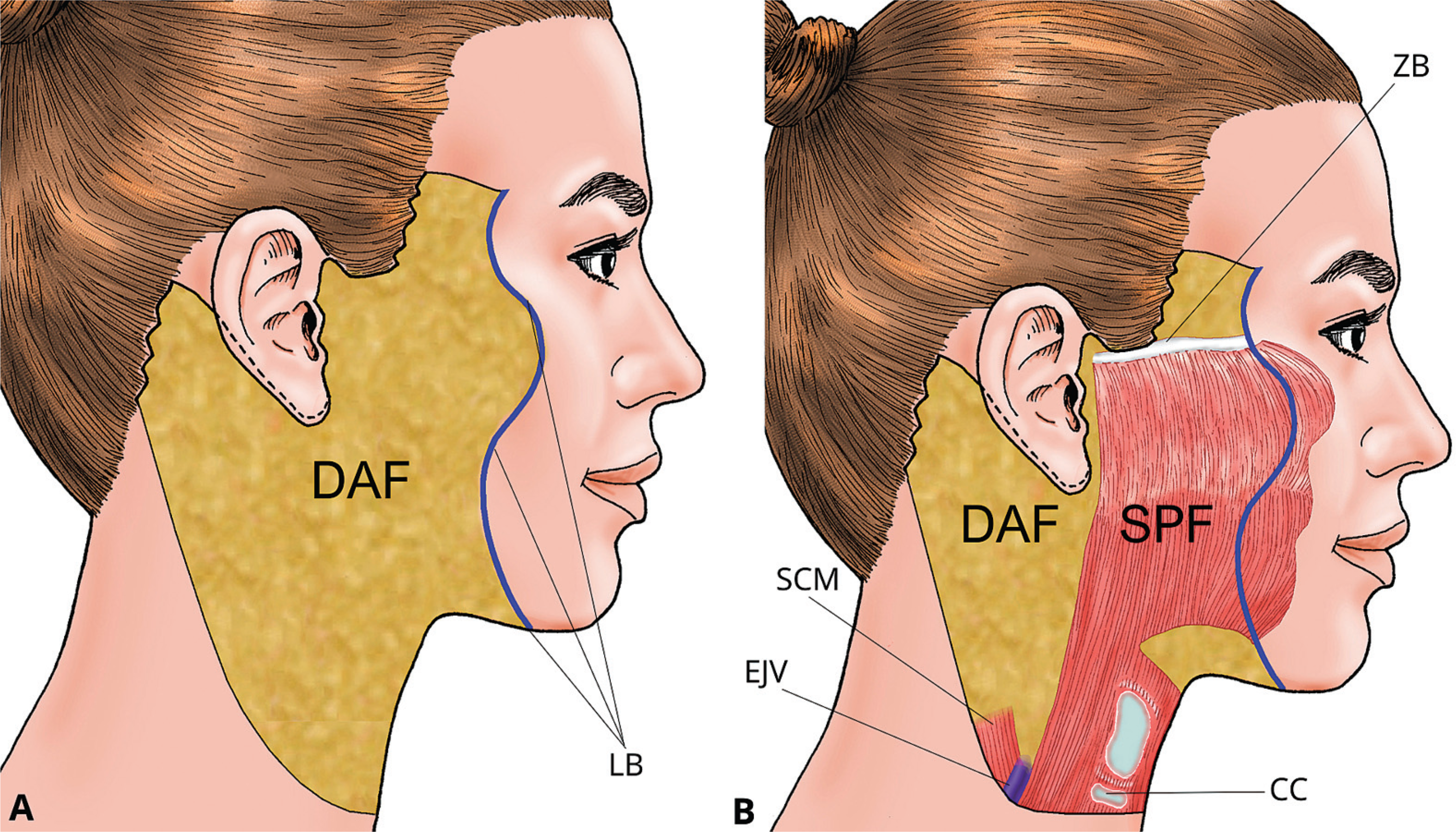
The SPF was initially created mimicking the actual Freelift surgery (Fig. 1). It was then extended for a full exposure of all relevant anatomical structures. The undermining of the DAF on the middle and lower face and upper cervical segments was performed even further towards the midline to characterize all no-touch zones through a superior view. This extended dermoadipose release created two distinct flaps, differently from the composite (SMAS-dermoadipose) flat encountered in the Freelift. In this technique, whereas the DAF release ends at the ligamentous boundaries, the SMAS-platysma dissection advances further medially) (Fig. 2A). To gain access to the anterior face, standardized tunnels (dead-end caves") are created. At this stage, additional release is obtained by loosening the roofs of these tunnels, taking extra caution to preserve the important structures on their floors, and keeping the composite segment of the flap (Fig. 2B). We have named these undisturbed areas facial no-touch zones in the Freelift technique (Fig. 2A, B). Finally, to identify all relevant structures within each zone, cadaveric dissection proceeded to enter the deep fascia, and direct visualization was accomplished.

Schematic representation of the surgical flaps in the Freelift technique. Abbreviations: (A) DAF, dermoadipose flap; LB, ligamentous boundaries. (B) SPF, SMAS-platysma flap; ZB, zygomatic bone; SCM, sternocleidomastoid muscle; EJV, external jugular vein; CC, cricoid cartilage.

Standardized tunnels and no-touch zones in the Freelift technique. Abbreviations: (A) zygomatic, superior and inferior buccal and subplatysmal tunnels; TA, temporal artery; FTB, facial temporal branch; ZT, zygomatic tunnel; FZB, facial zygomatic branch; PT, parotid duct; SBT, superior buccal tunnel; BFP, buccal fat pad; SBL, superior buccal loop; IBL, inferior buccal loop; IBT, inferior buccal tunnel; FMMB, facial marginal mandibular branch; FSCB, facial superior cervical branch; SPT, subplatysmal tunnel. (B) Facial no-touch zones, zygomatic, superior and inferior buccal and cervical no touch zones; OOc, orbicularis oculi muscle; ZMa, zygomaticus major muscle; OOr, orbicularis oris muscle; RM, risorius muscle. Note the overlapping areas between zones where structures from both zones might coexist less frequently.
Landmarks and Measurements
The supratragal notch was used to measure the distance to the origin of the zygomaticus major muscle (ZMa) where the most superior no-touch zone starts, in a descent and medial fashion, continuing into all other zones in the Freelift.
To characterize the location of the superficial superior cervical nerve branches—the cervical no-touch zone—the gonial angle was utilized as a landmark. An imaginary parallel line, about 5 mm below the mandibular margin, was used as reference. The head circumference was measured.
Histological Analysis
The SMAS-platysma samples were obtained at four different locations for histological analysis as follows: temporal SMAS; parotid area (fixed SMAS); premasseteric area (mobile SMAS); and cervical SMAS-platysma.
Histological analysis was performed on tissue samples fixed in 10% neutral buffered formalin. Samples were dehydrated through graded alcohols, cleared in xylene, and embedded in paraffin. Sections were cut at a thickness of 4 μm using a microtome and mounted on glass slides. Transverse cuts were particularly performed on cervical SMAS-platysma samples to verify the muscle-nerve-vessel-ligament junction.
Slides were stained with hematoxylin and eosin (H&E) using standard protocols. Stained sections were examined under an Olympus BX-41 light microscope (Olympus America) by an experienced pathologist. Images were captured using a digital camera system.
Demographic and Facial Anatomy Data
Age, sex, race, and time of death were obtained from social workers' records. The Freelift flaps, facial no-touch zones (zygomatic, superior and inferior buccal, and cervical), and relevant topographic characteristics and structures were documented by photography and videos and then analyzed.
Statistical Analysis
The rate of SDM consent was reported in percentages. Demographic data were expressed in terms of mean ± standard deviation or median (interquartile range, IQR) values and percentages, when appropriate. The measurements were expressed as mean ± standard deviation values in millimeters. Because it was an exploratory study of facial anatomy, no a priori sample size calculation was required.
Results
The study was conducted between November 2023 and January 2025. Out of 74 SDMs approached, 40 consented (54% consent rate). In three consented cases, we were unable to perform the dissections: participation in the study was withdrawn in one case due to other family members' desire; one, due to time constraint because of the funeral time; and another one had to be transferred to the State Forensic Medicine Institute because of head trauma being the suspected cause of death according to the pathologist in charge. Therefore, the hemifaces of 37 cadavers were dissected.
The average time from demise to dissection was 14.7 ± 5 hours. The median age of the cadaveric specimens was 70 (IQR: 58–77) years, and 54% of them were of the male sex. Most specimens were of mixed race (62%), followed by White (32%), and a small percentage of Black (6%). The mean head circumference was 53.4 ± 3 cm.
The upper and lower limits for the sub-SMAS-platysma flap are demonstrated in Video 2. For the medial release of the SPF, the undermining goes beyond the facial ligamentous boundaries of the DAF flap, and the four tunnels were created in a standardized fashion (Video 2).
Video 2
Freelift flaps and their landmarks, the standardized tunnels, and no-touch zones. zones. Online content including video sequences viewable at: https://www.thiemeconnect.com/products/ejournals/html/10.1055/s-0046-1817054.
The uppermost tunnel corresponds largely to the previously described facial prezygomatic space.11 This tunnel is superficial to the ZMa and other mimetic muscle, such as the zygomatic minor, levator anguli oris, levator labii superioris, and levator labii superioris alaeque nasi. The most superior "facial no-touch zone", named the "zygomatic no-touch zone", starts on average at 4.5 ± 0.3 cm from the supratragal notch (Fig. 3). Within this zone (on its floor), we found the ZMa, the zygomatic branch of the facial nerve, and the parotid gland duct (Video 3). Of note, in all cases, the zygomatic branch travelled under the ZMa giving out smaller branches over the muscle in 30% of the cases.

Cadaveric specimen and its schematic representation demonstrating that the zygomatic no-touch zone starts on average at 4.5 ± 0.3 cm from the supratragal notch; and that the cervical no-touch zone starts on average at 3.6 ± 0.3 cm from the gonial angle. Double arrows, indicate the distance from landmarks; purple arrow, points to the origin of the zygomaticus major muscle on the zygomatic bone; top blue sticker, indicates the zygomaticus major muscle; bottom blue sticker, shows the mandibular angle; note, the masseter muscle was retracted for exposure of the mandible; red arrow, signals the start of the cervical no-touch zone.
Video 3
Mimetic muscles and zygomaticus branch of the facial nerve within the zygomatic no-touch zone. Online content including video sequences viewable at: https://www.thieme-connect.com/products/ejournals/html/10.1055/s-0046-1817054.
The second and third tunnels are created anteriorly to the traditional premasseteric spaces.11 The second tunnel contained the superior loop of the buccal branch of the facial nerve and its connections to the zygomatic branch, and the upper segment of the buccal fat pad. Its matching no-touch zone (its floor) is the superior buccal no-touch zone. The third tunnel holds the inferior loop of the buccal branch and, when present, its communicating branches with the marginal mandibular branch; and its corresponding buccal fat pad. Its conforming no touch zone (its floor) was described as the inferior buccal no-touch zone. The risorius muscle, when present, lies within the boundaries of these two buccal zones, and is also preserved during dissections (Videos 3–4).
Video 4
Buccal loops of the facial nerve within the buccal no-touch zone cervical no-touch zone, facial marginal mandibular branch and facial superior cervical branch. Online content including video sequences viewable at: https://www.thieme-connect.com/products/ejournals/html/10.1055/s-0046-1817054.
Different from the facial no-touch zones, which are located on their cave's floors, the cervical no-touch zone is situated between the side walls of the lower buccal and subplatysmal tunnels (tridimensional zone). This zone agrees with the area where the superior cervical branches of the facial nerve become superficial to enter the platysma (Video 4). From the gonial angle, the first superficial branch could be encountered at 3.6 ± 0.3 cm in an area of approximately 2.5 cm (Fig. 3). Usually, 2 to 3 superficial branches were isolated, running alongside a tiny artery and vein within a connective tissue structure in this area (Video 4). The transverse cuts of histologic samples confirmed this trias neurovascularis entering the posterior surface of the platysma (Fig. 4). In several cases, a rich net of anastomosis between the marginal mandibular branch and the main superior cervical branch was found (Video 4).

Histology of a superior cervical nerve branch entering the undersurface of the platysma. (A) transverse cut of a cervical nerve branch within the platysma muscle fibers. (B) the cadaveric dissection of a superior cervical branch entering the platysma and its corresponding histologic sample demonstrating the trias neurovascularis entering the muscle.
The described topographic anatomy was identified after division of the deep fascia. The structures described underneath the deep fascia were correlated with the area corresponding to the zygomatic and buccal tunnels' floors during their creation. For the cervical no-touch zone, there is no elevation of the platysma flap.
In the microscopic analyses, the anterior SMAS, also known as mobile SMAS, particularly in the regions between the zygomatic nerve branch and parotid duct and buccal zones, showed almost total predominance of fat cells with scarce connective tissue and no definitive muscle layer. Over the parotid area, where the SMAS-platysma (fixed SMAS) is found, the histology sample evidenced a denser layer of connective tissue and fat cells. The temporal SMAS was characterized by an organized, but thin layer of collagen, representing the temporal fascia with a thinner fat layer. In the cervical region, the SMAS-platysma sample demonstrated total prevalence of muscle fibers with thinner connective layers and little fat cells (Fig. 5).

Microscopic analyses of the SMAS-platysma flap samples. Temporal, organized and thick layer of collagen, representing the temporal fascia with a thinner fat layer. Mobile, predominance of fat cells with scarce connective tissue and no definitive muscle layer. Fixed, denser layer of connective tissue and fat cells. Platysma, predominance of muscle fibers with thinner connective layers and little fat cells.
In the Freelift technique, the SPF is secured at the zygomatic bone periosteum. This uppermost location, between the temporal branch of the facial nerve and the superficial temporal artery, is exposed. The temporal branch nerve runs deeply at this level. The superficial temporal artery is easily felt by palpation during surgery. This safe area, measuring on average 1.5 cm, between these two structures, is recorded (Video 5).
Video 5
The safe location for anchoring the SMAS-platysma at the zygomatic bone, between the temporal branch of the facial nerve and the superficial temporal artery. The subcutaneous mandibular ligament. The marginal mandibular branch branching out before crossing the facial artery and vein. Complex anastomotic net between the superior cervical branch and the marginal mandibular branch. Footage of the Freelift technique shown in an actual patient. Online content including video sequences viewable at: https://www.thieme-connect.com/products/ejournals/html/10.1055/s-0046-1817054.
Although not a primary endpoint of this analysis, the subcutaneous mandibular ligament was observed in several cases in our dissections (Video 5). We also found that the marginal mandibular branch usually branches out before crossing the facial artery and vein. It can either bifurcate or trifurcate at this level, sending smaller branches posteriorly to the facial vessels (Video 5). It can also form a complex anastomotic net with the cervical branch (Video 5).
Discussion
Our facial anatomy study was able to demonstrate the topographic characteristics of the facial no-touch zones and their important anatomical structures, encountered in the recently described Freelift surgery. Furthermore, we were able to perform very timely dissections utilizing fresh human cadavers (15 hours after the demise) after informed consent was obtained. Finally, the present study describes two cadaveric dissection techniques that can preserve face appearance for funeral rites.
The Freelift technique, which involves an extensive undermining of the SPF, requires advanced surgical skills and facial anatomy knowledge to avoid injuries to important facial structures.10 Like other facial rejuvenation procedures that mobilize the SPF, such as the deep plane and the high SMAS techniques, the risk of complications, particularly facial nerve injuries, although rare, is worrisome.12–14 A national survey of American plastic surgeons reported that surgeons newly in practice (≤ 5 years) are more prone to perform skin-only procedures; and only 21% of all surgeons preform extended SMAS dissection.15 In a 2022 follow-up survey, 20 years later, this figure has not changed significantly.16 Thus, most plastic surgeons might not feel comfortable performing wide SMAS-platysma releases during facelift procedures.
The current study standardizes the surgical steps in the Freelift technique by characterizing the facial no-touch zones It correlates anatomical structures found underneath the deep fascia with the tunnels' floors, gliding planes on the face, during critical surgical dissection. In the Freelift technique, after creation of the tunnels (zygomatic and buccal) and once the no-touch zones are preserved, the connecting bridges are released. The standardization of surgical procedures aims at preventing complications and improving outcomes. In 1994, Seckel17 introduced the facial danger zones with the intent to avoid peripheral nerve injury. The most susceptible anatomic locations for nerve injury and its boundaries were described.18 Then, Stuzin and Rohrich have further contributed to the characterization of such zones.19 Codner et al. also described relevant facial anatomy applied to face rejuvenation surgery to prevent complications.5,20 In our study, we have not only described the topography of relevant peripheral facial nerves, but also of other anatomical structures, like the facial expression muscles, the parotid duct, the buccal fat pad, the superficial temporal artery, the facial artery and vein, the external jugular vein, and the submandibular gland.
The ZMa muscle origin and its path downwards were well characterized in our specimens. The distance from a fixed point (superior tragus notch), along the easily palpable zygomatic bone, to the origin of this muscle is extremely important, as a landmark, for a safe dissection during the Freelift. This point marks the superior limit of the facial no-touch zones. Others have described the relevance of thorough attention to the zygomaticus muscles during sub-SMAS approaches. Hamra, building on the first described deep plane approach (the Skoog flap) and aiming to address periorbital rejuvenation during rhytidectomy, incorporated the zygorbicular dissection. It relies on the origin of the ZMa to undermine the orbicularis oculi muscle laterally and enter the area medial do the zygomatic muscles, creating a segment of composite zygomaticus-orbicular flap.21–24 In the high SMAS facelift, the ZMa origin represents the transition point, from deep to the SMAS, up to over the lateral border of the ZMa, piercing through the overlaying fascia to access subcutaneous plane.25,26 Finally, the ZMa is also used as a landmark for the finger-assisted malar elevation (FAME) technique and the extended deep-plane facelift.27,28
The cervical no-touch zone is particularly important due to the potential risk of injuries to the superficializing superior cervical branches, usually 2 to 3, within this area. Although considered a rare event, most transient labial depressor dysfunction might be in fact due to cervical branch injuries, instead of marginal mandibular injuries. This condition is also known as marginal mandibular nerve pseudo-paralysis, and can be distinguished from the actual marginal mandibular injury by the patient's preserved ability to evert the lower lip.29 Our cadaveric dissections enabled us to determine a practical landmark, the gonial angle, and its distance to this cervical branch area that should be circumvented. Our histological samples additionally confirmed the presence of these small branches along tiny vessels within ligamentous structures entering the posterior surface of the platysma. Lindsey Jr. et al., in an anatomical study of 15 cadaveric dissections, also used the mandibular angle as a referent point for describing an area where cervical branches can be found.30 However, they drew a cervical line: connecting a point 5 cm below the mandibular angle at the anterior border of the SCM to the point where the facial artery crosses the inferior mandibular border. This cervical line was the boundary distal to which the cervical branches penetrated the deep cervical fascia to continue their superficializing path towards the posterior surface of the platysma. Instead of using the gonial angle, Daane and Owsley29 described an imaginary parallel line 3 cm below the inferior border of the mandible as the "danger zone" for injuries to the marginal mandibular and cervical branches. Even though these studies applied different methods, the superficial superior cervical branches were in the vicinity of ours cervical no-touch zone.
We demonstrated the unique histology of the SMAS-platysma at different areas. Of note, the mobile SMAS is practically inexistent. In keeping with our findings, recently, a cadaveric study, using complex histologic analysis, demonstrated that the SMAS is present only where there are platysma-auricular fascia and flat mimetic muscles, but not between these muscles.31 Therefore, the authors questioned the existence of the SMAS as a specific anatomical entity. This emphasizes the need for a meticulous dissection, particularly for the creation of the composite flap segment during sub-SMAS-platysma approaches, such as the Freelift, for its adequate mobilization and traction.
Although not the primary outcome of the present study, we observed in some specimens, a noticeable connective tissue formation, compatible to the mandibular ligament at the subcutaneous plane. The anatomical description of the mandibular ligament has varied significantly during history, and no consensus exists yet. Most recently, Minelli at al. observed the lack of a definitive subcutaneous component of the mandibular ligament.32 Whether this issue might be a matter of individual variation or nomenclature, we do not underestimate the need of its release to address substantial jowling in some patients.
Finally, the safe uppermost area for SPF anchoring was detailed to prevent injuries, particularly to the temporal branch at the zygomatic bone level. Securing the mobilized SPF in this area, where the bone periosteum offers a firm structure for fixation, tension across the entire flap is avoided, and the location of original ligament attachments is restored.33,34 In the Freelift, the upper segment of the SPF can be folded and secured in place, providing additional volumization of the malar region.
Limitations
The present was a small study performed in fresh specimens before funeral rituals. Therefore, the facial appearance had to be preserved as much as possible and the dissections executed in a time-constrained fashion. However, the focused standardized dissections allowed efficient identification and documentation of study outcomes. Also, the short time between death and our dissections, in actual fresh cadavers—not even refrigerated—created the "near-perfect" model for human anatomy studies. Finally, the facial no-touch zones and their described structures may vary due to possible discrepancy in surgical dissection and individual anatomy.
Conclusion
The present study of fresh specimens enabled the description of the facial no-touch zones in the Freelift approach for esthetic facial and neck surgery. These zones contain important structures and must be undisturbed while creating the composite segment of the Freelift flap. The identification of the origin of the zygomaticus major muscle on the zygomatic bone plays an important role in guiding the safe dissection plane. Sections of the zygomatic, the superior and inferior buccal, the marginal mandibular, superior cervical branches, as well as the parotid duct, the buccal fat pad and risorius muscle are encountered underneath the deep fascia in the no-touch zones. Finally, surgical standardization is crucial for safely performing the Freelift technique for cervico-facial rejuvenation.
Financial Support
Data Availability
Data will be available upon request to the corresponding author.
References
-
1 Triana L, Huatuco RMP, Campilgio G, Liscano E. Trends in surgical and nonsurgical aesthetic procedures: a 14-year analysis of the International Society of Aesthetic Plastic Surgery—ISAPS. Aesthetic Plast Surg 2024;48(20):4217–4227. Doi: 10.1007/s00266-024-04260-2
» https://doi.org/10.1007/s00266-024-04260-2 -
2 International Society of Aesthetic Plastic Surgery. Global statistics. West Lebanon, NH: ISAPS; [citado Ago 1, 2025]. Disponível em: https://www.isaps.org/discover/about-isaps/global-statistics/
» https://www.isaps.org/discover/about-isaps/global-statistics/ -
3 Aesthetic Plastic Surgery National Databank Statistics 2023. Aesthet Surg J 2024;44(Suppl 2):1–25. Doi: 10.1093/asj/sjae188
» https://doi.org/10.1093/asj/sjae188 -
4 Barrett DM, Casanueva FJ, Wang TD. Evolution of the rhytidectomy. World J Otorhinolaryngol Head Neck Surg 2016;2(01):38–44. Doi: 10.1016/j.wjorl.2015.12.001
» https://doi.org/10.1016/j.wjorl.2015.12.001 -
5 Derby BM, Codner MA. Evidence-based medicine: face lift. Plast Reconstr Surg 2017;139(01):151e–167e. Doi: 10.1097/PRS.0000000000002851
» https://doi.org/10.1097/PRS.0000000000002851 -
6 Rohrich RJ, Sinno S, Vaca EE. Getting better results in facelifting. Plast Reconstr Surg Glob Open 2019;7(06):e2270. Doi: 10.1097/GOX.0000000000002270
» https://doi.org/10.1097/GOX.0000000000002270 -
7 Luu NN, Friedman O. Facelift surgery: history, anatomy, and recent innovations. Facial Plast Surg 2021;37(05):556–563. Doi: 10.1055/s-0040-1715616
» https://doi.org/10.1055/s-0040-1715616 -
8 Jacono AA, Malone MH, Lavin TJ. Nonsurgical facial rejuvenation procedures in patients under 50 prior to undergoing facelift:habits, costs, and results. Aesthet Surg J 2017;37(04):448–453. Doi: 10.1093/asj/sjw217
» https://doi.org/10.1093/asj/sjw217 -
9 Sweis L, DeRoss L, Raman S, Patel P. Potential effects of repetitive panfacial filler injections on facelift surgery and surgical outcomes: survey results of the members of The Aesthetic Society. Aesthet Surg J Open Forum 2023;5:ojad010. Doi: 10.1093/asjof/ojad010 Errata em: Aesthet Surg J Open Forum. 2023 Mar 27;5:ojad025. doi:10.1093/asjof/ojad025. PMID: 36860684; PMCID:PMC9969530
» https://doi.org/10.1093/asjof/ojad010 - 10 Araujo M, Schwartz M, Melo G, Gama LJM. Freelift. In: Araujo M, Boggio R, Maino M, Uebel C, Viterbo F, editores. Cirurgia plástica da face e cosmiatria. 1. ed. Rio de Janeiro: Thieme Revinter; 2024: 249–251
-
11 Mendelson BC, Muzaffar AR, Adams WP Jr. Surgical anatomy of the midcheek and malar mounds. Plast Reconstr Surg 2002;110(03):885–896, discussion 897–911. Doi: 10.1097/00006534-200209010-00026
» https://doi.org/10.1097/00006534-200209010-00026 -
12 Mortada H, Alkilani N, Halawani IR, et al. Evolution of superficial muscular aponeurotic system facelift techniques: a comprehensive systematic review of complications and outcomes. JPRAS Open 2023;39:166–180. Doi: 10.1016/j.jpra.2023.06.003
» https://doi.org/10.1016/j.jpra.2023.06.003 -
13 Namin A, Shokri T, Vincent A, Saman M, Ducic Y. Complications in facial esthetic surgery. Semin Plast Surg 2020;34(04):272–276. Doi: 10.1055/s-0040-1721764
» https://doi.org/10.1055/s-0040-1721764 -
14 Roostaeian J, Rohrich RJ, Stuzin JM. Anatomical considerations to prevent facial nerve injury. Plast Reconstr Surg 2015;135(05):1318–1327. Doi: 10.1097/PRS.0000000000001244
» https://doi.org/10.1097/PRS.0000000000001244 -
15 Matarasso A, Elkwood A, Rankin M, Elkowitz M. National plastic surgery survey: face lift techniques and complications. Plast Reconstr Surg 2000;106(05):1185–1195, discussion 1196. Doi: 10.1097/00006534-200010000-00033
» https://doi.org/10.1097/00006534-200010000-00033 -
16 Sinclair NR, Kochuba A, Coombs DM, et al. Face lift practice patterns: an American Society of Plastic Surgeons member survey, 2000 and 2020. How much have we changed? Plast Reconstr Surg 2022;149(06):1096e–1105e. Doi: 10.1097/PRS.0000000000009097
» https://doi.org/10.1097/PRS.0000000000009097 -
17 Seckel BR. Facial danger zones: avoiding nerve injury in facial plastic surgery. Can J Plast Surg 1994;2(02):59–66. Doi: 10.1177/229255039400200207
» https://doi.org/10.1177/229255039400200207 - 18 Seckel BR. Facial Danger Zones: Avoiding Nerve Injury in Facial Plastic Surgery. 2. ed. Nova York: Thieme; 2010
-
19 Stuzin JM, Rohrich RJ. Facial nerve danger zones. Plast Reconstr Surg 2020;145(01):99–102. Doi: 10.1097/PRS.0000000000006401
» https://doi.org/10.1097/PRS.0000000000006401 -
20 Codner MA, Kikkawa DO, Korn BS, Pacella SJ. Blepharoplasty and brow lift. Plast Reconstr Surg 2010;126(01):1e–17e. Doi: 10.1097/PRS.0b013e3181dbc4a2
» https://doi.org/10.1097/PRS.0b013e3181dbc4a2 - 21 Skoog T. Plastic Surgery: New Methods and Refinements. Filadélfia: Saunders; 1974
- 22 Hamra ST. The deep-plane rhytidectomy. Plast Reconstr Surg 1990;86(01):53–61, discussion 62–63
-
23 Hamra ST. The zygorbicular dissection in composite rhytidectomy: an ideal midface plane. Plast Reconstr Surg 1998;102(05):1646–1657. Doi: 10.1097/00006534-199810000-00051
» https://doi.org/10.1097/00006534-199810000-00051 -
24 Hamra ST. Building the composite face lift: a personal odyssey. Plast Reconstr Surg 2016;138(01):85–96. Doi: 10.1097/PRS.0000000000002310
» https://doi.org/10.1097/PRS.0000000000002310 -
25 Barton FE Jr. The "high SMAS" face lift technique. Aesthet Surg J 2002;22(05):481–486. Doi: 10.1067/maj.2002.128628
» https://doi.org/10.1067/maj.2002.128628 -
26 Marten T, Elyassnia D. High SMAS facelift: combined single flap lifting of the midface, cheek, and jawline. Facial Plast Surg 2022;38(06):593–612. Doi: 10.1055/s-0042-1757757
» https://doi.org/10.1055/s-0042-1757757 -
27 Aston SJ, Walden J. Facelift with SMAS technique and FAME. Clinical Gate; 2015. Disponível em: https://clinicalgate.com/facelift-with-smas-technique-and-fame/
» https://clinicalgate.com/facelift-with-smas-technique-and-fame/ -
28 Jacono AA. A novel volumizing extended deep-plane facelift: using composite flap shifts to volumize the midface and jawline. Facial Plast Surg Clin North Am 2020;28(03):331–368. Doi: 10.1016/j.fsc.2020.03.001
» https://doi.org/10.1016/j.fsc.2020.03.001 -
29 Daane SP, Owsley JQ. Incidence of cervical branch injury with "marginal mandibular nerve pseudo-paralysis" in patients undergoing face lift. Plast Reconstr Surg 2003;111(07):2414–2418. Doi: 10.1097/01.PRS.0000061004.74788.33
» https://doi.org/10.1097/01.PRS.0000061004.74788.33 -
30 Lindsey JT Jr, Lee JJ, Phan HTP, Lindsey JT Sr. Defining the cervical line in face-lift surgery: a three-dimensional study of the cervical and marginal mandibular branches of the facial nerve. Plast Reconstr Surg 2023;152(05):977–985. Doi: 10.1097/PRS.0000000000010369
» https://doi.org/10.1097/PRS.0000000000010369 -
31 Minelli L, Van der Lei B, Mendelson BC. The superficial musculoaponeurotic system: does it really exist as an anatomical entity? Plast Reconstr Surg 2024;153(05):1023–1034. Doi: 10.1097/PRS.0000000000010557
» https://doi.org/10.1097/PRS.0000000000010557 -
32 Minelli L, Yang HM, Van der Lei B, Mendelson B. The surgical anatomy of the jowl and the mandibular ligament reassessed. Aesthetic Plast Surg 2023;47(01):170–180. Doi: 10.1007/s00266-022-02996-3
» https://doi.org/10.1007/s00266-022-02996-3 - 33 Mendelson BC. Surgery of the superficial musculoaponeurotic system: principles of release, vectors, and fixation. Plast Reconstr Surg 2001;107(06):1545–1552, discussion 1553–1555, 1556–1557, 1558–1561
-
34 Minelli L, Brown CP, Van der Lei B, Mendelson B. Anatomy of the facial glideplanes, deep plane spaces, and ligaments: implications for surgical and nonsurgical lifting procedures. Plast Reconstr Surg 2024;154(01):95–110. Doi: 10.1097/PRS.0000000000011078
» https://doi.org/10.1097/PRS.0000000000011078