 Prostatic Leiomyosarcoma: Case Report
ABSTRACT
Introduction: Sarcoma is a rare tumor, representing less than 0.1% of primary prostate tumors in adults. Of these, leiomyosarcoma is the most common subtype. They are generally found in advanced stages, making the prognosis unfavorable. The approach is variable, with surgery being the preferred management.
Case report: Male patient, 65-year-old complaining of weak urinary stream and nocturia, presenting, upon rectal exam, a prostatic area of hardened consistency. Imaging tests revealed a large solid prostatic nodule, with transrectal biopsy showing that it was a stromal tumor with uncertain potential malignancy. He underwent radical retropubic prostatectomy, with anatomopathological and immunohistochemical examination confirming prostatic leiomyosarcoma. After surgical and adjuvant treatment, the patient evolved satisfactorily.
Conclusion: Prostatic leiomyosarcoma is a rare and aggressive tumor. Generally, the prognosis is unfavorable, but it can be improved in patients with localized disease submitted to multimodal treatment.
Key words:
Leiomyosarcoma; Prostatic Neoplasms/surgery; Prostatectomy; Radiotherapy; Adjuvant
RESUMO
Introdução: O sarcoma é um tumor raro, representando menos de 0,1% dos tumores prostáticos primários em adultos. Destes, o leiomiossarcoma é o subtipo mais frequente. Geralmente, são descobertos em estádios avançados, tornando o prognóstico desfavorável. A abordagem é variável, sendo a cirurgia o manejo preferencial.
Relato do caso: Paciente do sexo masculino, 65 anos, com queixa de jato urinário fraco e noctúria, apresentando, ao toque retal, área prostática de maior consistência. Exames de imagem evidenciaram volumoso nódulo sólido prostático, com a biópsia transretal mostrando se tratar de um tumor estromal com malignidade potencial incerta. O paciente foi submetido à prostatectomia radical retropúbica, com exames anatomopatológico e imuno-histoquímico confirmando leiomiossarcoma de próstata. Após tratamento cirúrgico e adjuvante, o paciente evoluiu satisfatoriamente.
Conclusão: O leiomiossarcoma de próstata é um tumor raro e agressivo. Geralmente, o prognóstico é desfavorável, mas pode ser melhorado em pacientes com doença localizada submetidos a tratamento multimodal.
Palavras-chave:
Leiomiossarcoma; Neoplasias da Próstata/cirurgia; Prostatectomia; Radioterapia adjuvante
RESUMEN
Introducción: El sarcoma es un tumor raro, representa menos del 0,1% de los tumores primarios de próstata en adultos. De estos, leiomiosarcoma es el subtipo más común. Generalmente se descubren en estadios avanzados, lo que hace que el pronóstico sea desfavorable. El abordaje es variable, siendo la cirugía el tratamiento preferido.
Informe del caso: Paciente masculino, 65 años que refiere flujo urinario débil y nicturia, presentando al tacto rectal área prostática de mayor consistencia. Las pruebas de imagen revelaron un gran nódulo prostático sólido, y la biopsia transrectal mostró que se trataba de un tumor estromal con potencial maligno incierto. Se le realizó prostatectomía radical retropúbica, confirmándose leiomiosarcoma prostático mediante examen anatomopatológico e inmunohistoquímico. Luego del tratamiento quirúrgico y adyuvante el paciente evolucionó satisfactoriamente.
Conclusión: Leiomiosarcoma de próstata es un tumor raro y agresivo. Generalmente el pronóstico es desfavorable, pero puede mejorar en pacientes con enfermedad localizada sometidos a tratamiento multimodal.
Palabras clave:
Leiomiosarcoma; Neoplasias de la Próstata/cirugía; Prostatectomía; Radioterapia Adyuvante
INTRODUCTION
Soft tissue sarcoma represents less than 1% of all cancer diagnosed yearly in the United States; less than 5% of this total occurs in the urinary tract. Prostatic sarcoma is a tumor that originates from non-epithelial mesenchymal components of the stroma, representing less than 0.1% of primary prostate tumors in adults2. Leiomyosarcoma is the most common subtype of the primary prostate sarcoma that occurs in adults, found in 38% to 52% of the cases3 and having been first described by Stambert in 18534. Rhabdomyosarcoma, on the other hand, is the histological subtype most frequently found in pediatric patients5. Sarcomas are generally found in advanced stages, making the prognosis unfavorable5. Due to the rarity of these tumors, the treatment approach varies1, with surgery being the preferred management2. The objective of this article is to present a case report of prostatic leiomyosarcoma and a literature review about this rare disease.
This study has been approved by the institution's Research Ethics Committee, report number 6672753 (CAAE (submission for ethical review): 77794624.2.0000.5360), in compliance with Ordinance 466/20126 of the National Health Council.
CASE REPORT
Male patient, 65-years old, with a previous history of systemic hypertension (SH), dyslipidemia, and psoriasis, sought urological routine medical care. No family history of prostate cancer has been reported. Complained of weak urinary stream and nocturia (5 in the International Prostate Symptom Score)7, with no other symptoms. Physical rectal examination showed a prostate of fibroelastic consistency, of approximately 60 grams, with a more consistent area to the right, though not characterizing a nodule.
The prostate specific antigen (PSA) serum was 1.09 ng/ml. Transabdominal prostate ultrasound showed a 58.4 grams gland, with an adjacent hypoechoic cyst measuring 4.8 × 4.7 × 4.4 cm. The urinary tract ultrasound showed no abnormalities, except for a 2 cm cortical cyst in the middle third of the right kidney.
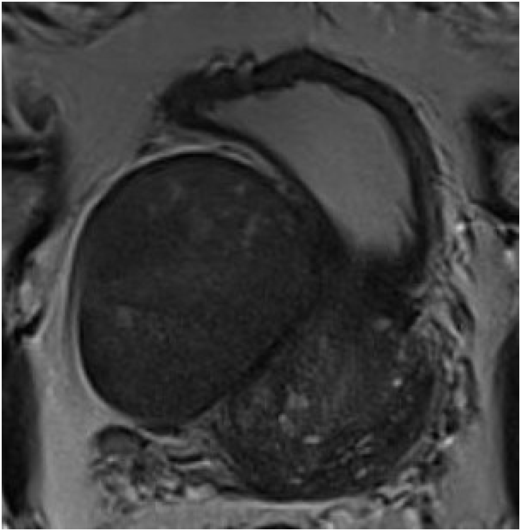
The patient then underwent a multiparametric prostate magnetic resonance (MRI) that revealed a solid voluminous nodule (5.0 × 4.0 cm) in the anterior periphery of the mid-basal third to the right of the prostate, predominantly exophytic, with acute restriction to diffusion and progressive contrast enhancement (PIRADS 3) (Figure 1).

Considering the image exams, the patient was submitted to transrectal ultrasound-guided prostatic biopsy (TRUS). The anatomopathological analysis of the specimens conducted in November 2022 suggested the diagnosis of stromal tumor of uncertain malignant potential (STUMP) in the right peripheral zone.
After a negative systemic staging (chest tomography, total abdomen MRI and bone scintigraphy), the patient was submitted, in February 2023, to a radical retropubic prostatectomy with bilateral iliac-obturator lymphadenectomy. The procedure was conducted with no complications, in a total estimated time of 150 minutes with bleeding measured at 400 ml, with no need of blood transfusion. The patient remained hospitalized for two days, with no postoperative complications, and had the indwelling bladder catheter removed on the 12th day after surgery.
The anatomopathological exam of the surgical piece suggested the diagnosis of prostatic leiomyosarcoma (Figure 2), showing a well-delimited nodule measuring 5.0 × 4.5 cm in the right lobe, with increased mitotic activity and negative surgical margins. Isolated lymph nodes as well as the seminal vesicles showed no neoplasm.

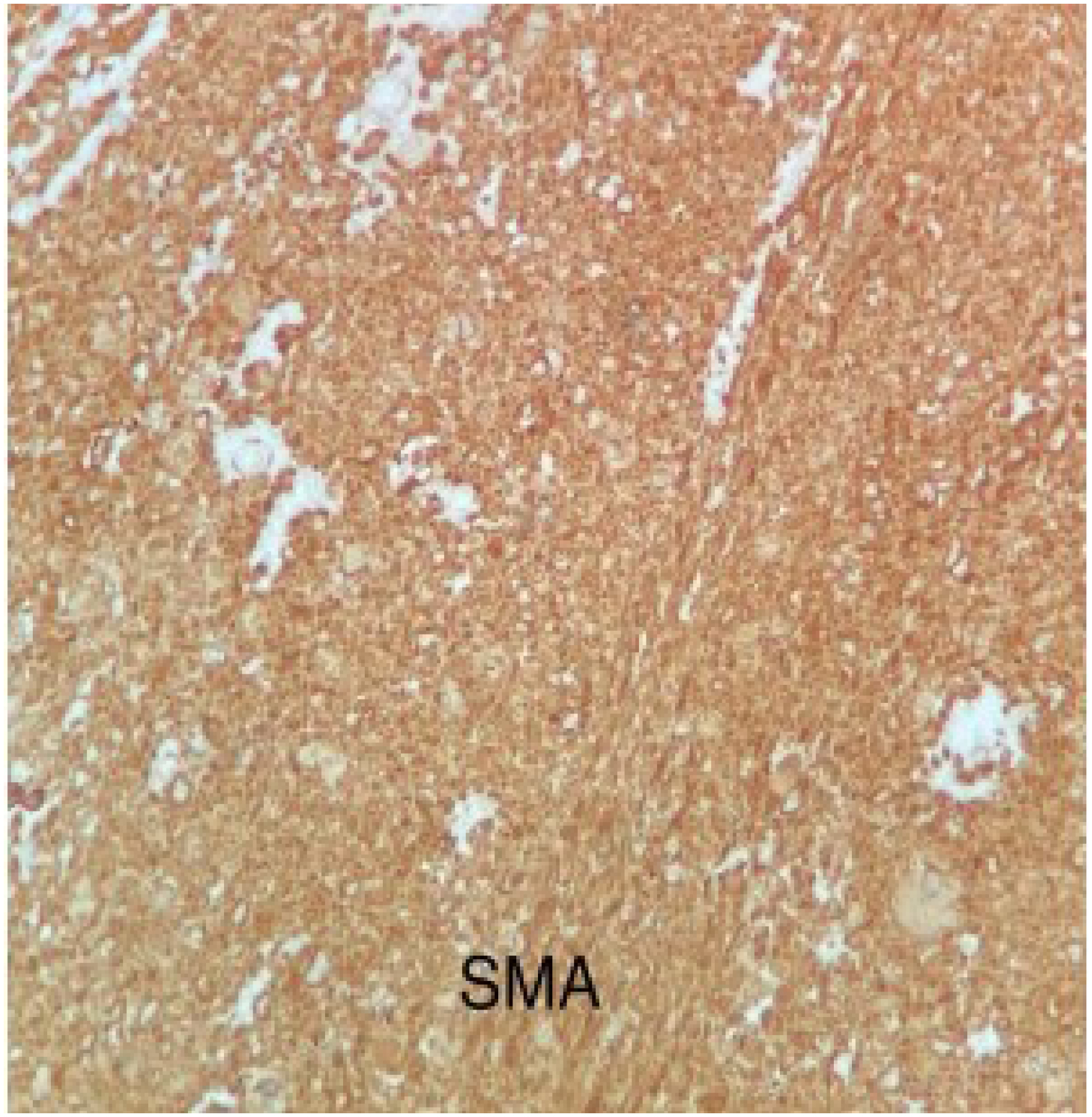
Complementation with immunohistochemistry confirmed the leiomyosarcoma diagnosis, with positivity for smooth muscle actin, desmin, caldesmon, vimentin and progesterone receptor, as well as negativity for CD34 and cytokeratins AE1/AE3 (Figure 3). After confirming the sarcoma diagnosis, the lactate dehydrogenase (LDH) dosage was found within normal parameters (132 U/1).

After surgery, the patient went through a prostatic bed (66 Gy) adjuvant therapy (Intensity-modulated Radiotherapy – IMRT), concluded in July 2023.
Six months after the surgery, during the medical appointment, the patient reported continence and sexual potency, with 0.01 PSA and chest and abdomen tomography showing no signs of recurrence of metastatic disease. He is still being monitored, having been last examined in December 2023, with no recurrence.
DISCUSSION
Prostate cancer is the most common malignity among 50-year-old men and older, being the main cause of death by cancer in men over 70 years old5. Prostate primary sarcomas are extremely rare tumors1 that can be categorized as leiomyosarcoma, rhabdomyosarcoma, fibrosarcoma and spindle cell sarcoma3. Leiomyosarcoma represents less than 0.1% of all prostate malignant neoplasms and is the most common primary prostate sarcoma subtype in adults, accounting for 38% to 52% of the cases8. Prostatic stromal lesions that have no obvious diagnosis are called STUMP3. This acronym embodies a group of lesions whose biological behavior is, generally, hard to determine histologically, in contrast to obviously sarcomatous lesions, such as rhabdomyosarcoma, leiomyosarcoma or stromal sarcoma9. Most STUMP lesions are positive for CD34 and vimentin but vary regarding smooth muscle actin and desmin positivity9. Due to the prostatic stroma's origin, the immunohistochemical analysis is frequently positive for progesterone receptors, being less constant for estrogen receptors9.
Primary prostate leiomyosarcoma presents uncertain etiology, though previous pelvic radiotherapy may be related in some cases10. Patients age ranges from 41 to 78 upon presentation, with a mean of 61 years old3.
Clinically, individuals most commonly present signs and symptoms of lower urinary tract obtrusion (LUTO); associated symptoms include perineal pain, hematuria, burning sensation upon ejaculation, constipation, and weight loss3. Cheville et al.11 reported in their 14 cases series the presence of inferior urinary tract symptoms in 100% of patients.
The lack of typical symptoms generally results in the disease being diagnosed in advanced stage – one third of the patients present metastatic disease, usually in the lungs or liver10. In the absence of typical clinical symptoms, the sarcoma may be incorrectly diagnosed as benign prostatic hyperplasia5.
Considering the non-specificity of tumoral markers, the diagnosis of these tumors is challenging. The physical rectal exam can reveal an increase in the prostate size with benign characteristics or even a firm tumor that extends to other pelvic structures10. Laboratory exams (circulating tumor markers) are not specific, which means PSA is generally normal (as in the case reported), due to the non-epithelial origin of the leiomyosarcoma2.
The prostatic leiomyosarcoma diagnosis is usually done through a transrectal ultrasound-guided biopsy, which is quite tolerable and provides adequate tissue to perform the histological diagnosis2. Macroscopically, tumors vary in size from 3 to 21 cm and are highly infiltrative8. The exam reveals an ill-defined mass with a firm consistency and yellow-pink appearance with focal areas of bleeding, necrosis and/or cystic degeneration3. Regarding the immunohistochemical profile, the majority of cases express vimentin, actin, progesterone receptor and CD34, while there is negativity for S-100 and CD11710. Cheville et al.11 showed that the tumoral cells of leiomyosarcomas were positive for vimentin in 100% of the cases, actin in 63%, desmin in 20%, cytokeratins in 27%, and negative for S-100 in all cases.
For the purpose of clinical staging, computed tomography (CT scan) of chest and abdomen was considered essential to evaluate the local and regional extension, as well as distant metastases5. Generally, it shows a great solid mass with well or ill-defined margins and heterogeneous contrast enhancement, delimiting necrotic-cystic areas5. Abdomen and pelvis MRI is the main exam for assessing local disease due to its high contrast resolution for soft tissue – weighted images in T1 reveal homogeneous hypointense masses –, while weighted sequences in T2 show heterogeneous masses with areas of intermediate to high signal10. Scintigraphy is the exam of choice for bone assessment3. Unlike other prostate sarcomas, in which the first distant metastasis location are the lymph nodes, nodal involvement is uncommon in prostatic leiomyosarcoma, occurring in only 10% of the cases10. Lungs, liver, and bones are the most frequent metastasis locations; bone lesions tend to be osteolytic, which differs from the osteoblastic standard typical of bone metastasis observed in adenocarcinomas.
The treatment for prostatic sarcoma is not yet standardized, relying on multidisciplinary approach that includes surgery and radiotherapy and/or neoadjuvant or adjuvant chemotherapy5. In the last couple of years, surgery has been the basis of treatment, consisting of radical prostatectomy, cystoprostatectomy or pelvic exenteration10. Notwithstanding, recently published data favor multimodal therapies for treating prostatic sarcomas, particularly locally advanced diseases10.
The general prognosis of prostatic leiomyosarcoma is unfavorable, with 50% to 75% of patients dying of cancer within two to five years3. However, the disease's outcome can be improved in patients with no evidence of distant metastases in the initial presentation and in those with localized disease in which full resection can be surgically achieved (negative margins)3. Sexton et al.2 showed that survival rate was significantly greater in patients with negative surgical margins when compared to those that presented positive and grossly positive margins2. Patients with great tumors involving adjacent structures must be considered for radiotherapy and/or adjuvant chemotherapy to increase the probability of full resection3.
Due to high recurrence rates, particularly in positive surgical margins, long-term monitoring is required for thorough monitoring2.
CONCLUSION
Though it is an extremely rare disease, leiomyosarcoma is the most common primary prostate cancer in adults. In general, it follows an aggressive course, presenting a challenging diagnosis, mostly discovered in advanced stages. This case report is based on histopathological and immunohistochemical analysis, with specimens obtained through prostate transrectal biopsy. Multimodal regimens are recommended. The disease prognosis is usually negative, but it can be improved in patients with no
evidence of distant metastases in the initial presentation and in those with localized disease in which full resection can be surgically achieved. Due to high recurrence risk, long term monitoring is recommended for every patient.
In view of this, this study reported a case of a rare disease such as prostate leiomyosarcoma diagnosed and treated with benefit, given the patient's positive outcome after the treatments implemented.
References
-
1 Janet NL, May AW, Akins RS. Sarcoma of the prostate: a single institutional review. Am J Clin Oncol. 2009;32(1):27-9. doi: https://doi.org/10.1097/coc.0b013e31817b6061
» https://doi.org/10.1097/coc.0b013e31817b6061 -
2 Sexton WJ, Lance RE, Reyes AO, et al. Adult prostate sarcoma: the M. D. Anderson Cancer Center experience. J Urol. 2001;166(2):521-5. doi: https://doi.org/10.1016/s0022-5347(05)65974-5
» https://doi.org/10.1016/s0022-5347(05)65974-5 -
3 Zazzara M, Divenuto L, Scarcia M, et al. Leiomyosarcoma of prostate: case report and literature review. Urol Case Rep. 2017;17:4-6. doi: https://doi.org/10.1016/j.eucr.2017.11.024
» https://doi.org/10.1016/j.eucr.2017.11.024 -
4 Venyo AK. A review of the literature on primary leiomyosarcoma of the prostate gland. Adv Urol. 2015;2015:485786. doi: https://doi.org/10.1155/2015/485786
» https://doi.org/10.1155/2015/485786 -
5 Jaouani L, Zaimi A, Al Jarroudi O, et al. Undifferentiated sarcoma: a rare tumor of the prostate. Cureus. 2023;15(6):e41056. doi: https://doi.org/10.7759/cureus.41056
» https://doi.org/10.7759/cureus.41056 - 6 Conselho Nacional de Saúde (BR). Resolução n° 466, de 12 de dezembro de 2012. Aprova as diretrizes e normas regulamentadoras de pesquisas envolvendo seres humanos. Diário Oficial da União, Brasília, DF. 2013 jun 13; Seção I:59.
-
7 Barry MJ, Fowler FJ Jr, O’Leary MP, et al. The American Urological Association symptom index for benign prostatic hyperplasia. The Measurement Committee of the American Urological Association. J Urol. 1992;148(5):1549-57. doi: https://doi.org/10.1016/s0022-5347(17)36966-5
» https://doi.org/10.1016/s0022-5347(17)36966-5 -
8 Miedler JD, Maclennan GT. Leiomyosarcoma of the prostate. J Urol. 2007;178(2):668. doi: https://doi.org/10.1016/j.juro.2007.05.036
» https://doi.org/10.1016/j.juro.2007.05.036 -
9 Herawi M, Epstein JI. Specialized stromal tumors of the prostate: a clinicopathologic study of 50 cases. Am J Surg Pathol. 2006;30(6):694-704. doi: https://doi.org/10.1097/00000478-200606000-00004
» https://doi.org/10.1097/00000478-200606000-00004 -
10 Alves LJ, Gameiro CD, Lopes SP, et al. Prostate leiomyosarcoma: a rare misleading tumor. Urology. 2019;130:13-16. doi: https://doi.org/10.1016/j.urology.2019.04.014v
» https://doi.org/10.1016/j.urology.2019.04.014v -
11 Cheville JC, Dundore PA, Nascimento AG, et al. Leiomyosarcoma of the prostate. report of 23 cases. Cancer. 1995;76(8):1422-7. doi: https://doi.org/10.1002/1097-0142(19951015)76:8%3C1422::aid-cncr2820760819%3E3.0.co;2-l
» https://doi.org/10.1002/1097-0142(19951015)76:8%3C1422::aid-cncr2820760819%3E3.0.co;2-l
Edited by
-
Scientific-Editor:
Anke Bergmann. Orcid iD: https://orcid.org/0000-0002-1972-8777