 Survival of Patients with Primary Cutaneous Melanoma: a Population-based Study in Goiânia
ABSTRACT
Introduction: Although it is a significant cause of death from neoplasms in Brazil, the survival profile of patients with primary cutaneous melanoma in areas considered of low incidence requires a more recent description.
Objective: To describe the survival profile of patients with primary cutaneous melanoma in the city of Goiânia between the years 2003 and 2016.
Method: Population-based study conducted with data from the Population-Based Cancer Registry of Goiânia (RCBP-GO). New cases of cutaneous melanoma diagnosed during the period were included. Data were collected and analyzed according to sex, age group, skin color, occupation, ICD-10, date of diagnosis, vital status, location of the primary lesion, metastasis, and histological type. Statistical analysis was performed using the SPSS package. The distribution according to vital status was tested using the chi-square test. Survival curves were constructed and tested using the log rank test. The significance level adopted was 5%.
Results: 653 new cases of cutaneous melanoma were recorded in Goiânia during the period. Factors related to lower patient survival were: male sex, age equal to or greater than 60 years, brown skin color, outdoor activities, and metastasis.
Conclusion: The survival profile found reinforces the need for strategies to encourage brown males, older than 60 years, to be diagnosed earlier.
Key words:
Melanoma/epidemiology; Survival Analysis; Demography
RESUMO
Introdução: Embora seja causa importante de óbitos por neoplasia no Brasil, o perfil de sobrevida dos pacientes com melanoma cutâneo primário em locais considerados de baixa incidência necessita de descrição mais recente.
Objetivo: Descrever o perfil de sobrevida do paciente com melanoma cutâneo primário na cidade de Goiânia entre os anos de 2003 e 2016.
Método: Estudo de base populacional realizado com dados do Registro de Câncer de Base Populacional de Goiânia (RCBP-GO). Foram incluídos casos novos de melanoma cutâneo diagnosticados no período. Os dados foram coletados e analisados segundo sexo, faixa etária, cor da pele, profissão, CID-10, data do diagnóstico, status vital, localização da lesão primária, metástase e tipo histológico. A análise estatística foi realizada com o pacote SPSS. A distribuição segundo o status vital foi testada pelo teste qui-quadrado. As curvas de sobrevida foram realizadas e testadas por meio do teste de log rank. O nível de significância adotado foi de 5%.
Resultados: Foram registrados 653 novos casos de melanoma cutâneo em Goiânia no período. Os fatores relacionados à menor sobrevida dos pacientes foram: sexo masculino, idade igual ou superior a 60 anos, cor parda, atividades externas e metástase.
Conclusão: O perfil de sobrevida encontrado reforça a necessidade de estratégias que incentivem homens de cor parda com idade superior a 60 anos a realizarem o diagnóstico precoce.
Palavras-chave:
Melanoma/epidemiologia; Análise de Sobrevida; Demografia
RESUMEN
Introducción: Aunque sea una causa importante de muerte por neoplasia en el Brasil, el melanoma cutáneo primario carece de una descripción más reciente del perfil de sobrevida de los pacientes en lugares considerados de baja incidencia.
Objetivo: Describir el perfil de sobrevida del paciente con melanoma cutáneo primario en la ciudad de Goiânia entre los años 2003 y 2016.
Método: Estudio de base poblacional realizado con datos del Registro de Cáncer de Base Poblacional de Goiânia (RCBP-GO). Se incluyeron casos nuevos de melanoma cutáneo diagnosticados en el período. Los datos fueron recolectados y analizados según sexo, grupo de edad, color de piel, profesión, CID-10, fecha de diagnóstico, estado vital, ubicación de la lesión primaria, metástasis y tipo histológico. El análisis estadístico se realizó con el paquete SPSS. La distribución según el estado vital fue evaluada mediante la prueba de ji al cuadrado. Las curvas de sobrevida fueron elaboradas y probadas mediante la prueba de log rank. El nivel de significación adoptado fue del 5%.
Resultados: Se registraron 653 nuevos casos de melanoma cutáneo en Goiânia en el período. Los factores relacionados con una menor sobrevida de los pacientes fueron: sexo masculino, edad igual o superior a 60 años, color de piel pardo, profesión externa y metástasis.
Conclusión: El perfil de sobrevida encontrado refuerza la necesidad de estrategias que atraigan a individuos masculinos, con edad superior a 60 años y de color de piel pardo para realizarse el diagnóstico temprano.
Palabras clave:
Melanoma/epidemiología; Análisis de Supervivencia; Demografía
INTRODUCTION
Skin cancer is the most common neoplasm in the majority of the countries, including Brazil. This neoplasm is divided in two types of cancer: melanoma skin cancer and non-melanoma skin cancer1. Melanoma represents 3-4% of skin cancer cases in Brazil, however, it is responsible for a high rate of metastasis and death, being the cause of 65-80% of deaths by skin cancer in the country2,3. The greater incidences of cutaneous melanoma (CM) are found in Australia, New Zealand and Northern, Central and Eastern European countries4. In Brazil, the South region concentrates the greater incidence of CM, according to the National Cancer Institute (INCA). For each year of the 2023-2025 period, it is estimated that 4,640 new cases of melanoma in men and 4,340 in women will be reported. This corresponds to a risk of 4.37 new cases for each 100 thousand men and 3.90 new cases for each 100 thousand women5. Thus, cutaneous melanoma is a public health issue in the country. However, data on this issue in Brazil are still scarce3.
Melanoma is a malignant neoplasm of melanocytes and the cutaneous form represents around 90% of cases; while approximately 5% are linked to the ocular form, less than 2% to the mucosal form and the remainder are of unknown primary origin6. Cutaneous melanoma is generally characterized by the presence of hyperchromic macula, with color variation and irregular borders, being resistant to chemotherapy and radiotherapy7. Target therapies are more effective in treating this disease. Cutaneous melanoma is presented in the following subtypes: superficial spreading, nodular, lentigo maligna and acral-lentiginous. Less common types, such as amelanotic, spitzoid and desmoplastic melanoma may also occur8.
The main risk factors for cutaneous melanoma are: individual and/or family history, white skin color, greater amount of atypical or acquired melanocytic nevus, immunosuppression and excessive ultraviolet radiation exposure (UV), either natural or artificial, as in tanning booths9. The greater incidence of cutaneous melanoma happens between 40 and 60 years old. This is probably due to the immune system deterioration and cumulative effect of solar exposure10. Studies also show a greater incidence of malign melanoma in communities with lower poverty levels, higher educational levels and lower unemployment rates11. Open field work practices that demand long-term exposure to sunlight are also among the risk factors for cutaneous melanoma development12.
In addition to being the main risk factor for cutaneous melanoma, UV radiation exposure is the only factor subject to intervention13. Cutaneous melanoma prevention should contemplate care with exposure to sunlight. However, in Brazil, solar protection is still highly neglected14. Within primary prevention, it is imperative that people, especially children, receive guidance regarding the following: period of greater UVB exposure (10am-4pm); the need to use parasols, hats, long-sleeved shirts and pants in places of higher solar incidence; and the use of photoprotectors14. Regarding secondary prevention, early diagnose of cutaneous melanoma is essential to improve the patient’s prognosis as well as to reduce costs to the health services. This diagnosis can be done, for instance, through massive detection campaigns15.
In this regard, the Population-Based Cancer Registry (RCBP)16 develops an important work of monitoring cutaneous melanoma in Brazil, as well as other neoplasms. From their systematically collected data, it is possible to extract information about incidence, mortality, and survival of neoplasm carriers, as well as analyze the quality of prevention strategies adopted. In this way, quality data are provided for planning health actions and carrying out epidemiological studies16.
The literature shows gaps in studies that discuss profile and survival of primary cutaneous melanoma (pCM) patients in recent periods in the city of Goiânia. The main objective of this study therefore is to fill in that data gap.
METHOD
Population-based descriptive study that aimed at characterizing survival of patients with primary cutaneous melanoma (pCM) through retrospective analysis of the Goiânia RCBP (RCBP-GO) data. The study has been approved by the Hospital das Clínicas da Universidade Federal de Goiás Research Ethics Committee, approval report number 5.251.103 (CAAE (submission for ethical review): 48272121.2.0000.5078), and the Associação de Combate ao Câncer de Goiás (ACCG) approval report number 5.296.048 (CAAE (submission for ethical review): 48272121.2.3001.0031), in compliance with Resolution 466/201217 of the National Health Council.
Data collection was carried out using forms filled out by RCBP-GO, with the help of a questionnaire prepared by the authors. The study included patients that lived in Goiânia and were diagnosed with malign cutaneous melanoma, according to the tenth edition of the International Classification of Diseases and Related Health Problems (ICD-10)18, C43, from 2003 to 2016. The collected variables used to analyze the survival of patients with pCM in Goiânia were sex, skin color, profession, ICD-10, date of diagnosis, vital status, location of primary lesion, metastasis, and histological type.
The data were analyzed using the SPSS statistical package (version 26)19. The characterization of the demographic and clinical profile was carried out using absolute frequency (n), relative frequency (%); mean, standard deviation, median, minimum, and maximum. The association between age group and clinical profile was conducted based on Pearson/post-hoc chi-square test. Distribution of patients profile according to their vital status was tested through Pearson chi-square test followed by standardized residual analysis, post-hoc chi-square. Survival Kaplan-Meier curves were constructed and tested using the log rank test (Mantel-Cox). The significance level adopted for all analyses was 5% (p < 0.05).
RESULTS
From 2003 to 2016, 653 new cases of cutaneous melanoma (CM) were registered in the city of Goiânia. As shown in Table 1, most of these cases occurred on female individuals (356 records, 54.5%), aged between 0 and 59 years old (337 records, 51.6%), of white skin (323 records, 49.5%) and who worked indoors (310 records, 47.5%). Three hundred and sixteen (48.4%) new CM cases were found in individuals aged 60 or over. It is worth mentioning that the percentage of patients whose skin color and profession information were unavailable were, respectively, 36.9% and 40%. Considering the patients with vital status records, 152 (23.3%) were alive up to the last record in the RCBP-GO system. A hundred and thirty-four (20.5%) had died and six (0.9%) had loss-to-follow-up (LF). In 361 cases (55.3%), the vital status was not informed, being therefore treated as unknown.
Table 2 shows the clinical profile of patients included in the study. The most recorded ICD-10 in the period were C43.5, with 179 cases (27.4%) and C43.7, with 152 cases (23.3%). Localized disease was observed in 414 patients (63.4%) and metastasis found in 63 cases (9.6%). Diagnosed melanoma that were not classified according to histological type correspond to 64%. Superficial spreading melanoma was detected in 114 (17.5%) cases. Nodular melanoma corresponded to 56 (8.6%) of total cases.
According to Table 3, an association (p < 0.01) was found between male individuals and dead vital status, with 84 (67.7%) deaths in this population. Patients aged 60 or over showed an association (p < 0.01) with the dead vital status (82 patients, 61.2%) and LF (5 patients, 83.3%). As for patients aged 20 to 39, they presented an association (p < 0.01) with the alive vital status (29 patients, 19.1%). Regarding skin color, it was possible to note an association (p < 0.02) between white skin and alive vital status (108 patients, 71.1%), while brown skin was associated to the dead vital status (35 patients, 26.1%). The indoors profession was associated (p < 0.01) to the alive vital status (96 patients, 63.2%), while the outdoors profession was associated to the dead vital status (37 patients, 27.6%).
Table 4 shows the characterization of the clinical profile according to the vital status. Among the patients who died, the most recorded ICD-10 was C43.9 (20.1%). In the dead group, the predominant primary lesion location was the lower limbs (40 cases, 29.9%). Most individuals presented localized disease (52 cases, 38.8%) and 30.6% (41 cases) presented metastasis. The most predominant histological type in patients who died was nodular melanoma (15 cases, 11.2%), however, 71.6% of the cases had no histological classification.
Regarding patients that were alive, the most predominant ICD-10 was C43.7 (54 cases, 35.5%). Regarding the primary lesion location, lower limbs predominated (54 cases, 35.5%), while 69.1% of patients (105 cases) presented localized disease and 5.9% (9 cases) presented metastasis. Moreover, the most common histological type among the living patients were superficial spreading melanoma (28 cases, 18.4%), with 61.8% of melanoma cases not classified as to histological type. In the group of patients who had loss of follow-up, predominant ICD-10 was C43.7 (33.3%). Regarding localization, head and neck predominated (3 cases, 50%), and 50% of this group showed localized disease (3 cases). As to the histological type, acral-lentiginous melanoma and nodular melanoma showed the same prevalence (1 case, 16.7%), with histological type not informed in 66.7% of the patients.
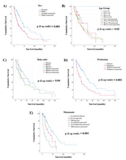
Figure 1 shows the curves related to the factors that presented significant statistical association with pCM patient survival, respectively: sex (p < 0.001), age group (p = 0.02), skin color (p = 0.04), profession (p < 0.001) and metastasis (p < 0.001).

Survival of patients diagnosed with pCM in Goiânia, from 2003 to 2016 A) Survival curve according to sex; B) Survival curve according to age group; C) Survival curve according to skin color; D) Survival curve according to profession; E) Survival curve according to metastasis
DISCUSSION
The demographic profile of patients diagnosed with primary cutaneous melanoma (pCM) in Goiânia showed patterns similar to national studies20,21,22,23,24. There was a greater occurrence of cases in female white skin individuals, as reported in the South and Southeast regions of the country in similar periods23,25,26. However, other studies that aimed to characterize pCM in the country showed greater occurrences in male individuals in most Brazilian cities12,27. A study in Rio Grande do Norte found a greater predominance of pCM cases in brown skin individuals, which can be related to the greater concentration of brown and black skin individuals in the country’s Northeast region2. In the present study, brown skin was the second most affected. This difference may be related to the characteristics of each analyzed region, considering that, in the global literature, white skin individuals are the most affected. The Brazilian South region presents a population with the phenotypic characteristics inherited from the great contingency of white skin European migrants who occupied the region from the 19th to the 20th century, which is related to the high local levels of pCM28. Other factors that can explain the divergence found are the greater racial miscegenation that characterizes the Brazilian population along with the number of patients with no skin color data (36.9% of cases) in the present study.
Most pCM cases were found in the 0 to 59 age group, which is in accordance with national researches that show a greater incidence in individuals between the fourth and sixth decade of life12,14,21,27. This finding may be related to the location of the city of Goiânia in the 16°40’S latitude, receiving high solar incidence all year long, with cumulative effects that predispose earlier appearance of initial lesions29. However, it is worth highlighting the number of pCM cases found in individuals aged 60 or over (316 records, 48.4%), which is in line with most of the published international literature on the subject. It is understood that the elderly did not occupy the first incidence position in this study due to the greater difficulty in diagnosing CM at more advanced ages, which is related to the presentation of lesions with characteristics different from the classic pattern and in places with compromised visibility, such as the scalp30.
A greater occurrence of pCM was observed in individuals with indoor professions. People who work in covered locations show intermittent acute exposure, which enhances risk of cutaneous melanoma30. A study conducted in Paraná observed that 24.2% of patients diagnosed with pCM showed risk of intense solar exposure, but most of them (61.3%) worked in activities with less risk of solar exposure12. Possible explanations for this phenomenon are the fact that most indoor professions generate more income when compared to outdoor professions, which enables more leisure opportunities with acute sun exposure and cumulative action of UV rays. It is also possible that the profile of patients under greater risk of developing cutaneous melanoma (white skin males with a family history of the disease) are more commonly directed to indoor than outdoor professions31.
In this study, the trunk was the most frequent location of pCM, followed by lower limbs, upper limbs and the head and neck areas. The same profile was observed in a study conducted in Uruguay, which found the back and limbs to be the most common locations of cutaneous melanoma32. Studies conducted in the city of Criciúma, Santa Catarina (South), and in the state of Rio Grande do Norte (Northeast), as well as a national multicenter study, reported a greater incidence of primary lesions in the trunk and lower limbs areas2,22,27. Although the head and neck areas were the least affected in the researched patients, other national studies and a research conducted in Colombia found greater incidence of primary lesions in this area24,33,34. The location of primary lesion suffers influence of sociodemographic factors that vary according to the sample found in each study and may explain these differences.
The most present histological type in this study was superficial spreading, which is the most frequent histological type in the literature, regardless of sex and age21,27,33,34,35,36. The second most frequent histological type was nodular melanoma. These results match the findings of some national and international researches32. They diverge, however, from a Colombian research that found it to be less frequent34. World literature presents divergences regarding the prevalence of nodular melanoma, though there is a consensus that this is the most frequent type in the elderly30,37. This may explain the findings of the present research, due to the significant number (48.4%) of cases in the age group of 60 years and over.
Most cases presented a diagnosis of localized disease. The metastasis cases correspond to 9.6% of the sample. The locoregional disease was observed in 35 patients (5.4%). There is, in the world literature, a trend towards stability in melanoma mortality rates due to advances in diagnostic techniques. These allow for the detection of increasingly thinner tumors, with early surgical removal38,39. Early detection contributes to decreasing metastasis cases and may explain these findings.
When comparing results to a research conducted in Goiânia based on RCBP-GO data, between 1988 and 200029, it was possible to observe maintenance and changes in the demographic and clinical profiles of patients diagnosed with pCM. The Sortino-Rachou et al.29 study found 260 cases of pCM in the period, with a greater occurrence in male patients aged 0 to 59 years old, similar to what was found in this study. The previous study also obtained similar results regarding primary lesion location, with initial lesions more frequently located in the following order: trunk, lower limbs, head and neck and upper limbs. Moreover, while the Sortino-Rachou et al.29 study reported 56.9% of invasive melanoma, 17.9% of metastatic melanoma, and 2.8% of in situ melanoma, the present research found 63.4% of localized disease, 9.6% of metastatic melanoma, 5.4% of locoregional melanoma and 0.2% in situ melanoma. The increase in the number of cases found between 2003 and 2016, with greater frequency in women, and the reduction in metastasis rates may be related to factors such as increased quality of RCBP-GO records, greater efficiency of local campaigns against skin cancer and greater accuracy of diagnostic techniques used by clinicians and dermatologists in the city.
Regarding survival of patients with pCM in the city of Goiânia, the aspects associated with lower survival were male sex, age equal to 60 years or over, brown skin, outdoor profession, and metastasis.
In Brazil, more men died of cutaneous melanoma than women40. Such finding can be explained by the male culture that considers a sign of manliness to tolerate pain and not worry about their body health40, which can delay diagnosis and lead to treatment in more advanced cancer stages, with fewer chances of healing9.
The initial treatment of cutaneous melanoma is the surgical removal of the lesion. Nevertheless, it is not unusual to use adjuvant treatments to help in the healing process9. In the elderly, however, these additional treatments are seldom used, due to the presence of comorbidities, inability to tolerate side effects, reduction of autonomy, psychosocial and financial dependency, in addition to histological types and more atypical location of tumors41. Patients aged between 20 to 39 years old are treated more broadly, considering the commonly less aggressive profile of tumors, in addition to presenting less comorbidities and better toleration to side effects of the several available therapies, with better healing rates42.
Regarding the primary cutaneous melanoma type, acral melanoma is the most common type in dark skin individuals and presents the worse prognosis43. Failure to perform plantar physical examinations by many clinicians and dermatologists impairs the early diagnosis of this histological type. Moreover, access to public health still shows signs of racism and negligence against the darker skin population, represented by the higher rotation of these patients in the basic care units of the National Health System (SUS)44. This prevents diagnosis and worsens the patient’s prognosis, which may partially explain results found regarding brown skin.
Regarding work activities, patients who worked in outdoor environments were more likely to die, as they show less-favorable social and economic conditions, which limits their access to health care and increases their chances of developing new melanocytic lesions31. Moreover, these workers are mainly black or brown skinned31, condition which is more related to primary tumor location of worse prognosis, such as acral43. The opposite occurs with patients who worked indoors, presenting greater survival rates, which is in line with the research results.
Primary lesion location is a key factor in the prognosis of cutaneous melanoma cases. While worse prognoses are associated to melanomas with primary lesions of axial location (trunk, head and neck), melanomas with primary lesion in the upper and lower limbs are associated to a better prognosis45. It is noteworthy that, within the locations of lower and upper limbs, the extremities (soles of the feet, palms of the hands and nail bed) are included, which can be a confounding factor in the analysis of the prognosis of limb lesions, since acral melanoma is considered to have the worst prognosis in the literature43. In the present study, it was not possible to determine a relationship between primary lesion location and survival time, in spite of this prognostic factor’s relevancy in the literature46.
The presence of metastasis is a variable that significantly influences the prognosis of patients with cutaneous melanoma13. Patients with localized disease show a good prognosis in most cases. Patients with locoregional disease show an uncertain prognosis according to the presence or absence of ulceration and the number of lymph nodes involved41. Patients with metastasis show unfavorable prognosis and a lower survival rate among the mentioned primary locations8. Results found in the study are therefore in line with the literature.
Histological classification of primary cutaneous melanoma is important for diagnosis and defining a course of action, though it is not significantly relevant to the prognosis of melanoma patients46. The present study did not identify an association between histological type and survival time of patients with melanoma. Thus, the findings of this study are in line with the literature.
The limitations to this study included using data that were primarily collected by RCBP-GO employees, which narrowed the researcher’s control over the quality of that process. The great number of cases with unavailable information regarding the chosen variables also limited extrapolation of results obtained for the Goiânia population.
The strengths of this study include the great number of primary cutaneous melanoma (pCM) cases found (n = 653), which allowed for a robust analysis of the epidemiological profile of pCM in the city of Goiânia, despite the great amount of unavailable information. Moreover, the use of data from a RCBP, which receives patient data from several information sources in the city, helped reduce the selection bias in the study.
CONCLUSION
Primary cutaneous melanoma (pCM) is a disease of great relevance in the city of Goiânia. The evidenced factors that worsen survival of patients with pCM were male individuals, aged over 60 years old, brown skin, outdoor profession, and presence of metastasis, while the factors that improved survival of these patients were female individuals, age group from 20 to 39 years old, white skin, indoor profession, and localized disease. No significant difference was observed between survival time and location of the primary lesion and between histological type in the present study.
Results show the need for expanding pCM mass detection campaigns in the city of Goiânia and the creation of strategies that increasingly attract male, elder and brown skin individuals for assessment, to early diagnose this population that is already less likely to survive. Moreover, the most common locations for primary lesion in this study reinforce the precarious adoption of solar protection measures by the Goiânia population, which should be encouraged through campaigns mainly targeted at children and young people, so they grow up to be conscious adults.
Finally, considering the great number of uninformed data found in the RCBP-GO records, better guidance for doctors is suggested in relation to filling out notification forms in institutions that supply the RCBP-GO with data on cutaneous melanoma in the city of Goiânia. Another key suggestion is to include education and socioeconomic condition fields in the RCBP-GO notification forms, as these variables are known to interfere in the survival time of patients with pCM and could help further studies.
ACKNOWLEDGMENTS
To ACCG and the employees of RCBP-GO who provided the data analyzed in this study; to the statistic professional Macks Wendhell Gonçalves who performed the statistical analysis of the current study.
References
-
1 Diepgen TL, Mahler V. The epidemiology of skin cancer. Br J Dermatol. 2002;146(s61):1-6. doi: https://doi.wiley.com/10.1046/j.1365-2133.146.s61.2.x
» https://doi.org/10.1046/j.1365-2133.146.s61.2.x - 2 Lima RLF, Nóbrega MQR, Moraes MDLSA, et al. Estudo retrospectivo de melanomas cutâneos e mucosos na população do estado do Rio Grande do Norte, Brasil. J Bras Patol e Med Lab. 2009;45(6):493-9.
-
3 Steglich RB, Coelho KMPA, Cardoso S, et al. Epidemiological and histopathological aspects of primary cutaneous melanoma in residents of Joinville, 2003-2014. An Bras Dermatol. 2018;93(1):45-53. doi: https://doi.org/10.1590/abd1806-4841.20185497
» https://doi.org/10.1590/abd1806-4841.20185497 -
4 Bray F, Ferlay J, Soerjomataram I, et al. Global cancer statistics 2018: GLOBOCAN estimates of incidence and mortality worldwide for 36 cancers in 185 countries. CA Cancer J Clin. 2018;68(6):394-424. doi: http://doi.wiley.com/10.3322/caac.21492
» https://doi.org/10.3322/caac.21492 -
5 Instituto Nacional de Câncer. Estimativa 2023: incidência de câncer no Brasil [Internet]. Rio de Janeiro: INCA; 2022. [acesso em 2023 nov 13]. Disponível em: Disponível em: https://www.inca.gov.br/sites/ufu.sti.inca.local/files/media/document/estimativa-2023.pdf
» https://www.inca.gov.br/sites/ufu.sti.inca.local/files/media/document/estimativa-2023.pdf -
6 Chang AE, Karnell LH, Menck HR. The National Cancer Data Base report on cutaneous and noncutaneous melanoma. Cancer. 1998;83(8):1664-78. doi: https://doi.org/10.1002/(SICI)1097-0142(19981015)83:8%3C1664::AID-CNCR23%3E3.0.CO;2-G
» https://doi.org/10.1002/(SICI)1097-0142(19981015)83:8%3C1664::AID-CNCR23%3E3.0.CO;2-G - 7 Bakos L. Melanoma cutâneo: estudos de base populacional no Brasil. An Bras Dermatol. 2006;81(5):402.
-
8 Soong SJ, Ding S, Coit D, et al. Predicting Survival outcome of localized melanoma: an electronic prediction tool based on the AJCC melanoma database. Ann Surg Oncol. 2010;17(8):2006-14. doi: https://doi.org/10.1245/s10434-010-1050-z
» https://doi.org/10.1245/s10434-010-1050-z -
9 Schadendorf D, Fisher DE, Garbe C, et al. Melanoma. Nat Rev Dis Prim. 2015;1(1):15003. doi: https://doi.org/10.1038/nrdp.2015.3
» https://doi.org/10.1038/nrdp.2015.3 -
10 Minocha R, Damian DL, Halliday GM. Melanoma and nonmelanoma skin cancer chemoprevention: a role for nicotinamide? Photodermatol Photoimmunol Photomed. 2018;34(1):5-12. doi: http://doi.wiley.com/10.1111/phpp.12328
» https://doi.org/10.1111/phpp.12328 - 11 Eide MJ, Weinstock MA, Clark MA. Demographic and socioeconomic predictors of melanoma prognosis in the United States. J Health Care Poor Underserved. 2009;20(1):227-45.
-
12 Purim KSM, Sandri CO, Pinto NT, et al. Perfil de casos de melanoma em um hospital universitário, 2003 a 2007. Rev Bras Cancerol. 2013;59(2):193-9. doi: https://doi.org/10.32635/2176-9745.RBC.2013v59n2.523
» https://doi.org/10.32635/2176-9745.RBC.2013v59n2.523 -
13 Armstrong BK, Cust AE. Sun exposure and skin cancer, and the puzzle of cutaneous melanoma. Cancer Epidemiol. 2017;48:147-56. doi: https://doi.org/10.1016/j.canep.2017.04.004
» https://doi.org/10.1016/j.canep.2017.04.004 - 14 Battisti R, Nunes DH, Weber AL, et al. Evaluation of the epidemiological profile and the mortality rate of the patients with primary cutaneous melanoma in Florianopolis - SC, Brazil. An Bras Dermatol. 2009;84(4):335-42.
-
15 Lages RB, Barbosa PB, Almeida IP, et al. Detecção precoce do câncer de pele: experiência de campanha de prevenção no Piauí-Brasil. Rev Bras em Promoção da Saúde [Internet]. 2012 [acesso 2023 nov 23];25(2):221-7. Disponível em: Disponível em: http://www.unifor.br/images/pdfs/rbps/2012.2_artigo13.pdf
» http://www.unifor.br/images/pdfs/rbps/2012.2_artigo13.pdf -
16 Moura L, Curado MP, Simões EJ, et al. Avalição do Registro de Câncer de Base Populacional do Município de Goiânia, Estado de Goiás, Brasil. Epidemiol serv saúde. 2006;15(4):7-17. doi: http://dx.doi.org/10.5123/S1679-49742006000400002
» https://doi.org/10.5123/S1679-49742006000400002 - 17 Conselho Nacional de Saúde (BR). Resolução n° 466, de 12 de dezembro de 2012. Aprova as diretrizes e normas regulamentadoras de pesquisas envolvendo seres humanos. Diário Oficial da União, Brasília, DF. 2013 jun 13; Seção I:59.
- 18 Organização Mundial da Saúde. CID-10: Classificação Estatística Internacional de Doenças com disquete. Vol. 3, Índice Alfabético. São Paulo: Edusp; 2008.
-
19 SPSS®: Statistical Package for Social Science (SPSS) [Internet]. Versão 26.0. [Nova York]. International Business Machines Corporation. [acesso 2023 mar 9]. Disponível em: Disponível em: https://www.ibm.com/br-pt/spss?utm_content=SRCWW&p1=Search&p4=43700077515785492&p5=p&gclid=CjwKCAjwgZCoBhBnEiwAz35Rwiltb7s14pOSLocnooMOQh9qAL59IHVc9WP4ixhNTVMjenRp3-aEgxoCubsQAvD_BwE&gclsrc=aw.ds
» https://www.ibm.com/br-pt/spss?utm_content=SRCWW&p1=Search&p4=43700077515785492&p5=p&gclid=CjwKCAjwgZCoBhBnEiwAz35Rwiltb7s14pOSLocnooMOQh9qAL59IHVc9WP4ixhNTVMjenRp3-aEgxoCubsQAvD_BwE&gclsrc=aw.ds - 20 Vilanova CMA, Lages RB, Ribeiro SM, et al. Perfil epidemiológico e histopatológico do melanoma cutâneo em um centro do nordeste brasileiro de 2000 a 2010. An Bras Dermatol. 2013;88(4):545-53.
- 21 Costa NF, Fernandes NC, Borges MRMM. Study of the histopathological types of cutaneous melanoma in palmas-TO from 2001 to 2011. An Bras Dermatol. 2015;90(5):638-45.
- 22 Melo AC, Wainstein AJA, Buzaid AC, et al. Melanoma signature in Brazil: Epidemiology, incidence, mortality, and trend lessons from a continental mixed population country in the past 15 years. Melanoma Res. 2018;28(6):629-36.
-
23 Lima AS, Stein CE, Casemiro KP, et al. Epidemiology of melanoma in the south of Brazil: study of a city in the Vale do Itajaí from 1999 to 2013. An Bras Dermatol. 2015;90(2):185-9. doi: https://doi.org/10.1590/abd1806-4841.20153076
» https://doi.org/10.1590/abd1806-4841.20153076 -
24 Veneziano DB. Estudo do perfil epidemiológico do melanoma cutâneo na cidade de Jaú-SP através do registro de base populacional [Internet]. Botucatu: Universidade Estadual Paulista Júlio de Mesquita Filho, Faculdade de Medicina de Botucatu; 2014. [acesso 2023 nov 23]. Disponível em: Disponível em: http://repositorio.unesp.br/handle/11449/108855
» http://repositorio.unesp.br/handle/11449/108855 - 25 Moreno M, Schmitt RL, Lang MG, et al. Epidemiological profile of patients with cutaneous melanoma in a region of southern Brazil. J Skin Cancer. 2012;2012:1-8.
- 26 Wainstein AJA, Duprat Neto JP, Enokihara MY, et al. Demographic, clinical, and pathologic features of patients with cutaneous melanoma: final analysis of the brazilian melanoma group database. JCO Glob Oncol. 2020;(6):575-82.
- 27 Konrad P, Fabris MR, Melao S, et al. Perfil epidemiológico e histopatológico dos casos de melanoma cutâneo primário diagnosticados em Criciúma no período entre 2005 e 2007. An Bras Dermatol. 2011;86(3):457-61.
-
28 Bakos L, Masiero N, Bakos R, et al. European ancestry and cutaneous melanoma in Southern Brazil. J Eur Acad Dermatology Venereol. 2009;23(3):304-7. doi: https://doi.org/10.1111/j.1468-3083.2008.03027.x
» https://doi.org/10.1111/j.1468-3083.2008.03027.x - 29 Sortino-Rachou AM, Curado MP, Latorre MRDO. Melanoma cutâneo: estudo de base populacional em Goiânia, Brasil, de 1988 a 2000. An Bras Dermatol. 2006;81(5):449-55.
-
30 Iglesias-Pena N, Paradela S, Tejera-Vaquerizo A, et al. Cutaneous melanoma in the elderly: review of a growing problem. Actas Dermosifiliogr. 2019;110(6):434-47. doi: http://dx.doi.org/10.1016/j.adengl.2019.05.012
» https://doi.org/10.1016/j.adengl.2019.05.012 -
31 Modenese A, Korpinen L, Gobba F. Solar radiation exposure and outdoor work: an underestimated occupational risk. Int J Environ Res Public Health. 2018;15(10):2063. doi: https://doi.org/10.3390/ijerph15102063
» https://doi.org/10.3390/ijerph15102063 - 32 Amarillo D, Boni D, Nantes F, et al. Melanoma cutáneo en Uruguay: características clínico-epidemiológicas y sobrevida en cohorte de pacientes de dos centros de referencia en el período 2008-2017. Rev Medica Del Uruguay. 2020;36(2):146-54.
-
33 Bertoldi MB, Augusto C, Bastos S, et al. Melanoma cutaneo em um hospital universitario, 2001-2016. Rev Bras Cancerol. 2020;66(3):e-03911. doi: https://doi.org/10.32635/2176-9745.RBC.2020v66n3.911
» https://doi.org/10.32635/2176-9745.RBC.2020v66n3.911 - 34 Pozzobon FC, Acosta AE. Epidemiological profile of primary cutaneous melanoma over a 15-year period at a private skin cancer center in Colombia. Rev Salud Publica. 2018;20(2):226-31.
-
35 Ferreira T, Santos IDAO, Oliveira AF, et al. Estudo retrospectivo dos pacientes portadores de melanoma cutâneo atendidos na Universidade Federal de São Paulo. Rev Col Bras Cir. 2018;45(4):e1715. doi: https://doi.org/10.1590/0100-6991e-20181715
» https://doi.org/10.1590/0100-6991e-20181715 -
36 Mélo KC, Santos AGG, Amorim JR, et al. Análise temporal da mortalidade por cancer de pele melanona no Brasil: 2000 a 2016. Surg Cosmet Dermatology. 2019;11(4):287-94. doi: http://www.dx.doi.org/10.5935/scd1984-8773.20191141486
» https://doi.org/10.5935/scd1984-8773.20191141486 -
37 Tragos C, Hieken TJ. Optimizing the management of cutaneous melanoma in the elderly. Surgery. 2011;150(4):828-35. doi: http://dx.doi.org/10.1016/j.surg.2011.07.054
» https://doi.org/10.1016/j.surg.2011.07.054 -
38 Garbe C, McLeod GRC, Buettner PG. Time trends of cutaneous melanoma in Queensland, Australia and Central Europe. Cancer. 2000;89(6):1269-78. doi: https://doi.org/10.1002/1097-0142(20000915)89:6%3C1269::AID-CNCR11%3E3.0.CO;2-C
» https://doi.org/10.1002/1097-0142(20000915)89:6%3C1269::AID-CNCR11%3E3.0.CO;2-C -
39 MacKie RM, Bray CA, Hole DJ, et al. Incidence of and survival from malignant melanoma in Scotland: an epidemiological study. Lancet. 2002;360(9333):587-91. doi: https://doi.org/10.1016/s0140-6736(02)09779-9
» https://doi.org/10.1016/s0140-6736(02)09779-9 -
40 Separavich MA, Canesqui AM. Saúde do homem e masculinidades na Política Nacional de Atenção Integral à Saúde do Homem: uma revisão bibliográfica. Saúde e Soc. 2013;22(2):415-28. doi: https://doi.org/10.1590/S0104-12902013000200013
» https://doi.org/10.1590/S0104-12902013000200013 - 41 Schadendorf D, van Akkooi ACJ, Berking C, et al. Melanoma. Lancet. 2018;392(10151):971-84.
-
42 Purim KSM, Bonetti JPC, Silva JYF, et al. Características do melanoma em idosos. Rev Col Bras Cir. 2020;47. Disponível em: https://doi.org/10.1590/0100-6991e-20202441
» https://doi.org/10.1590/0100-6991e-20202441 -
43 Maia M, Russo C, Ferrari N, et al. Melanoma acrolentiginoso: um desafio ao diagnóstico precoce. An Bras Dermatol. 2003;78(5):553-60. doi: https://doi.org/10.1590/S0365-05962003000500004
» https://doi.org/10.1590/S0365-05962003000500004 -
44 Massignam FM, Bastos JLD, Nedel FB. Discriminação e saúde: um problema de acesso. Epidemiol Serv Saúde. 2015;24(3):544-1. doi: http://dx.doi.org/10.5123/S1679-49742015000300020
» https://doi.org/10.5123/S1679-49742015000300020 -
45 Strudel M, Festino L, Vanella V, et al. Melanoma: prognostic factors and factors predictive of response to therapy. Curr Med Chem. 2020;27(17):2792-813. doi: http://dx.doi.org/10.2174/0929867326666191205160007
» https://doi.org/10.2174/0929867326666191205160007 -
46 Bobos M. Histopathologic classification and prognostic factors of melanoma: a 2021 update. Ital J Dermatology Venereol. 2021;156(3):300-21. doi: https://doi.org/10.23736/S2784-8671.21.06958-3
» https://doi.org/10.23736/S2784-8671.21.06958-3