 Esophageal Neoplasm with Breast Metastasis: Case Report
ABSTRACT
Introduction: Esophageal cancer is the seventh most frequently diagnosed cancer and the sixth leading cause of cancer-related death worldwide. In Brazil, it ranks 13th among the most common types of cancer. The most usual sites of metastatic esophageal cancer are lungs, liver, and bones. Metastases from esophageal cancer to the breast are extremely rare and poorly documented in the literature.
Case report: A 45-year-old man was diagnosed with esophageal adenocarcinoma after presenting with dyspepsia and epigastralgia. Investigation via upper gastrointestinal endoscopy revealed esophageal adenocarcinoma with no distant metastases. He underwent esophagectomy and adjuvant chemotherapy. Post-treatment positron emission tomography (PET-CT) detected local recurrence, leading to the initiation of palliative therapy. Shortly before, a nodule in the right breast was initially diagnosed as a primary breast lesion, triple-negative carcinoma of no special type. However, a review of the slide revealed poorly differentiated metastatic adenocarcinoma from the esophagus, resulting in two discordant diagnoses. A third analysis of the breast material by immunohistochemistry confirmed a secondary metastatic lesion from the esophagus. During treatment, the patient had a seizure, with diagnosis of secondary brain implant. He underwent radiotherapy for the central nervous system lesion and mastectomy. PET-CT revealed no signs of abnormal metabolic activity. He is currently asymptomatic with therapeutic maintenance immunotherapy associated with HER2 blockade.
Conclusion: This case highlights the rarity of breast metastasis in esophageal cancer and the crucial importance of immunohistochemistry for an accurate differential diagnosis and adequate therapeutic planning.
Key words:
Esophageal Neoplasms; Adenocarcinoma; Neoplastic Metastasis; Breast Neoplasms; Immunohistochemistry
RESUMO
Introdução: A neoplasia de esôfago é o sétimo câncer mais frequentemente diagnosticado e a sexta principal causa de morte relacionada ao câncer no mundo. No Brasil, ocupa a 13ª posição entre os tipos de câncer mais frequentes. Os sítios mais comuns de disseminação do câncer esofágico são pulmão, fígado e ossos. As metástases do câncer de esôfago para a mama são extremamente raras e pouco documentadas na literatura.
Relato do caso: Homem, 45 anos, diagnosticado com adenocarcinoma de esôfago após manifestar dispepsia e epigastralgia. A investigação por endoscopia digestiva alta revelou adenocarcinoma de esôfago, sem metástases a distância. Foi submetido à esofagectomia e quimioterapia adjuvante. Na tomografia por emissão de pósitrons (PET-CT), realizada pós-tratamento, foi detectada recidiva local, iniciando terapia paliativa. Pouco antes, uma nodulação na mama direita foi diagnosticada inicialmente como lesão primária da mama, carcinoma do tipo não especial, triplo-negativo. Contudo, uma revisão da lâmina revelou um adenocarcinoma pouco diferenciado metastático do esôfago, resultando em dois diagnósticos discordantes. Uma terceira análise do material da mama por imuno-histoquímica confirmou a lesão metastática secundária ao esôfago. Durante o tratamento, o paciente apresentou crise convulsiva, com diagnóstico de implante cerebral secundário. Realizou radioterapia em lesão do sistema nervoso central e mastectomia. O PET-CT revelou ausência de sinais de atividade metabólica anormal. Atualmente, encontra-se assintomático com programação terapêutica de imunoterapia de manutenção associada a bloqueio do HER2.
Conclusão: Este caso sublinha a raridade da metástase mamária no câncer de esôfago e a importância crucial da imuno-histoquímica para um diagnóstico diferencial preciso e planejamento terapêutico adequado.
Palavras-chave:
Neoplasias Esofágicas; Adenocarcinoma; Metástase Neoplásica; Neoplasias da Mama; Imuno-histoquímica
RESUMEN
Introducción: La neoplasia de esófago es el séptimo cáncer más frecuentemente diagnosticado y la sexta principal causa de muerte relacionada con el cáncer en el mundo. En el Brasil, ocupa el 13º lugar entre los tipos de cáncer más frecuentes. Los sitios más comunes de diseminación del cáncer esofágico son los pulmones, el hígado y los huesos. Las metástasis del cáncer de esófago hacia la mama son extremadamente raras y poco documentadas en la literatura.
Informe del caso: Hombre, 45 años, diagnosticado con adenocarcinoma de esófago después de manifestar dispepsia y epigastralgia. La investigación a través de una endoscopia digestiva alta reveló adenocarcinoma de esófago, sin metástasis a distancia. Se sometió a esofagectomía y quimioterapia adyuvante. En la tomografía por emisión de positrones (PET-CT), realizada después del tratamiento, se detectó recidiva local, iniciando terapia paliativa. Poco antes, una nodulación en la mama derecha fue diagnosticada inicialmente como lesión primaria de la mama, carcinoma del tipo no especial, triple negativo. Sin embargo, una revisión de la lámina reveló adenocarcinoma pobremente diferenciado metastásico del esófago, resultando en dos diagnósticos discordantes. Un tercer análisis del material mamario mediante inmunohistoquímica confirmó lesión metastásica secundaria al esófago. Durante el tratamiento, el paciente desarrolló una convulsión, con diagnóstico de implantación cerebral secundaria. Realizó radioterapia para lesiones del sistema nervioso central y mastectomía. La PET-CT no reveló signos de actividad metabólica anormal. Actualmente se encuentra asintomático con esquema terapéutico de inmunoterapia de mantenimiento asociada a bloqueo de HER2.
Conclusión: Este caso resalta la rareza de las metástasis mamarias en el cáncer de esófago y la importancia crucial de la inmunohistoquímica para un diagnóstico diferencial preciso y una planificación terapéutica adecuada.
Palabras clave:
Neoplasias Esofágicas; Adenocarcinoma; Metástasis Neoplásica; Neoplasias de la Mama; Inmunohistoquímica
INTRODUCTION
Esophageal cancer is the seventh most frequently diagnosed cancer and the sixth leading cause of cancer-related death worldwide1. In Brazil, 10,990 new cases of esophageal cancer are estimated for 2023-2025, an incidence rate of 5.97 per 100 thousand inhabitants, 8,200 in men and 2,790 in women. Except non-melanoma skin cancer, esophageal cancer ranks 13th among the most frequent types with elevated rates in the South Region1.
Two histological types of neoplasms with different biological features (squamous cell carcinoma and adenocarcinoma) and diverse etiology, pathology, therapeutic and prognosis are diagnosed on the esophagus2,3. It is more common in 60-70 years old Black men, mostly affecting the middle and upper portions of the esophagus, whose major risk factors are alcohol abuse and smoking. Esophageal adenocarcinoma, on its turn, is more frequent on the lower portion, the main risk factors are gastroesophageal reflux disease and obesity4,5.
Dysphagia is one of the first symptoms of esophageal cancer due to obstruction caused by tumor growth making deglutition difficult and weight loss6. The most common metastases occur on lungs, liver and bones7, but to the breast is extremely rare, particularly in men.
Extramammary metastases account for 0.4% to 1.3% among extramammary tumors, the most common are melanoma, lung cancer, renal cell carcinoma and ovarian tumors in case of breast metastasis8. Case reports highlight that breast metastasis from esophageal cancer can be confounded with breast primary tumor, making differential diagnosis crucial9.
The present case report addresses a case of a 45-year old man diagnosed with esophageal carcinoma in 2022 that metastasized to the right breast. The Institutional Review Board of “Universidade São Francisco” approved the study, report number 6802217 (CAAE (submission for ethical review): 79103524.0.0000.5514), in compliance with Directive 466/1210 of the National Health Council. The patient signed the Informed Consent Form (ICF) after being briefed about the study.
CASE REPORT
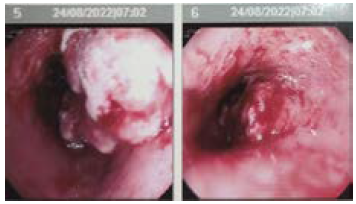
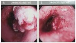
45 years-old man, without comorbidities, presented symptoms of dyspepsia and epigastric pain in the first semester of 2022. Upper digestive endoscopy in August of the same year revealed distal esophageal vegetating lesion suggestive of malignancy (Figure 1). Biopsy confirmed distal esophageal adenocarcinoma and local disease was ratified by complementary thoracic, abdominal and pelvis computed tomography (cT2N0M0 - ECII).The attending surgeon opted for upfront surgery and the patient waited three months for the insurer’s authorization. In November 2022, he was submitted to video-laparoscopy esophageal gastrectomy with reconstruction of the digestive tract without complications.
Initial clinical staging was cT2N0M0, with no indication of neoadjuvant chemotherapy. Anatomopathology, however, upstaged to poor differentiated adenocarcinoma G3, pT3N2M0 - ECIIIa, with compromise of five of the 23 lymph nodes investigated. The patient was referred for the first clinical oncological assessment.
Surgery delay and lack of pre-operative restaging negatively impacted the implementation of neoadjuvant chemotherapy, the treatment of choice for advanced stages, followed by surgery and adjuvant therapy with or without immunotherapy depending on the pathological response.
During the first consult in January 2023, adjuvant chemotherapy was prescribed from February to July 2023 with eight cycles of FOLFOX (folinic acid, fluorouracil and oxaliplatin). However, a positron emission tomography-computed tomography (PET-CT) in August 2023 revealed local relapse with increased metabolism in esophageal anastomosis and regional lymph nodes (SUV 11).
Immunohistochemistry of the material was performed to define best strategy of palliative chemotherapy, which revealed positivity of receptors HER2 (3+) and PDL1 (CPS 6). Based on the study KEYNOTE 811 a first-line palliative chemotherapy with pembrolizumab, trastuzumab, cisplatin and fluorouracil was initiated.
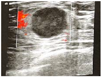
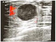
A physical exam in September 2023 identified right breast nodes confirmed by ultrasound with percutaneous biopsy (Figure 2). The initial biopsy suggested triple-negative no special grade 3 invasive carcinoma with negative hormone receptors and HER2 low (HER2 ++, FISH negative). However, another laboratory reviewed the exam and indicated metastatic poorly differentiated esophageal adenocarcinoma.
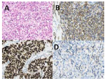
A third breast immunohistochemistry in January 2024 confirmed breast nodes as metastatic esophageal adenocarcinoma (Figure 3); it also revealed expression of pan-cytokeratin, confirming the epithelial origin of the neoplasm. The mammary markers tested, GATA3, SOX10 and estrogen receptor were negative. CDX2 was positive, strong and diffuse. In addition, the marker chromogranin A associated with neuroendocrine origin was negative.
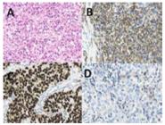
A) HE (Hematoxylin/eosin), magnified 40x; B) Positive Pan-cytokeratin (AE1/AE3), magnified 40x; C) Positive CDX2, magnified 40x; D) Negative GATA3, magnified 40x
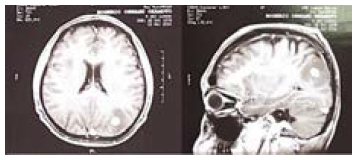
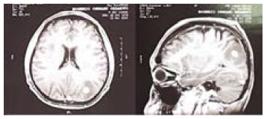
In March 2024, the patient attended the emergency due to a convulsive crisis and was submitted to cranial CT which revealed nodes on brain parenchyma suggestive of metastasis. For better delimitation of brain lesion and therapeutic plan, cranial magnetic resonance showed 16 x 13 x 14 mm nodular lesion at the left angular gyrus (Figure 4).
On March 28, 2024, PET-CT revealed abnormal neoplastic activity exclusively on mammary lesion without increased metabolism in esophageal anastomosis and local lymph nodes. Radiotherapy treatment for central nervous system (CNS) lesion and surgery to resect the mammary lesion were planned with temporary interruption of chemo and immunotherapy.
In May 2024, the patient was submitted to radiotherapy for CNS lesion and new cranial magnetic resonance imaging which revealed reduction of the lesion to 10 x 10 x 9 mm and perilesional edema. In June 2024, free-margins mastectomy was performed (8.0 x 6.0 x 5.5 cm) with negative biopsy of sentinel lymph node.
The last PET-CT on July 19, 2024 showed absence of abnormal signs of metabolic activity. So far, the patient is asymptomatic and without neurological sequelae. As no signs of active systemic disease were detected, only a remaining CNS injury, the therapeutic approach is to keep maintenance immunotherapy associated with HER 2 block.
DISCUSSION
The progression of esophageal adenocarcinoma regardless of the initial surgery and adjuvant chemotherapy is quite common. Accurate staging and early treatment are essential for successful outcomes. Early detection of metastases is crucial for effective therapeutic adjustments. Immunotherapy and target-therapy based palliative chemotherapy reflects the most recent treatment strategies for specific molecular markers tumors as positive HER2 and PDL111.
Breast metastases from esophageal tumors are extremely rare and little understood. This complex process involves several steps that allow carcinogenic cells to migrate from the esophageal primary tumor and circulate through the body towards new breast cancer foci. Metastatic spread from esophageal tumors occurs by lymphatic, hematogenous and intraluminal pathways, however, the specific route that facilitates breast metastases is not well established yet due to its rarity. Most likely, the spread occurs through the blood allowing esophageal carcinogenic cells to metastasize to the breast12.
The ability of esophageal tumor to metastasize to breast involves a complex interaction between molecular characteristics of carcinogenic cells, tumor microenvironment and host systemic factors. Studies have suggested that metastases are facilitated by genetic and epigenetic alterations on tumor cells and adaptative capacity to the new breast environment12.
The accurate identification of the origin of a breast lesion in patients with history of esophageal cancer is pivotal for proper therapeutic management. Immunohistochemistry plays a key role in this process allowing the characterization of molecular markers that help to differentiate a primary breast cancer and an esophageal metastasis9,13.
The final biopsy review of the right breast revealed fragments of mammary tissue infiltratred by atypical epythelioid cells neoplasm. The morphological findings suggested poorly differentiated adenocarcinoma.
Hormone receptors markers (estrogen and progesterone), GATA3 and CDX2 are frequently utilized to guide the differential diagnosis in this situation14,15. GATA3 is a DNA binding protein which plays a key role in various tissues differentiation and cellular proliferation, including the breast. It is a marker to evaluate mammary tumors, being positive in many primary mammary carcinomas.
GATA 3 negativity rules out the mammary origin of the lesion and suggests the possible metastasis to another organ16,17. CDX2 is a transcription factor that regulates the genic expression during the embryonic development and maintenance of intestinal adult function. It is frequently positive in colorectal adenocarcinomas and gastrointestinal tract. Its positivity in mammary lesion suggests possible gastrointestinal origin as esophageal adenocarcinoma metastasis18.
Thorough evaluation with advanced techniques as immunohistochemistry plays a crucial role in differentiation between breast primary tumor and esophageal metastasis as in the present case. Correct identification of the mammary lesion directly impacts therapeutic decisions leading to better decisions among local and systemic treatments19,20.
CONCLUSION
The case portrays the challenging management of esophageal cancer, particularly if metastasized to uncommon sites as the breast. The rarity and complexity of these events emphasize the necessity of a customized and integrated approach to treat these patients.
It contributed to understand the uncommon metastization of esophageal cancer and points out the necessity of cautious surveillance. Patient-centered therapeutic approaches should be adopted due to clinical challenges of this complex condition.
References
- 1 Instituto Nacional de Câncer José Alencar Gomes da Silva. Estimativa 2023: Incidência de câncer no Brasil. Rio de Janeiro: INCA; 2023.
-
2 Rustgi AK, El-Serag HB. Esophageal carcinoma. N Engl J Med. 2014;371:2499-509. doi: https://doi.org/10.1056/NEJMra1314530
» https://doi.org/10.1056/NEJMra1314530 -
3 Zhang Y. Epidemiology of esophageal cancer. World J Gastroenterol. 2013;19(34):5598-606. doi: https://doi.org/10.3748/wjg.v19.i34.5598
» https://doi.org/10.3748/wjg.v19.i34.5598 -
4 Lagergren J, Lagergren P. Oesophageal cancer. BMJ. 2010;341:c6280. doi: https://doi.org/10.1136/bmj.c6280
» https://doi.org/10.1136/bmj.c6280 -
5 Pennathur A, Gibson MK, Jobe BA, et al. Oesophageal carcinoma. Lancet. 2013;381(9864):400-12. doi: https://doi.org/10.1016/S0140-6736(12)60643-6
» https://doi.org/10.1016/S0140-6736(12)60643-6 -
6 Kelsen DP, Ahuja R, Hopfan S, et al. Combined modality therapy of esophageal carcinoma. Cancer. 1981;48(1):31-7. doi: https://doi.org/10.1002/1097-0142(19810701)48:1%3C31::aid-cncr2820480108%3E3.0.co;2-4
» https://doi.org/10.1002/1097-0142(19810701)48:1%3C31::aid-cncr2820480108%3E3.0.co;2-4 -
7 Ai D, Zhu H, Ren W, et al. Patterns of distant organ metastases in esophageal cancer: a population-based study. J Thorac Dis. 2017;9(9):3023-30. doi: https://doi.org/10.3389%2Ffonc.2018.00302
» https://doi.org/10.3389%2Ffonc.2018.00302 -
8 Williams SA, Ehlers RA II, Hunt KK, et al. Metastases to the breast from nonbreast solid neoplasms. Cancer. 2007;110(4):731-7. doi: https://doi.org/10.1002/cncr.22835
» https://doi.org/10.1002/cncr.22835 -
9 Ghibour A, Shaheen O. Breast metastasis in esophageal cancer: literature review and report on a case. Case Rep Surg. 2016;2016:8121493. doi: https://doi.org/10.1155/2016/8121493
» https://doi.org/10.1155/2016/8121493 - 10 Conselho Nacional de Saúde (BR). Resolução n° 466, de 12 de dezembro de 2012. Aprova as diretrizes e normas regulamentadoras de pesquisas envolvendo seres humanos. Diário Oficial da União, Brasília, DF. 2013 jun 13; Seção I:59.
-
11 Shah MA, Kojima T, Hochhauser D, et al. Efficacy and safety of pembrolizumab for heavily pretreated patients with advanced, metastatic adenocarcinoma or squamous cell carcinoma of the esophagus: The phase 2 KEYNOTE-180 study. JAMA Oncol. 2019;5(4):546-50. doi: https://doi.org/10.1001/jamaoncol.2018.5441
» https://doi.org/10.1001/jamaoncol.2018.5441 -
12 Tornos C, Silva EG, el-Naggar A, et al. Metastatic tumors to the breast. Ann Surg Oncol. 1994;1(4):319-24. doi: https://doi.org/10.1007/BF02449546
» https://doi.org/10.1007/BF02449546 -
13 Tse GM, Tan PH, Putti TC, et al. Metastatic tumors to the breast: a clinicopathological study of 78 cases in a chinese population. Histopathology. 2006;49(2):127-34. doi: https://doi.org/10.1111/j.1365-2559.2006.02464.x
» https://doi.org/10.1111/j.1365-2559.2006.02464.x -
14 Rakha EA, Ellis IO. An overview of assessment of prognostic and predictive factors in breast cancer needle core biopsy specimens. J Clin Pathol. 2007;60(12):1300-06. doi: https://doi.org/10.1136/jcp.2007.048207
» https://doi.org/10.1136/jcp.2007.048207 - 15 Dabbs DJ. Breast pathology. 3 ed. Philadelphia: Elsevier Health Sciences; 2009.
-
16 Abreu FB, Liu X, Tsongalis GJ. GATA3 expression in breast carcinoma: utility in triple-negative, sarcomatoid, and metastatic carcinomas. Am J Clin Pathol. 2015;144(5):665-71. doi: https://doi.org/10.1309/AJCP1Q5URFTTFNYI
» https://doi.org/10.1309/AJCP1Q5URFTTFNYI -
17 Nonaka D, Chiriboga L, Rubin BP. SOX10: a pan-schwannian and melanocytic marker. Am J Surg Pathol. 2008;32(8):1291-8. doi: https://doi.org/10.1097/PAS.0b013e31816a005a
» https://doi.org/10.1097/PAS.0b013e31816a005a -
18 Werling RW, Yaziji H, Bacchi CE, et al. CDX2, a highly sensitive and specific marker of adenocarcinomas of intestinal origin: an immunohistochemical survey of 476 primary and metastatic carcinomas. Am J Surg Pathol. 2003;27(3):303-10. doi: https://doi.org/10.1097/00000478-200303000-00005
» https://doi.org/10.1097/00000478-200303000-00005 -
19 Pennathur A, Gibson MK, Jobe BA, et al. Oesophageal carcinoma. Lancet. 2013;381(9864):400-12. doi: https://doi.org/10.1016/S0140-6736(12)60643-6
» https://doi.org/10.1016/S0140-6736(12)60643-6 -
20 Song Z, Zhang X, Yu X, et al. Mechanisms of chemoresistance in esophageal cancer. Curr Cancer Drug Targets. 2018;18(10):968-84. doi: https://doi.org/10.2174/1568009618666180116111715
» https://doi.org/10.2174/1568009618666180116111715