
 Identification of MicroRNAs Associated with the Diagnosis and Prognosis of Cholangiocarcinoma
ABSTRACT
Introduction: Cholangiocarcinoma (CHOL) is a malignant neoplasm of the biliary epithelium and represents the second most usual form of liver cancer. Its high aggressiveness and late diagnosis hinder the implementation of effective therapies. The lack of reliable prognostic biomarkers also impairs proper clinical management. In this context, microRNAs (miRNAs) emerge as post-transcriptional regulators of gene expression, with diagnostic and prognostic potential in various tumors.
Objective: Identify key miRNAs associated with the diagnosis and prognosis of CHOL, with a particular focus on lymph node metastasis, through bioinformatic analysis of data from The Cancer Genome Atlas (TCGA).
Method: A cross-sectional, descriptive, and exploratory study based on bioinformatics was conducted. A total of 45 samples (36 tumoral and 9 normal) were analyzed from TCGA. The CancerMIRNome, OncomiR, and UALCAN platforms were used for screening, filtering, and differential expression analysis of miRNAs in relation to clinical and demographic variables.
Results: Two hundred forty-five miRNAs with statistical significance were identified, of which nine were associated with lymph node metastasis: let-7c-5p, miR-1258, miR-182-5p, miR-183-5p, miR-194-3p, miR-301a-3p, miR-378a-3p, miR-92b-3p, and miR-96-5p. Notably, miR-194-3p was associated with all clinical variables analyzed. No significant correlation was found between miRNA expression and overall survival.
Conclusion: The identified miRNAs demonstrate potential prognostic value in CHOL, particularly for stratifying patients with lymph node metastasis. Further studies involving experimental validation and functional analyses are necessary to confirm their role in CHOL tumor progression.
Key words:
Liver Neoplasms/diagnosis; Cholangiocarcinoma/diagnosis; MicroRNAs; Prognosis; Computational Biology/statistics & numerical data
RESUMO
Introdução: O colangiocarcinoma (CHOL) é uma neoplasia maligna do epitélio biliar, sendo a segunda principal forma de câncer hepático. Sua alta agressividade e diagnóstico tardio dificultam terapias eficazes. A falta de biomarcadores prognósticos confiáveis também impede o manejo clínico adequado. Nesse contexto, os microRNA (miRNA) surgem como reguladores pós-transcricionais da expressão gênica, com potencial diagnóstico e prognóstico em diversos tumores.
Objetivo: Identificar os principais miRNA associados ao diagnóstico e prognóstico do CHOL, com enfoque especial na metástase linfonodal, por meio da análise bioinformática de dados obtidos do The Cancer Genome Atlas (TCGA).
Método: Estudo transversal, descritivo e exploratório baseado em ferramentas de bioinformática. Foram analisadas 45 amostras (36 tumorais e 9 normais) provenientes do TCGA. Para triagem, filtragem e análise da expressão diferencial dos miRNAs em relação às variáveis clínicas e demográficas, utilizaram-se as plataformas CancerMIRNome, OncomiR e UALCAN.
Resultados: Identificaram-se 245 miRNA com significância estatística, dos quais 9 demonstraram associação com a presença de metástase linfonodal: let-7c-5p, miR-1258, miR-182-5p, miR-183-5p, miR-194-3p, miR-301a-3p, miR-378a-3p, miR-92b-3p e miR-96-5p. O miR-194-3p destacou-se por sua correlação com todas as variáveis clínicas avaliadas. Não foi observada associação estatisticamente significativa entre os miRNA e a sobrevida global dos pacientes.
Conclusão: Os miRNA identificados apresentam potencial para serem utilizados como biomarcadores prognósticos no CHOL, especialmente na estratificação dos pacientes com metástase linfonodal. Entretanto, são necessários estudos adicionais com validação experimental e análises funcionais para confirmar o papel desses miRNAs na progressão tumoral do CHOL.
Palavras-chave:
Neoplasias Hepáticas/diagnóstico; Colangiocarcinoma/diagnóstico; MicroRNAs; Prognóstico; Biologia Computacional/estatística & dados numéricos
RESUMEN
Introducción: El colangiocarcinoma (CHOL) es una neoplasia maligna del epitelio biliar y constituye la segunda forma más común de cáncer hepático. Su alta agresividad y diagnóstico tardío dificultan la implementación de terapias eficaces. La falta de biomarcadores pronósticos confiables también impide un manejo clínico adecuado. En este contexto, los microARN (miARN) surgen como reguladores postranscripcionales de la expresión génica, con potencial diagnóstico y pronóstico en diversos tumores.
Objetivo: Identificar los principales miARNs asociados al diagnóstico y pronóstico del CHOL, con énfasis en la metástasis ganglionar, mediante análisis bioinformático de datos del The Cancer Genome Atlas (TCGA).
Método: Estudio transversal, descriptivo y exploratorio basado en bioinformática. Se analizaron 45 muestras (36 tumorales y 9 normales) extraídas del TCGA. Se utilizaron las plataformas CancerMIRNome, OncomiR y UALCAN para la selección, filtrado y análisis de la expresión diferencial de miARNs en relación con variables clínicas y demográficas.
Resultados: Se identificaron 245 miARNs con significación estadística, de los cuales nueve presentaron asociación con metástasis ganglionar: let-7c-5p, miR-1258, miR-182-5p, miR-183-5p, miR-194-3p, miR-301a-3p, miR-378a-3p, miR-92b-3p y miR-96-5p. El miR-194-3p se destacó por su asociación con todas las variables clínicas analizadas. No se observó una correlación significativa con la supervivencia global de los pacientes.
Conclusión: Los miARNs identificados presentan un valor pronóstico potencial en el CHOL, especialmente en la estratificación por metástasis ganglionar. No obstante, se requieren nuevos estudios con validación experimental y análisis funcionales para confirmar su papel en la progresión tumoral del CHOL.
Palabras clave:
Neoplasias Hepáticas/diagnóstico; Colangiocarcinoma/diagnóstico; MicroARNs; Pronóstico; Biología Computacional/estadística & datos numéricos
INTRODUCTION
Cholangiocarcinoma (CHOL) is a malignant neoplasm originating from the epithelium that covers the biliary ducts and constitutes the second main form of liver cancer, only behind hepatocellular carcinoma1. Although rare, its incidence has increased over the past decades, especially in the intrahepatic form2. This growth is partly attributed to improvements in diagnostic methods and to greater exposure to risk factors, such as primary sclerosing cholangitis, hepatolithiasis, liver parasitic infections (Clonorchis sinensis, Opisthorchis viverrini), and congenital anomalies of the biliary tract2,3. The global incidence of CHOL varies significantly within regions. In Western countries, the age-standardized rate sits within 0.3 and 3.5 cases per 100 thousand people/year, in 1990-2018 series, while endemic Asian regions can reach up to 85 cases per 100 thousand people/year4.
Clinically, CHOL is extremely aggressive and usually asymptomatic in the initial stages, which impairs early detection. Most diagnoses occur in advanced stages, when surgical resection with clear margins, the main healing strategy, is no longer viable3. Even with systemic therapies, overall survival remains limited5,6. In intra-hepatic cholangiocarcinoma (iCCA), the five-year rate is around 9%, reaching up to 40% in cases that can undergo resection5. After surgery, however, recurrence rates remained high; in a cohort with 169 patients, the five-year rate was 74.1%, with recurrence-free survival of only 26.1%6. The presence of lymph node metastasis is one of the main prognostic factors, consistently associated with lower overall survival and higher risk of recurrence7. These data reinforce the need for new diagnostic and prognostic biomarkers to support more effective and individualized strategies.
The clinical CHOL staging, usually based on the American Joint Committee on Cancer modified TNM system, considers the tumoral extension (T), lymph node affliction (N), and the presence of distant metastases (M), classifying patients from stages I to IV8. The absence of effective biomarkers for screening, risk stratification, and therapeutic monitoring still represents a central obstacle for managing this disease9.
In this scenario, microRNAs (miRNAs) stand out as post-transcriptional regulators of gene expression, modulating processes such as cellular differentiation, apoptosis, metabolism, and oncogenesis10,11. These small non-codifying RNAs (17–25 nucleotides), when deregulated by genetic or epigenetic mechanisms, may activate tumoral pathways or inhibit suppressor genes, acting as oncomiR or tumoral suppressors10,12. Their stability in tissues and fluids, and the specific-tissue expression, confer a high potential as diagnostic and prognostic biomarkers13. In the CHOL context, previous studies suggest that miR-21 and miR-25 are differently expressed, the latter being superexpressed in malignant cells and associated with resistance to apoptosis for inhibiting death receptor 4 (DR4)14,15. Although these results are promising, further studies are still needed to validate these findings and explore new miRNAs with clinical potential.
The advancement of bioinformatic tools applied to large databases, such as The Cancer Genome Atlas (TCGA), allowed the identification of differently expressed miRNAs in CHOL, favoring their molecular characterization and opening opportunities for precision medicine16. Despite these advancements, there is still a scarcity of reliable biomarkers for early detection and prognostic stratification of CHOL, limiting effective clinical interventions. In this scenario, the present study aimed at identifying new miRNAs with diagnosis and prognosis potential through integrated analysis of miRNA expression data and clinical information from TCGA-CHOL, with emphasis on the association with lymph node metastasis. In this way, the aim was to contribute to understanding tumor biology and to identify potential targets for future clinical applications.
METHOD
Cross-sectional, descriptive, and exploratory study with a bioinformatic approach, based on publicly available and anonymized data from the TCGA repository17. A total of 45 biological samples were analyzed, of which 36 came from tumoral tissues of patients with CHOL and 9 from normal control tissues with no evidence of neoplasm, also made available by the TCGA. The molecular data employed in this study corresponded to miRNA (RNA-seq) RNA sequencing, previously normalized as Reads Per Million (RPM), which enables us to compare samples with different sequencing depths. The same set of tumoral and normal data was used in all the platforms employed in this study.
The analytical flow began with a wide screening conducted in CancerMIRNome (version 2.0, accessed in July 2024)18, a database that integrates TCGA and circulating miRNome data from independent studies, allowing the identification of differently expressed miRNA and the exploration of associations with clinical outcomes and survival. In this step, we assessed differences in expression between tumoral and normal tissues, considering clinical variables such as staging and histological grade, with a significance threshold of p<0.01, which resulted in the identification of 245 differently expressed miRNAs associated with relevant clinical variables.
Next, we used OncomiR (version 1.0, accessed in August 2024)19, a tool that analyzes miRNA cancer expressions based on TCGA data. The initial screening was conducted through the CancerMIRNome and OncomiR platforms. Through CancerMIRNome, 245 miRNAs with statistical significance (p<0.01) were identified that correlated with clinical variables and survival profile. Then, specific filtering was conducted on OncomiR, incorporating previously assessed and additional clinical variables, like patients’ sex, body mass index, race, and overall survival data. OncomiR was employed in two distinct steps: first, to validate the findings obtained in CancerMIRNome, ensuring consistency of the differential expression; and then, for more detailed stratified analyses of the CHOL subtype. In this phase, statistical tests were applied, including ANOVA and multivariate log-rank, adjusted by False Discovery Rate (FDR), considering significant only the miRNAs with adjusted p<0.01. Only the miRNAs that presented statistically significant differences and overlapped in both platforms were selected, reducing the initial set from 245 to 20 miRNAs with statistical and clinical relevance for the following analyses.
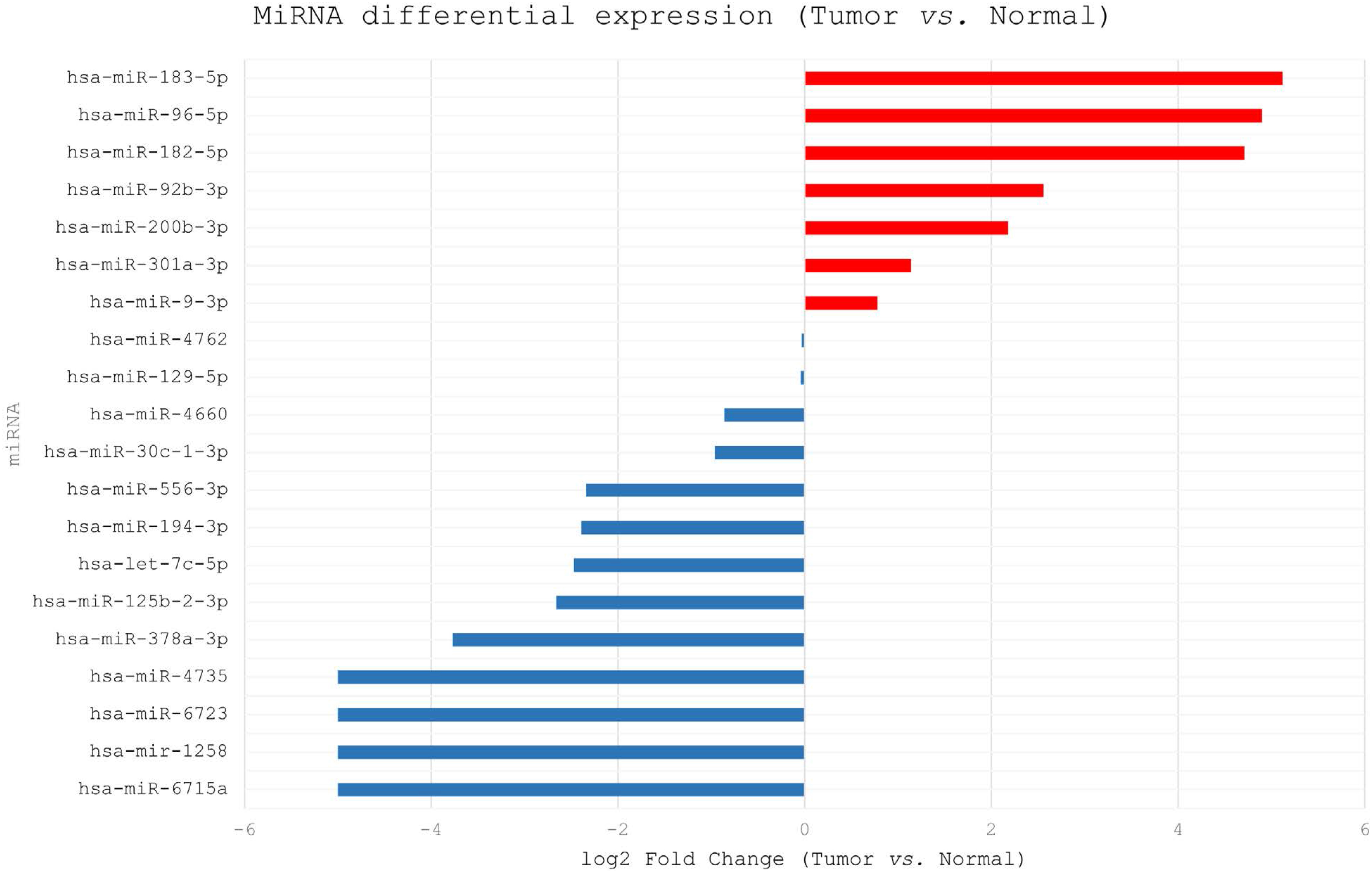
The last step in the analytical flow was conducted on UALCAN (version 2.0, accessed in September 2024)20, a platform that enables stratified analyses of gene and miRNA expressions in TCGA data. The 20 intermediate miRNAs were assessed regarding robustness of differential expression and their association with multiple clinical and prognostic parameters, including sample type (normal tissue versus primary tumor), clinical staging, histological grade, lymph node metastasis status, age, sex, body mass index, and race, in addition to exploratory analysis on overall survival. The relative expression difference was calculated by log2 fold change (log2FC), adopting |log2FC|>1 as a biological relevance criterion, associated with p<0.01. Positive values indicated tumoral superexpression, while negative values reflected greater expression in the normal tissue. After this validation, nine final miRNAs were selected, consistent across platforms and robustly associated with the assessed clinical and prognostic parameters, especially lymph node metastasis.
Finally, an integrative analysis was conducted, in which the selected miRNAs were confronted with previously described data in the literature, aiming to consolidate biological and clinical interpretation, particularly regarding the presence of lymph node metastasis, an important prognostic marker in cholangiocarcinoma.
Considering the investigation was exclusively based on secondary, publicly available, and anonymized data, there was no need for submission to a Research Ethics Committee, in compliance with Resolution N. 510/201621 of the National Health Council and Law N. 12.527/201122.
RESULTS
Figure 1 presents the sample composition, with the proportion of tumoral and normal tissues (A), as well as the distribution of patients with CHOL following clinical staging (B), including stages I to IV.

Graphic representation of the sample composition analyzed in this study. (A) Proportion between tumoral and normal control samples. (B) Distribution of patients with CHOL following clinical staging
The initial screening identified 245 differently expressed miRNAs among tumoral and normal tissues, selected from the CancerMIRNome and OncomiR platforms. The analyses employed internal tests of differential expression and ANOVA with FDR correction, considering p<0.01 as a threshold and, when applicable, |log2 Fold Change|>1. These miRNAs presented a significant statistical association with relevant clinical variables.
After specific filtering for the CHOL subtype (p<0.01), 20 miRNAs were selected: let-7c-5p, miR-1258, miR-125b-2-3p, miR-129-5p, miR-182-5p, miR-183-5p, miR-194-3p, miR-200b-3p, miR-30c-1-3p, miR-301a-3p, miR-378a-3p, miR-4660, miR-4735, miR-4762, miR-556-3p, miR-6715a, miR-6723, miR-9-3p, miR-92b-3p, and miR-96-5p. To improve the flow of reading, the miRNAs are referred to in the text without the "hsa" prefix.
The differential expression analysis conducted through the UALCAN platform revealed distinct gene regulation profiles. Figure 2 presents a bar graph with miRNAs’ log2 fold change values for tumoral and normal tissues.

Log2 Fold Change bar graph (Tumor vs. Normal) for the analyzed microRNAs (miRNAs). Positive values indicated tumor superexpression, while negative values reflected greater expression in normal tissue. |log2FC|>1 was adopted as a biologically relevant cutoff
Based on the analysis of differential expressions associated with the presence of lymph node metastasis, nine miRNAs with prognostic relevance potential were identified in CHOL. For that end, the platforms CancerMIRNome, OncomiR, and UALCAN were used, with the application of ANOVA with FDR (OncomiR) correction and Student's T test (UALCAN), considering p<0.01 as a threshold. All the selected miRNAs presented statistically significant association with the presence or absence of lymph node metastases (p<0.01); however, no significant correlation was observed between their expression levels and overall patient survival(p<0.05).
Table 1 presents in detail the nine identified miRNAs, accompanied by the respective p values associated with different clinical, demographic, and pathological variables. They all expressed significant differential expression between tumoral and normal tissues (p<0.01) and demonstrated a correlation with relevant prognostic parameters, including clinical staging, histological grade, and lymph node metastasis status. Between the analyzed miRNAs, superexpression was observed in tumoral tissues for miRNAs 182-5p, 183-5p, 301a-3p, 92b-3p, and 96-5p, while let-7c-5p, 1258, 194-3p, and 378a-3p presented higher levels in normal tissues.
Additionally, the miRNAs’ differential expression was consistent across different population subgroups, encompassing both sexes, 41-100 years age groups, body mass index categories, and, for miR-194-3p specifically, also between individuals from different ethnicities (Caucasians and Asians).
The significant association of miRNAs with multiple histological grades and clinical stages reinforces the hypothesis of their participation in different phases of biliary carcinogenesis, from initial stages to more advanced phases of the disease. Among the assessed biomarkers, miR-194-3p stood out for presenting statistical significance in every clinical and demographic variable analyzed, being a promising candidate to be considered a wide and robust prognostic marker in the context of CHOL.
DISCUSSION
MiRNA differential expression is widely recognized as a central epigenetic mechanism in the regulation of gene expression, influencing essential cellular processes, like proliferation, apoptosis, differentiation, and cellular migration10-12. In this context, miRNAs have stood out as crucial epigenetic regulators in several neoplasms, including CHOL23,24. These molecules act predominantly through post-transcriptional genes, interfering in the translation or stability of the messenger RNA, directly modulating tumoral progression and the biological behavior of neoplasms10,11.
The findings in the present study reveal a set of nine differently expressed miRNAs with statistical significance, associated mainly with lymph node metastasis, a prognosis marker of high clinical relevance. Among the nine explored, seven (let-7c-5p, miR-182-5p, miR-183-5p, miR-194-3p, miR-378a-3p, miR-92a-3p, and miR-96-5p) had already been correlated with CHOL, and the results of this study are in line with the data previously described in literature25-30. The other two miRNAs addressed in the article (miR-1258 and miR-301a-3p) are new, despite their relationship with other cancer types, mainly hepatocellular carcinoma31,32.
In this sense, such miRNAs can be more directly implicated in specific events of local tumoral progression and lymphatic dissemination, exerting limited influence on clinical outcomes in the long term, for instance, mortality. Among them, we highlight let-7c-5p, a member of the let-7 family, known for its oncogenic suppression function25. Previous studies demonstrated that a reduction in the let-7c-5p expression in CHOL tumoral tissues is associated with higher tumoral aggressiveness and inhibition of tumoral self-renewal and growth, although it can also promote invasion and growth in extra-hepatic sites, suggesting a dual role that relies on the cellular context and the molecular targets involved33. This duality can reflect the interaction with distinct targets, like EZH2 and the DVL3/β-catenin axis, underscoring the complexity of its therapeutic application33.
MiR-1258 has been characterized as a tumoral suppressor, inhibiting the translation of RTA protein, a regulator of the reactivation of oncogenic herpes viruses, such as the one associated with Kaposi's sarcoma34. In gastrointestinal cancers, reduced levels of miR-1258 have been associated with a higher tumoral stage, lymphovascular commitment, and pathological progression34, which confers prognostic value and therapeutic potential.
MiR-182-5p also stands out as a therapeutic and diagnostic biomarker, associated with mutations in critical genes like BRCA1, BCR-ABL1, and HPGD35,36. Exosomal miRNA mir-182-5p present in the bile of patients with cholangiocarcinoma presented superexpression, promoting tumoral progression by inhibiting the HPGD gene and consequently increasing PGE2, highlighting their potential as a biomarker and therapeutic target26.
MiR-183 regulates crucial cellular cycle pathways, apoptosis, and differentiation37,38. In liver, lung, prostate, and colorectal carcinomas, elevated levels of miR-183 were associated with DNA hypermethylation, greater invasiveness, and worse prognosis37,38, suggesting their utility as a biomarker and therapeutic target.
In this study, miR-194-3p stood out for its association with all the clinical and demographic variables analyzed. Recognized as a tumoral suppressor in several neoplasms, it regulates the epithelial-mesenchymal transition (EMT), the expression of FoxM1, PD-L1/PD-L2, and other pro-tumoral genes39-44. Specifically, in CHOL, miR-194-3p inhibits tumoral growth by negatively regulating the transformation sequence of epithelial cells 2 (ECT2) and blocking the Rho pathway, thus configuring as a potential therapeutic target27. The reintroduction of these miRNAs demonstrated the ability to inhibit proliferation, migration, immune evasion, and EMT in different models, indicating their promising potential for therapeutic strategies, including in chemo-resistant tumors39-42.
MiR-301a, in turn, acts predominantly as an oncomiR, inhibiting tumoral suppressors like RUNX3, compromising the function of NK cells, and favoring tumoral proliferation45-47. Despite its contextual effects on the prognosis, this study observed no significant correlation between its expression and patient survival.
MiR-378a-3p demonstrated to be associated with normal suppression, inhibiting RAB31 and the Hedgehog pathway, reducing proliferation, migration, and formation of tumoral stem cells48,49. Its performance as a diagnostic and prognostic biomarker has been promising in prostate cancer, for instance.
MiR-92b-3p presented an oncomiR profile, being associated with a worse prognosis in hepatocarcinoma, breast cancer, and other neoplasms, modulating targets like CPEB3, ACADL, and Smad750,51. Similarly, miR-96-5p, a member of the miR-183 cluster, exhibited tumoral superexpression in several types of cancer, repressing suppressor genes, like FOXO3a and PDCD4, and promoting cellular proliferation and survival52-57. Specifically, in CHOL, recent studies demonstrated that miR-96 exerts an oncogenic function, favoring tumoral progression and metastasis by inhibiting the MTSS1 gene, suggesting its potential as a prognostic biomarker or therapeutic target, after external validation30.
Collectively, the results reinforce the relevance of an integrative approach that combines bioinformatic analyses and experimental validation to clarify the functional role of these miRNAs. The nine differently expressed miRNAs seem to work in processes like EMT, extracellular matrix remodeling, immune evasion, and proliferation, central mechanisms in the lymphatic dissemination of CHOL. Thus, this study supports the hypothesis that these miRNAs modulate tumoral aggressiveness, presenting potential for prognostic stratification of patients with CHOL. Although they were not correlated with overall survival, they demonstrated a strong association with lymph node metastasis, one of the main determinants for prognosis and eligibility for surgical resection. Thus, even with no direct impact on mortality, these miRNAs can be more directly implicated in specific events of local tumoral progression and lymphatic dissemination, helping in post-surgical stratification and therapeutic decision-making.
Among the study's limitations, we highlight the absence of experimental validation in vitro or in vivo, restricting the immediate translation of the findings to clinical practice. This is thus an exclusively bioinformatic analysis, with an exploratory character and no external validation. On the other hand, the transdemographic stability suggests that the analyzed miRNAs present biological and clinical robustness, being little influenced by individual characteristics, which reinforces their potential as prognostic markers. Furthermore, the data derived exclusively from TCGA are subject to sample bias and gaps in detailed clinical information. Further investigations shall contemplate the manipulation of miRNA, like miR-194-3p and miR-1258, in cellular and murine models, as well as the validation in independent cohorts, to confirm their applicability as non-invasive biomarkers or therapeutic targets.
CONCLUSION
This study identified nine differently expressed miRNAs associated with the presence of lymph node metastasis in CHOL, highlighting miR-194-3p for its broad correlation with clinical and demographic variables. Although no significant associations with overall survival have been observed, the identified miRNAs demonstrate a potential diagnostic and prognostic value, especially for tumoral stratification and understanding the underlying molecular mechanisms of disease local progression. Additional studies, with experimental validation and functional analysis, are needed to consolidate their utility as biomarkers and potential therapeutic targets to manage CHOL, considering the exploratory character of the present analysis.
DATA AVAILABILITY STATEMENT
All the contents associated with the article are included in the manuscript.
ACKNOWLEDGMENTS
To Dr. Ruan César Aparecido Pimenta for his valuable lecture on "Introduction to Bioinformatics for Biomedical Research Using Online Platforms", ministered at the Faculdade Atenas Passos’ Oncology Academic League (Liga Acadêmica de Oncologia da Faculdade Atenas Passos), which contributed to delving into the knowledge applied in this study on miRNA in the diagnosis and prognosis of cholangiocarcinoma.
References
-
1 Massarweh NN, El-Serag HB. Epidemiology of hepatocellular carcinoma and intrahepatic cholangiocarcinoma. Cancer Control. 2017;24(3):1073274817729245. doi: https://doi.org/10.1177/1073274817729245
» https://doi.org/10.1177/1073274817729245 -
2 Gomes RV. Expressão do receptor do fator de crescimento epidérmico (EGFR) como fator prognóstico no colangiocarcinoma [dissertação na Internet]. Belo Horizonte: Universidade Federal de Minas Gerais; 2016 [acesso 2025 jan 15]. 64 p. Disponível em: http://hdl.handle.net/1843/BUBD-AMNLLH
» http://hdl.handle.net/1843/BUBD-AMNLLH -
3 Banales JM, Cardinale V, Carpino G, et al. Cholangiocarcinoma: current knowledge and future perspectives consensus statement from the European Network for the Study of Cholangiocarcinoma (ENS-CCA). Nat Rev Gastroenterol Hepatol. 2016;13(5):261-80. doi: https://doi.org/10.1038/nrgastro.2016.51
» https://doi.org/10.1038/nrgastro.2016.51 -
4 Qurashi M, Vithayathil M, Khan SA. Epidemiology of cholangiocarcinoma. Eur J Surg Oncol. 2025;51(2):107064. doi: https://doi.org/10.1016/j.ejso.2023.107064
» https://doi.org/10.1016/j.ejso.2023.107064 -
5 Tsung C, Quinn PL, Ejaz A. Management of intrahepatic cholangiocarcinoma: a narrative review. Cancers (Basel). 2024;16(4):739. doi: https://doi.org/10.3390/cancers16040739
» https://doi.org/10.3390/cancers16040739 -
6 Maki H, Kawaguchi Y, Nagata R, et al. Conditional recurrence analysis of intrahepatic cholangiocarcinoma: Changes in recurrence rate and survival after recurrence resection by disease-free interval. Hepatol Res. 2023;53(12):1224-34. doi: https://doi.org/10.1111/hepr.13951
» https://doi.org/10.1111/hepr.13951 -
7 Brindley PJ, Bachini M, Ilyas SI, et al. Cholangiocarcinoma. Nat Rev Dis Primers. 2021;7(1):65. doi: https://doi.org/10.1038/s41572-021-00300-2
» https://doi.org/10.1038/s41572-021-00300-2 -
8 Zhang XF, Xue F, Dong DD, et al. Proposed modification of the eighth edition of the AJCC staging system for intrahepatic cholangiocarcinoma. HPB (Oxford). 2021;23(5):581591. doi: https://doi.org/10.1016/j.hpb.2020.08.016
» https://doi.org/10.1016/j.hpb.2020.08.016 -
9 Magri Júnior JE. Características anatomoclínicas e análise da sobrevida na neoplasia intraductal papilífera dos ductos biliares (IPNB) [dissertação na Internet]. Belo Horizonte: Universidade Federal de Minas Gerais; 2020 [acesso 2025 jan 15]. 45 p. Disponível em: http://hdl.handle.net/1843/46582
» http://hdl.handle.net/1843/46582 -
10 Saliminejad K, Khorram Khorshid HR, Soleymani Fard S, et al. An overview of microRNAs: biology, functions, therapeutics, and analysis methods. J Cell Physiol. 2019;234(5):5451-65. doi: https://doi.org/10.1002/jcp.27486
» https://doi.org/10.1002/jcp.27486 -
11 Jorge AL, Pereira ER, Oliveira CS, et al. MicroRNAs: understanding their role in gene expression and cancer. Einstein (São Paulo). 2021;19:eRB5996. doi: https://doi.org/10.31744/einstein_journal/2021RB5996
» https://doi.org/10.31744/einstein_journal/2021RB5996 -
12 Hill M, Tran N. miRNA interplay: mechanisms and consequences in cancer. Dis Model Mech. 2021;14(4):dmm047662. doi: https://doi.org/10.1242/dmm.047662
» https://doi.org/10.1242/dmm.047662 -
13 Di Cosimo S, Appierto V, Pizzamiglio S, et al. Early modulation of circulating microRNAs levels in HER2-positive breast cancer patients treated with trastuzumab-based neoadjuvant therapy. Int J Mol Sci. 2020;21(4):1386. doi: https://doi.org/10.3390/ijms21041386
» https://doi.org/10.3390/ijms21041386 -
14 Bautista-Sánchez D, Arriaga-Canon C, Pedroza-Torres A, et al. The promising role of miR-21 as a cancer biomarker and its importance in RNA-based therapeutics. Mol Ther Nucleic Acids. 2020;20:409-20. doi: https://doi.org/10.1016/j.omtn.2020.03.003
» https://doi.org/10.1016/j.omtn.2020.03.003 -
15 Hussen BM, Hidayat HJ, Salihi A, et al. MicroRNA: a signature for cancer progression. Biomed Pharmacother. 2021;138:111528. doi: https://doi.org/10.1016/j.biopha.2021.111528
» https://doi.org/10.1016/j.biopha.2021.111528 -
16 Liu X, Zhao H, Liu J, et al. Identification of tumor-suppressive miRNAs that target LAT1 expression in cholangiocarcinoma cells. Biochem Biophys Res Commun. 2024;713:1-8. doi: https://doi.org/10.1016/j.bbrc.2024.03.045
» https://doi.org/10.1016/j.bbrc.2024.03.045 -
17 The Cancer Genome Atlas Program (TCGA) [Internet]. Bethesda: NIH; 2006 [acesso 2025 set 1]. Disponível em: https://www.cancer.gov/ccg/research/genome-sequencing/tcga
» https://www.cancer.gov/ccg/research/genome-sequencing/tcga -
18 Li R, Qu H, Wang S, et al. CancerMIRNome: an interactive analysis and visualization database for miRNome profiles of human cancer. Nucleic Acids Res. 2022;50(D1):D1139-46. doi: https://doi.org/10.1093/nar/gkab784
» https://doi.org/10.1093/nar/gkab784 -
19 OncomiR: WashU pan-cancer miRNome atlas [Internet]. St. Louis (MO): WashU; 2018 [acesso 2025 jul 1]. Disponível em: https://www.oncomir.org
» https://www.oncomir.org -
20 UALCAN: TCGA miRNA analysis portal [Internet]. Birmingham: University of Alabama at Birmingham, Department of Pathology; 2017 [acesso 2025 jul 1]. Disponível em: https://ualcan.path.uab.edu
» https://ualcan.path.uab.edu -
21 Conselho Nacional de Saúde (BR). Resolução n° 510, de 7 de abril de 2016. Dispõe sobre as normas aplicáveis a pesquisas em Ciências Humanas e Sociais cujos procedimentos metodológicos envolvam a utilização de dados diretamente obtidos com os participantes ou de informações identificáveis ou que possam acarretar riscos maiores do que os existentes na vida cotidiana, na forma definida nesta Resolução [Internet]. Diário Oficial da União, Brasília, DF. 2016 maio 24 [acesso 2025 abr 7]; Seção 1:44. Disponível em: http://bvsms.saude.gov.br/bvs/saudelegis/cns/2016/res0510_07_04_2016.html
» http://bvsms.saude.gov.br/bvs/saudelegis/cns/2016/res0510_07_04_2016.html -
22 Presidência da República (BR). Lei nº 12.527, de 18 de novembro de 2011. Regula o acesso a informações previsto na Constituição Federal. Diário Oficial da União [Internet], Brasília, DF. 2011 nov 18 [acesso 2025 abr 7]; Edição 221-A; Seção 1:1-4. Disponível em: https://pesquisa.in.gov.br/imprensa/jsp/visualiza/index.jsp?data=18/11/2011&jornal=1000&pagina=1&totalArquivos=12
» https://pesquisa.in.gov.br/imprensa/jsp/visualiza/index.jsp?data=18/11/2011&jornal=1000&pagina=1&totalArquivos=12 -
23 Alkhazaali-Ali Z, Sahab-Negah S, Boroumand AR, et al. MicroRNA (miRNA) as a biomarker for diagnosis, prognosis, and therapeutics molecules in neurodegenerative disease. Biomed Pharmacother. 2024;177:116899. doi: https://doi.org/10.1016/j.biopha.2024.116899
» https://doi.org/10.1016/j.biopha.2024.116899 -
24 He B, Zhao Z, Cai Q, et al. miRNA-based biomarkers, therapies, and resistance in cancer. Int J Biol Sci. 2020;16(14):2628-47. doi: https://doi.org/10.7150/ijbs.47203
» https://doi.org/10.7150/ijbs.47203 -
25 Xie Y, Zhang H, Guo XJ, et al. Let-7c inhibits cholangiocarcinoma growth but promotes tumor cell invasion and growth at extrahepatic sites. Cell Death Dis. 2018;9(2):249. doi: https://doi.org/10.1038/s41419-018-0286-6
» https://doi.org/10.1038/s41419-018-0286-6 -
26 Shu L, Li X, Liu Z, et al. Bile exosomal miR-182/183-5p increases cholangiocarcinoma stemness and progression by targeting HPGD and increasing PGE2 generation. Hepatology. 2024;79(2):307-22. doi: https://doi.org/10.1097/HEP.0000000000000437
» https://doi.org/10.1097/HEP.0000000000000437 -
27 Gao J, Dai C, Yu X, et al. Upregulated microRNA-194 impairs stemness of cholangiocarcinoma cells through the Rho pathway via inhibition of ECT2. J Cell Biochem. 2020;121(10):4239-50. doi: https://doi.org/10.1002/jcb.29648
» https://doi.org/10.1002/jcb.29648 -
28 Zhou Z, Ma J. miR-378 serves as a prognostic biomarker in cholangiocarcinoma and promotes tumor proliferation, migration, and invasion. Cancer Biomark. 2019;24(2):173-81. doi: https://doi.org/10.3233/CBM-181980
» https://doi.org/10.3233/CBM-181980 -
29 Han HS, Kim MJ, Han JH, et al. Bile-derived circulating extracellular miR-30d-5p and miR-92a-3p as potential biomarkers for cholangiocarcinoma. Hepatobiliary Pancreat Dis Int. 2020;19(1):41-50. doi: https://doi.org/10.1016/j.hbpd.2019.10.009
» https://doi.org/10.1016/j.hbpd.2019.10.009 -
30 Yin X, Chai Z, Sun X, et al. Overexpression of microRNA-96 is associated with poor prognosis and promotes proliferation, migration and invasion in cholangiocarcinoma cells via MTSS1. Exp Ther Med. 2020;19(4):2757-65. doi: https://doi.org/10.3892/etm.2020.8502
» https://doi.org/10.3892/etm.2020.8502 -
31 Lin W, Lin J, Li J, et al. Kindlin-2-miR-1258-TCF4 feedback loop promotes hepatocellular carcinoma invasion and metastasis. J Gastroenterol. 2022;57(5):372-86. doi: https://doi.org/10.1007/s00535-022-01866-8
» https://doi.org/10.1007/s00535-022-01866-8 -
32 Peng X, Yang R, Wang C, et al. The YTHDF3-DT/miR-301a-3p /INHBA axis attenuates autophagy-dependent ferroptosis in lung adenocarcinoma. Cancer Lett. 2025;613:217503. doi: https://doi.org/10.1016/j.canlet.2025.217503
» https://doi.org/10.1016/j.canlet.2025.217503 -
33 Xie Y, Zhang H, Guo XJ, et al. Let-7c inhibits cholangiocarcinoma growth but promotes tumor cell invasion and growth at extrahepatic sites. Cell Death Dis. 2018;9:249. doi: https://doi.org/10.1038/s41419-018-0286-6
» https://doi.org/10.1038/s41419-018-0286-6 -
34 Shi J, Chen P, Sun J, et al. MicroRNA-1258: an invasion and metastasis regulator that targets heparanase in gastric cancer. Oncol Lett. 2017;13(5):3739-3745. doi: https://doi.org/10.3892/ol.2017.5886 Erratum in: Oncol Lett. 2021;22(6):842. doi: https://doi.org/10.3892/ol.2021.13103
» https://doi.org/10.3892/ol.2017.5886 -
35 Yacob AM, Muhamad NA, Chang KM, et al. Hsa-miR-181a-5p, hsa-miR-182-5p, and hsa-miR-26a-5p as potential biomarkers for BCR-ABL1 among adult chronic myeloid leukemia treated with tyrosine kinase inhibitors at the molecular response. BMC Cancer. 2022;22(1):332. doi: https://doi.org/10.1186/s12885-022-09396-5
» https://doi.org/10.1186/s12885-022-09396-5 -
36 Elias K, Smyczynska U, Stawiski K, et al. Identification of BRCA1/2 mutation female carriers using circulating microRNA profiles. Nat Commun. 2023;14(1):3350. doi: https://doi.org/10.1038/s41467-023-38925-4
» https://doi.org/10.1038/s41467-023-38925-4 -
37 Oliveira-Rizzo C, Ottati MC, Fort RS, et al. Hsa-miR-183-5p Modulates Cell Adhesion by Repression of ITGB1 Expression in Prostate Cancer. Noncoding RNA. 2022;8(1):11. doi: https://doi.org/10.3390/ncrna8010011
» https://doi.org/10.3390/ncrna8010011 -
38 Li M, Xu DM, Lin SB, et al. Transcriptional expressions of hsa-mir-183 predicted target genes as independent indicators for prognosis in bladder urothelial carcinoma. Aging (Albany NY). 2022;14(9):3782-800. doi: https://doi.org/10.18632/aging.204040
» https://doi.org/10.18632/aging.204040 -
39 Zhu X, Li D, Yu F, et al. miR-194 inhibits the proliferation, invasion, migration, and enhances the chemosensitivity of non-small cell lung cancer cells by targeting forkhead box A1 protein. Oncotarget. 2016;7(11):13139-52. doi: https://doi.org/10.18632/oncotarget.7545
» https://doi.org/10.18632/oncotarget.7545 -
40 Fan F, Chen K, Lu X, et al. Dual targeting of PD-L1 and PD-L2 by PCED1B-AS1 via sponging hsa-miR-194-5p induces immunosuppression in hepatocellular carcinoma. Hepatol Int. 2021;15(2):444-58. doi: https://doi.org/10.1007/s12072-020-10101-6
» https://doi.org/10.1007/s12072-020-10101-6 -
41 Cui Z, Wang J, Chen G, et al. The upregulation of CLGN in hepatocellular carcinoma is potentially regulated by hsa-miR-194-3p and associated with patient progression. Front Oncol. 2023;12:1081510. doi: https://doi.org/10.3389/fonc.2022.1081510
» https://doi.org/10.3389/fonc.2022.1081510 -
42 Vajen B, Greiwe L, Schäffer V, et al. MicroRNA-192-5p inhibits migration of triple negative breast cancer cells and directly regulates Rho GTPase activating protein 19. Genes Chromosomes Cancer. 2021;60(11):733-42. doi: https://doi.org/10.1002/gcc.22982
» https://doi.org/10.1002/gcc.22982 -
43 Wang Y, Huang L, Shan N, et al. Establishing a three-miRNA signature as a prognostic model for colorectal cancer through bioinformatics analysis. Aging (Albany NY). 2021;13(15):19894-907. doi: https://doi.org/10.18632/aging.203400
» https://doi.org/10.18632/aging.203400 -
44 Su L, Zhang J, Zhang X, et al. Identification of cell cycle as the critical pathway modulated by exosome-derived microRNAs in gallbladder carcinoma. Med Oncol. 2021;38(12):141. doi: https://doi.org/10.1007/s12032-021-01594-8
» https://doi.org/10.1007/s12032-021-01594-8 -
45 Zhang J, Yang Y, Wei Y, et al. Hsa-miR-301a-3p inhibited the killing effect of natural killer cells on non-small cell lung cancer cells by regulating RUNX3. Cancer Biomark. 2023;37(4):249-59. doi: https://doi.org/10.3233/cbm-220469
» https://doi.org/10.3233/cbm-220469 -
46 Öztemur Islakoğlu Y, Noyan S, Gür Dedeoğlu B. Hsa-miR-301a- and SOX10-dependent miRNA-TF-mRNA regulatory circuits in breast cancer. Turk J Biol. 2018;42(2):103-12. doi: https://doi.org/10.3906/biy-1708-17
» https://doi.org/10.3906/biy-1708-17 -
47 Pliakou E, Lampropoulou DI, Dovrolis N, et al. Circulating miRNA expression profiles and machine learning models in association with response to irinotecan-based treatment in metastatic colorectal cancer. Int J Mol Sci. 2022;24(1):46. doi: https://doi.org/10.3390/ijms24010046
» https://doi.org/10.3390/ijms24010046 -
48 Xu X, Li Y, Liu G, et al. MiR-378a-3p acts as a tumor suppressor in gastric cancer via directly targeting RAB31 and inhibiting the Hedgehog pathway proteins GLI1/2. Cancer Biol Med. 2022;19(12):1662-82. doi: https://doi.org/10.20892/j.issn.2095-3941.2022.0337
» https://doi.org/10.20892/j.issn.2095-3941.2022.0337 -
49 Zhang Y, Ding N, Xie S, et al. Identification of important extracellular vesicle RNA molecules related to sperm motility and prostate cancer. Extracell Vesicles Circ Nucleic Acids. 2021;2(2):104-26. doi: https://doi.org/10.20517/evcna.2021.02
» https://doi.org/10.20517/evcna.2021.02 -
50 Deng R, Cui X, Dong Y, et al. Construction of circRNA-Based ceRNA network to reveal the role of circRNAs in the progression and prognosis of hepatocellular carcinoma. Front Genet. 2021;12:626764. doi: https://doi.org/10.3389/fgene.2021.626764
» https://doi.org/10.3389/fgene.2021.626764 -
51 Manzanarez-Ozuna E, Flores DL, Gutiérrez-López E, et al. Model based on GA and DNN for prediction of mRNA-Smad7 expression regulated by miRNAs in breast cancer. Theor Biol Med Model. 2018;15(1):24. doi: https://doi.org/10.1186/s12976-018-0095-8
» https://doi.org/10.1186/s12976-018-0095-8 -
52 Hong Y, Liang H, Uzair-ur-Rehman, et al. miR-96 promotes cell proliferation, migration and invasion by targeting PTPN9 in breast cancer. Sci Rep. 2016;6:37421. doi: https://doi.org/10.1038/srep37421
» https://doi.org/10.1038/srep37421 -
53 Mendes DCC, Calvano Filho CMC, Garcia N, et al. Could FOXO3a, miR-96-5p, and miR-182-5p be useful for Brazilian women with luminal a and triple-negative breast cancers prognosis and target therapy? Clinics (Sao Paulo). 2023;78:e100155. doi: https://doi.org/10.1016/j.clinsp.2022.100155
» https://doi.org/10.1016/j.clinsp.2022.100155 -
54 Kandhavelu J, Subramanian K, Khan A, et al. Computational analysis of miRNA and their gene targets significantly involved in colorectal cancer progression. Microrna. 2019;8(1):68-75. doi: https://doi.org/10.2174/2211536607666180803100246
» https://doi.org/10.2174/2211536607666180803100246 -
55 Gujrati H, Ha S, Wang BD. Deregulated microRNAs involved in prostate cancer aggressiveness and treatment resistance mechanisms. Cancers (Basel). 2023;15(12):3140. doi: https://doi.org/10.3390/cancers15123140
» https://doi.org/10.3390/cancers15123140 -
56 Ma Y, Liang AJ, Fan YP, et al. Dysregulation and functional roles of miR-183-96-182 cluster in cancer cell proliferation, invasion and metastasis. Oncotarget. 2016;7(27):42805-25. doi: https://doi.org/10.18632/oncotarget.8715
» https://doi.org/10.18632/oncotarget.8715 -
57 Zheng Y, Sukocheva O, Tse E, et al. MicroRNA-183 cluster: a promising biomarker and therapeutic target in gastrointestinal malignancies. Am J Cancer Res [Internet]. 2023 [acesso 2025 nov 17];13(12):6147-75. Disponível em: https://pubmed.ncbi.nlm.nih.gov/38187051/
» https://pubmed.ncbi.nlm.nih.gov/38187051/
Edited by
-
Associate editor:
Tatiana de Almeida Simão. Orcid iD: https://orcid.org/0000-0001-8509-2247
-
Scientific-editor:
Anke Bergmann. Orcid iD: https://orcid.org/0000-0002-1972-8777

 Source: Elaborated with data from the TCGA portal
 Source: Elaborated based on data from the UALCAN (TCGA) database