 Effect of Supplementation of Organic Selenium on Physiological and Production Parameters of Ghagus hens during the Early Laying Period
ABSTRACT
Trace mineral organic selenium is known to increase egg production significantly in exotic breeds. The present study was conducted with the Ghagus native hen breed to observe the effect of organic-selenium-enriched yeast on body weight, plasma hormones, and expression of genes that influence egg production. The control (C) group was offered a corn and soybean-based basal feed without organic selenium supplementation. The other two treatment groups (T1 and T2) were offered 0.2 and 0.4 ppm of organic selenium, respectively. The results indicated that, in T1, plasma levels of ghrelin and melatonin increased (p<0.05), while progesterone levels decreased significantly (p<0.05); furthermore, the expression of melatonin, and ghrelin hormone receptors in jejunum and magnum tissues also increased (p<0.001). When compared to C and T2, the fold change expression of all amino acid transporters increased (p<0.001) in jejunum tissue, whereas in magnum tissue, the expression of only three transporters (CAT, LAT2, and LAT4) increased (p<0.001) in T1. Treatment of chickens with 0.2 ppm also increased egg production by 6±1%, and egg weight by 3± 0.5g. The study indicated that there is potential for increases in egg production in Ghagus hens with the supplementation of organic selenium during the early laying period.
Keywords:
Early laying period; egg; Ghagus; gene expression; hormones; organic selenium
INTRODUCTION
Ghagus hens are primarily reared for meat and egg purposes in backyard systems in the districts of Karnataka and Andhra Pradesh in India. The annual egg production reported is 60-80 eggs, and adult female body weight is 2.0 kg by 36 weeks of age (Vij et al., 2006). Different physiological parameters influence egg parameters, and the present study aimed to observe the efficacy of organic selenium supplementation on the modulation of some physiological parameters influencing egg production. There is a relationship between gut hormones and reproduction (Comninos et al., 2014), but their relation in chicken is not clear. MET has the potential to mitigate oxidative stress and inflammation in chickens and humans, whether administered by implantation or through feed. Reports also indicate that treatment with this substance increases egg parameters in chickens (Jain, 2012; Hao et al., 2021; Yong et al., 2021), protecting the organisms by scavenging free radicals, which helps in regulating various reproductive and growth aspects (Shao et al., 2024; Khan et al., 2024). When administered intraperitoneally in aged hens, melatonin increased plasma levels of LH, estradiol, and the number of immature follicles (Hao et al., 2020). Ghrelin (GHL), another gut hormone, is secreted by the proventriculus of chickens (Murugesan & Nidamanuri, 2022). In vitro studies with chicken granulosa cells treated with human and chicken GHL fragments showed reduced secretion of progesterone (P4) (Sirotkin & Grossman, 2015). Reports from Sirotkin & Grossman (2015) and Sirotkin et al. (2017) suggest that GHL has a role in integrating nutrition, metabolism and egg production. Ovarian Estradiol -17b (EST) is the most abundant estrogen and an important gonadal steroid that is involved in oviduct development and yolk precursor production (Williams et al., 2004). Its concentration increases gradually until twenty weeks of age and remains high during next few weeks (Beck & Hansen, 2004). Estrogen regulates levels of gonadotropins through negative feedback effect, and is produced by the cells of less mature follicles (Porter et al., 1989). Contradictory regulation of EST by MET has been reported in humans and rats (Chuffa et al., 2011; Cheng et al., 2020). In hens, P4 is produced by granulosa cells obtained from preovulatory follicles (Woods et al., 2009) and is regulated by LH (Yang et al., 1997). These studies confirm the relation between gut hormones steroids and ovarian functions.
Reports indicate that treatment with organic selenium (O Se) for naked neck chicks subjected to heat stress increased body weight, FCR, breast and thigh weight (Khan et al., 2023). O Se has been demonstrated to be one of the essential trace minerals required for reproduction in Vanaraja and Aseel chickens (Nidamanuri et al., 2023; 2024), as it influenced key physiological parameters. Supplementation of O Se in layers is associated with a higher efficacy in Se transfer to the egg (Tufarelli et al., 2016), which is beneficial for internal egg quality. A beneficial effect of supplementation of higher doses of O Se in layers has been observed (Cobonova et al., 2011; Zia et al., 2018), and reports indicate that inorganic forms of Se are poorly stored and absorbed in chickens (Anizoba et al., 2024). Hence the present study aimed to investigate the effect of O Se supplementation on endocrine and gene expression parameters which may affect the egg production of Ghagus hens during the early laying period (EP). Till date no work has been published with respect to supplementation of O Se in this breed.
MATERIALS AND METHODS
Approval to conduct the experiments was obtained from the Institutional Animal Ethics committee (IAEC/DPR/19/3) Regn. No.355/GO/RBi/S/01/CPCSEA).
One hundred and fifty Ghagus hens with a mean body weight of 1858 ± 11.5 g were selected and reared at the farm of the Directorate of Poultry Research, Hyderabad, India. The experiment was conducted during the months of February and March, 2021. At 22 weeks of age, hens were placed in individual California type cages, with floor space per bird of around 450-525 sq cm (0.6-0.75 sq ft). They were housed in an open side housing system. The mean shed temperature and relative humidity were 28±1 ºC and 46%, respectively. All birds were allowed to acclimate towards an amount of basal feed of 90g/bird/d, based on maize and soybean, and containing 0 ppm of organic Se (O Se, selenium yeast) (Table1) for 2 weeks. Water was provided ad libitum. For the present experiment, experimental tenure was from 24-28 weeks of age (EP). The experiment was conducted in a Completely Randomized Design (CRD). At the beginning of 24 weeks, the hens were divided into three equal groups: Control (C), Treatment1(T1) and Treatment 2(T2), each with 50 birds and five replicates, with ten birds in each replicate. A commercially available organic product, selenium enriched yeast (O Se, Seleno Source TM AF 2000, Nurture Organics, New Delhi), was added to the basal feed (Table 1). The Control group was offered basal feed only (0ppm O Se), and groups T1 and T2 were offered basal feed mixed with 0.2ppm and 0.4ppm of O Se, respectively. All other components of the feed were supplemented according to the requirements stated in the Nutrient Requirements of Poultry -3 (ICAR, 2012), under a 16:8-h light dark cycle. During the course of the experiment, temperature ranged between 29-32 ºC, and relative humidity between 60-71%.
Treatment group (T1) - Basal feed + 0.2ppm Yeast enriched selenium. (T2)- Basal feed + 0.4ppm Yeast enriched selenium. Control group (C)- Basal feed containing 0.05ppm Se (As analyzed from feed ingredients).
Blood samples were collected from the wing veins of eight birds selected at random from each group, and were place in EDTA coated tubes for the estimation of hormones. The same eight birds were chosen for blood collection at weekly intervals for four weeks. After collecting blood samples, tubes were kept in ice and transported to the laboratory. The samples were further centrifuged at 3000 rpm for 15 min. The plasma obtained as supernatant was stored at -20 oC for analysis of MET, GHL, P4 and EST hormones.
Birds from each group were sacrificed at 28 weeks of age by cervical dislocation for the collection of jejunum (from the pancreatic loop to Meckel’s diverticulum) and magnum tissues (portion of oviduct) (Reicher et al., 2020). After collection, they were transported to the lab in saline. The tissues were rinsed in PBS, placed on moistened paper towels, and any adhering fat and connective tissue were removed. They were immediately stored at -80 oC for further analysis. Gene expression studies for MET, GHL hormone receptors (MNTR, GHLR), and four amino acid transporters were conducted for both jejunum and magnum tissues of the birds.
The blood samples collected at weekly intervals were analyzed for different hormones separately. Mean±SE values for each hormone per bird were calculated for each week, and then Mean±SEM was calculated for the EP phase. Hormones MET (E12M0005), Estradiol (EST, E12E0023), Progesterone (P4, E12P0200) were assayed in the plasma using commercial Chicken ELISA kits (BlueGene Biotech, Shanghai). The Ghrelin (BC-ECh 040174) hormone was also estimated using commercial chicken ELISA kits (Biocodon Technologies, Kansas, USA) according to manufacturer’s instructions. The ELISA kits used for the estimation of the first three hormones were based on the competitive enzyme immunoassay technique, while those used for GHL measurements were based on a non-competitive assay. At the final step, the absorbance’s of the color developed in the samples was measured at 450nm in a ELISA plate reader (BioTek Instruments, Inc.). From the standard curve, the concentration of hormones was estimated. The intra and inter coefficients of variation were <7 and 8% respectively. Each plasma test samples and standards were run in duplicate.
Total RNA was extracted from approximately 100mg of tissues, using Trizol reagent (Invitrogen, Carlsbad, CA) purified with RNeasy Mini Kit (Qiagen, Valencia, CA), and treated with RNAse-Free DNAse kit (Qiagen, Valencia, CA) according to the manufacturer’s instructions. The RNA was dissolved in diethyl pyro carbonate (DEPC) treated water. The sample purity and concentration were measured on a NanoDrop 2000 Spectrophotometer (Thermo Scientific, Wilmington, DE). The purity of the extracted RNA was assessed by taking the ratio (OD 260/280) for all samples. Values ranged between 1.9 -2.0, while the RNA concentrations ranged from 2.0 to 2.5 μg /μl. RNA samples were stored at -80°C until further assay. A total of 2 μg of total RNA was reverse transcribed with a cDNA reverse transcription kit according to manufacturer’s protocol (Thermo Scientific, Verso cDNA synthesis kit), using a Gradient Master cycler (Eppendorf, Hauppauge, NY). Further cDNA samples were stored at -20°C. cDNA samples were diluted 1: 1 prior to RT-PCR analysis with the help of an Applied Biosystems RT-PCR machine. Each reaction consisted of 1 μL diluted cDNA, 1.0 μL forward primer, 1.0 μL reverse primer, 7uL DEPC water, and 10 μL Maxima SYBR Green/ROX qPCR Master Mix (2x) (Thermo Fisher SCIENTIFIC). The genes and primer sequences used for the RT-PCR assays are listed in Table 2. The primers were synthesized by Chromous Co. Bangalore, India. The qPCR conditions were 95°C for 5 min, followed by 40 cycles of 95 °C for 30 s and 55 °C for 30 s, followed by 72 ºC for 30 s. All samples were run in triplicate. Both control and treatment (T1, T2) values were normalized against the b actin reference gene. The relative quantification (RQ) was expressed as a ratio of the target gene to the control gene using the delta-delta Ct method (Rao et al., 2013). The difference in values was considered significant at the level of p<0.05.
The total Se content of the basal diets (Fresh) and treatment diet was analyzed using inductively coupled plasma MS (ICP-MS; Agilent 7500cx, Agilent Technologies, Tokyo, Japan) (Ohki et al., 2016).
Body weight (BW) was recorded at a fortnight interval during the study. Mean ±SEM values were estimated for twenty birds from each group.
Percentage of weekly egg production (EP%) was calculated as Total number of eggs for a week/Actual no. of hen days X 100.
Actual no. of hen days = Total number of hens in a week - loss of hens in that week.
For the estimation of egg weight (EW), 20 -30 eggs were randomly collected every day from each group. Eggs were weighed on an electronic balance to the nearest g, and mean and Mean±SEM values were subsequently calculated.
Statistical Analysis
Data are presented as Mean ± SEM. All data were analyzed using the SPSS statistical software (SPSS for Windows, version 16.0; SPSS Inc. Chicago, IL). One-way analysis of variance followed by Duncan’s multiple comparison test was used to identify differences in means among treatments. The hormonal data were analyzed using repeated measures ANOVA. Data were assumed to be statistically significant at p<0.05 or p<0.01 for gene expression parameters.
RESULTS AND DISCUSSION
The present study mainly aimed to investigate the effect of treatment with O Se on the modulation of egg production for the Ghagus breed. Furthermore, its effect in modulating the ovarian and gut hormones and gene expression of hormone receptors and amino acid transporters was also investigated.
Effect on BW, EW, EP%: While supplementation with 0.2 ppm (T1) and 0.4 ppm (T2) of organic selenium (O-Se) from 24 to 28 weeks of age did not significantly (p>0.05) affect body weight compared to the control group, the T1 group exhibited a significant increase in egg production percentage (6 ± 1%, p<0.01) and an increase in egg weight (3 ± 0.5 g, p<0.01) compared to both the control and T2 groups (Table 3). Previous studies have demonstrated that organic selenium supplementation can enhance growth and reproductive performance in poultry, with increased growth rates in birds supplemented with O-Se (Khan et al., 2018; Bora et al., 2024). In the present study neither of the treatments caused an increase in body weight or feed intake of the laying birds. Similar results have been reported for broiler chicks (Bakhshalinejad et al., 2019). When birds reach adult age, they maintain relatively stable body weight, and when they enter the egg production stage, the energy and nutrients are also diverted for egg production, which may explain these results. The two doses of O Se supplemented in the present study could not increase body weight, as the difference in the values for egg production and weight between control and treatment groups was significant. Most of previous studies have been restricted to amelioration of heat stress upon supplementation of SY (Mohamed et al., 2024). It has been reported that selenium doses have to be increased during the laying period for higher egg production (Gul et al., 2021). Further research is required to formulate the optimal dose of O Se in feed for different applications in chicken according to the state of physiological functions.
Effect on Hormones: Supplementation with O-Se (T1) significantly increased (p<0.05) plasma concentrations of MET and GHL while concurrently decreasing (p<0.05) plasma progesterone (P4) hormone levels compared to the control and T2 groups. No significant differences in plasma estradiol (EST) levels were observed among the treatment groups (Table 3). Hormones like LH and FSH have been associated with reproduction or egg production. It has been reported that in vivo treatment with exogenous MET increased egg production (Jia et al., 2016; Kahraman et al., 2022), and in vitro treatment with ghrelin on ovarian cells also modulated ovarian hormones (Schalla & Stengel, 2021; Sirotkin et al., 2023). Reports on the physiological levels of MET and GHL and their modulation by organic selenium (O-Se) supplementation are scarce in backyard native chickens, with limited data available specifically for Aseel and Vanaraja breeds, as reported in our previous studies (Nidamanuri et al., 2023; 2024). Compared to treatment with inorganic Se, the organic form proved to be safer, presenting less toxicity and a higher utilization (Liu et al., 2020; Kim et al., 2020). Hence, in the present study, O Se was used as a supplement. A study utilizing ghrelin analogs on ovarian granulosa cells in vitro indicated that ghrelin directly affected ovarian functions, especially steroid and peptide hormone secretions (Sirotkin & Grossman, 2007; Nidamanuri et al., 2023). In the present study, the increase in the levels of ghrelin upon treatment did not modulate the levels of steroid hormones significantly compared to the control and T2 groups (Table 3). Nevertheless, significant increases in egg production and weight were observed (Table 3). This indicates that treatment with 0.2ppm of O Se and higher ghrelin levels might have had a direct influence on egg production and jejunum and magnum tissues. Although ghrelin acts as an anorexigenic hormone in chickens, this study found that elevated ghrelin (GHL) levels did not reduce feed intake (data not presented) or body weight. Additionally, our results do not indicate a beneficial effect of organic selenium (O Se) on steroid hormones. Therefore, the improvements in production observed in the present study are likely to be a direct effect of ghrelin and other metabolic hormones.
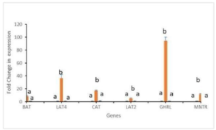
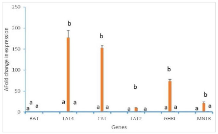
Gene Expression: Compared to the control and T2 groups, supplementation with O-Se (T1) significantly increased (p<0.001) the fold change in expression of the hormone receptors MNTR and GHRL in both jejunum and magnum tissues (Fig. 1 & 2). Similarly, in jejunum tissue, T1 significantly increased (p<0.001) the fold change in expression of all four amino acid transporters studied (BAT, LAT4, LAT2, and CAT) compared to the other groups (Fig. 1). However, in magnum tissue, only the expression of LAT4, LAT2, and CAT was significantly increased (p<0.001) in T1 compared to the control and T2 groups (Fig. 2). Treatment with 0.4ppm of O Se (T2) decreased the concentration of all the hormones, fold change in the expression of the receptors, and amino acid transporters when compared to control, but the difference was not significant (Fig 1 & 2). Moreover, it led to decreased egg production compared to the control (2±0.4%, p<0.01) and T1 group (8±0.6%, p<0.001) (Table 3). Similarly, upon comparison between groups, the weight of eggs only increased for the T1 group (Table 3) (p<0.05). The significant increase in the fold change expression of GHRL and MNTR in both jejunum and magnum tissues in the T1 group suggests that lower doses of O-Se may enhance egg production and weight by increasing plasma ghrelin and melatonin levels. However, higher doses of O-Se (0.4 ppm) significantly decreased egg production compared to both the T1 and control groups (p<0.001). The elevated levels of melatonin and the increased expression of its receptors in the ovary and jejunum, as reported by Holt et al. (2021) and Mandal (2023), may have contributed to improved nutrient absorption, jejunum integrity, and magnum function in the T1 group. Hence, supplementation of 0.2 ppm of O Se affected egg production and weight positively. Studies in aquaculture indicate that selenium supplement plays an important role in the composition of enzymes, leading to the synthesis and release of more nutrients from the intestinal epithelium (Zhu et al., 2014). Selenium (Se) is primarily absorbed in the small intestine and plays a crucial role in maintaining the integrity of vital tissues (Ozgul & Naziroglu, 2012). The observed increase in the expression of certain amino acid transporters in the T1 group suggests enhanced nutrient absorption, which may have contributed to the improved egg production. Furthermore, organic selenium (O-Se) is well-known for its antioxidant properties, effectively mitigating oxidative stress in poultry, a common challenge during the production period (Elgendy et al., 2022; Bora et al., 2024).
Fold change in the expression levels of mRNA of hormone receptors (MNTR and GHRL) and amino acid transporters (BAT, LAT4, CAT, LAT2) of jejunum tissue during the early laying period. Bars representing mean values with different superscripts are significantly different from each other at least at p<0.01, N=6.
Fold change in the expression levels of mRNA of hormone receptors (MNTR and GHRL) and amino acid transporters (BAT, LAT4, CAT, LAT2) of magnum tissue during the early laying period. Bars representing mean values with different superscripts are significantly different from each other at least at p<0.01, N=6.
O-Se supplementation can also positively influence reproductive capacity (Jlali et al., 2013). While the current study demonstrated beneficial effects of O-Se supplementation at lower doses, higher doses may have detrimental effects on egg production, suggesting the need for optimizing the supplementation level for optimal performance.
Estimation of Selenium in feed: The estimated selenium concentration in the basal diet was 0.05 ppm (from the feed constituents only), and it was observed to be respectively 0.19 ppm and 0.36 ppm in T1 and T2.
CONCLUSION
From the present study, it can be concluded that 0.2ppm O Se supplementation is beneficial for egg production in the native Ghagus poultry breed during the early laying period through modulation of physiological parameters, without adversely affecting bodyweight.
ACKNOWLEDGEMENTS
None.
REFERENCES
-
Acuña-Castroviejo D, Escames, Negas C, et al. Extrapineal melatonin: sources, regulation, and potential functions. Cell Molecular Life Sciences 2014;71(16):2997-3025. https://doi.org/10.1007/s00018-014-1579-2
» https://doi.org/10.1007/s00018-014-1579-2 -
Anizoba NW, Ikeh NE, Ezenwosu C, et al. Effect of dietary organic selenium on growth performance, antioxidant status and carcass characteristics of broiler chicken reared in the tropics. The Indian Journal of Animal Sciences 2024;94(6). https://doi.org/10.56093/ijans.v94i6.144852
» https://doi.org/10.56093/ijans.v94i6.144852 -
Bakhshalinejad R, Hassan Aabadi A, Swick RA, et al. Dietary sources and levels of selenium supplements affect growth performance, carcass yield, meat quality and tissue selenium deposition in broilers. Animal Nutrition 2019;5(3). https://doi.org/10.1016/j.aninu.2019.03.003
» https://doi.org/10.1016/j.aninu.2019.03.003 -
Beck MM, Hansen KK. Role of estrogen in avian osteoporosis. Poultry Science 2004;83 (2):200-6. https://doi.org/10.1093/ps/83.2.200
» https://doi.org/10.1093/ps/83.2.200 -
Bora S, Sonowal M, Baishya, P, et al. Effect of vitamin E and selenium supplementation on growth performance and heat shock protein 70 levels in broiler chickens exposed to summer heat stress. Indian Journal of Animal Research 2024;58(10). https://doi.org/10.18805/ijar.b-5261
» https://doi.org/10.18805/ijar.b-5261 - Bubenik GA. Thirty-four years since the discovery of gastrointestinal melatonin. Journal of Physiology and Pharmacology 2008;59 (Suppl 2):33-51.
-
Cheng JC, Fang LL. Melatonin stimulates aromatase expression and estradiol production in human granulosa-lutein cells: relevance for high serum estradiol levels in patients with ovarian hyper stimulation syndrome. Experimental Molecular Medicine 2020;52:1341-50. https://doi.org/10.1038/s12276-020-00491-w
» https://doi.org/10.1038/s12276-020-00491-w -
Chuffa A, Seiva LG, Fávaro FR, et al. Melatonin reduces LH, 17 beta-estradiol and induces differential regulation of sex steroid receptors in reproductive tissues during rat ovulation. Reproductive Biology and Endocrinology 2011;9(1). https://doi.org/10.1186/1477-7827-9-108
» https://doi.org/10.1186/1477-7827-9-108 -
Čobanová K, Petrovič V, Mellen, M, et al. Effects of dietary form of selenium on its distribution in eggs. Biological trace element research 2011;144 (1-3). https://doi.org/10.1007/s12011-011-9125-7
» https://doi.org/10.1007/s12011-011-9125-7 -
Comninos AN, Jayasena CN, Dhillo WS. The relationship between gut and adipose hormones, and reproduction. Human Reproduction 2014;20(2):153-74. https://doi.org/10.1093/humupd/dmt033
» https://doi.org/10.1093/humupd/dmt033 -
Elgendey F, Wakeel RA, Hemeda SA, et al. Selenium and/or vitamin E upregulate the antioxidant gene expression and parameters in broilers. BMC Veterinary Research 2022;18(1). https://doi.org/10.1186/s12917-022-03411-4
» https://doi.org/10.1186/s12917-022-03411-4 -
Gul F, Ahmad B, Afzal S, et al. Comparative analysis of various sources of selenium on the growth performance and antioxidant status in broilers under heat stress. Brazilian Journal of Poultry Science 2021;83. https://doi.org/10.1590/1519-6984.251004
» https://doi.org/10.1590/1519-6984.251004 -
Hao EY, Chen H, Wang DH, et al. Melatonin regulates the ovarian function and enhances follicle growth in aging laying hens via activating the mammalian target of rapamycin pathway. Poultry Science 2020;99(4). https://doi.org/10.1016/j.psj.2019.11.040
» https://doi.org/10.1016/j.psj.2019.11.040 - Holt EH, Lupsa B, Lee GS, et al. Goodman's basic medical endocrinology. 5th ed. Amsterdam: Eselvier; 2021. ISBN 978-0-12-815844-9
- Jain A. Studies on production performance of layers supplemented with dietary melatonin. Indian Journal of Poultry Science 2012;47(3).
-
Jia Y, Yang M, Zhu K, et al. Melatonin implantation improved the egg-laying rate and quality in hens past their peak egg-laying age. Scientific Reports 2016;6(1). https://doi.org/10.1038/srep39799
» https://doi.org/10.1038/srep39799 -
Jlali M, Briens M, Rouffineau F, et al. Effect of 2- hydroxy-4-methylselenobutanoic acid as a dietary selenium supplement to improve the selenium concentration of table eggs. Journal of Animal Science 2013;91(4). https://doi.org/10.2527/jas.2012-5825
» https://doi.org/10.2527/jas.2012-5825 - Kahraman O, Safa İnanç Z, Derya Arık H, et al. Use of melatonin as a feed additive. IntechOpen; 2022. https://doi.org/10.5772/intechopen.105999
-
Khan MT, Mahmud A, Javed K, et al. Organic and inorganic selenium in Aseel chicken diets: effect on production performance. Journal of Applied Poultry Research 2018;27(3). https://doi.org/10.3382/japr/pfx070
» https://doi.org/10.3382/japr/pfx070 -
Khan MT, Niazi AS, Arslan M, et al. Effects of selenium supplementation on the growth performance, slaughter characteristics, and blood biochemistry of naked neck chicken. Poultry Science 2023;1023(3). https://doi.org/10.1016/j.psj.2022.102420
» https://doi.org/10.1016/j.psj.2022.102420 -
Khan MT, Ahmed S, Azhar M, et al. Effects of selenium-enriched diets on the growth performance, slaughter characteristics, and blood biochemistry of Rhode Island Red chicken. Revista Brasileira de Ciência Avícola 2024;26(1). https://doi.org/10.1590/1806-9061-2022-1747
» https://doi.org/10.1590/1806-9061-2022-1747 -
Kim JH, Kil DY. Comparison of toxic effects of dietary organic or inorganic selenium and prediction of selenium intake and tissue selenium concentrations in broiler chickens using feather selenium concentrations. Poultry Science 2020;99(12):6462-73 https://doi.org/10.1016/j.psj.2020.08.061
» https://doi.org/10.1016/j.psj.2020.08.061 -
Liu H, Yu, Q, Fang C, et al. Effect of selenium source and level on performance, egg quality, egg selenium content, and serum biochemical parameters in laying hens. Foods 2020;9(1). https://doi.org/10.3390/foods9010068
» https://doi.org/10.3390/foods9010068 -
Ma N, Zhang J, Reiter RJ, et al. Melatonin mediates mucosal immune cells, microbial metabolism, and rhythm crosstalk: a therapeutic target to reduce intestinal inflammation. Medical Research Review 2020;40(2). https://doi.org/10.1002/med.21628
» https://doi.org/10.1002/med.21628 - Mandal A. Hormone interactions with receptors. Manchester: News-Medical Life Science; 2023. Available from: https://www.news-medical.net/health/Hormone-Interactions-with-Receptors.aspx
-
Mohamed ASA, Milošević M, Mohany M, et al. Heat stress relief for broiler chickens: organic selenium and a vitamin C and E blend can enhance growth, nutrient digestibility, and blood parameters. Italian Journal of Animal Science 2024;23(1). https://doi.org/10.1080/1828051X.2023.2301446
» https://doi.org/10.1080/1828051X.2023.2301446 -
Murugesan S, Nidamanuri AL, et al. Role of leptin and ghrelin in regulation of physiological functions of chicken. World's Poultry Science Journal 2022;78. https://doi.org/10.1080/00439339.2022.2119917
» https://doi.org/10.1080/00439339.2022.2119917 -
Nidamanuri AL, Konadaka SRR, Sivalingam J, et al. Differential level of hormones amino acids expression of amino acid transporters and hormone receptors during early and mid laying period and their modulation by organic selenium in vanaraja chickens. Acta Scientific Veterinary Sciences 2023;5(7). https://doi.org/10.31080/asvs.2023.05.0700
» https://doi.org/10.31080/asvs.2023.05.0700 -
Nidamanuri AL, Murugesan S, Sivalingam J, et al. Plasma hormone amino acid expression of amino acid transporters hormone receptors and their modulation by organic selenium in laying hens. Acta Scientific Veterinary Sciences 2024;6(9). https://doi.org/10.31080/ASVS.2024.06.0917
» https://doi.org/10.31080/ASVS.2024.06.0917 - Nutrient Requirements of Poultry. 10th ed. New Delhi: ICAR; 2012.
-
Ohki A, Nakajima T, Hirakawa S, et al. A simple method of the recovery of selenium from food samples for the determination by ICP-MS. Microchemical 2016;124. https://doi.org/10.1016/j.microc.2015.10.012
» https://doi.org/10.1016/j.microc.2015.10.012 -
Özgül C, Naziroglu M. TRPM2 channel protective properties of N-acetylcysteine on cytosolic glutathione depletion dependent oxidative stress and Ca2+ influx in rat dorsal root ganglion. Physiology of Behaviour 2012;106 (2):122-8. https://doi.org/10.1016/j.physbeh.2012.01.014
» https://doi.org/10.1016/j.physbeh.2012.01.014 -
Porter TE, Hargis BM, Silsby JL, et al. Differential steroid production between theca interna and theca externa cells: a three-cell model for follicular steroidogenesis in avian species. Endocrinology 1989;125(1). https://doi.org/10.1210/endo-125-1-109
» https://doi.org/10.1210/endo-125-1-109 - Rao X, Huang X, Zhou Z, et al. An improvement of the 2ˆ (-delta delta CT) method for quantitative real-time polymerase chain reaction data analysis. Biostatistics Bioinformatics Biomathematics 2013;3(3):71-85.
-
Reicher N, Epstein T, Gravitz D. From broiler breeder hen feed to the egg and embryo: the molecular effects of guanidinoacetate supplementation on creatine transport and synthesis. Poultry Science 2020;99(7). https://doi.org/10.1016/j.psj.2020.03.052
» https://doi.org/10.1016/j.psj.2020.03.052 -
Schalla MA, Stengel A. The role of the gastric hormones ghrelin and nesfatin-1 in reproduction. International Journal of Molecular Science 2021;22(20):11059. https://doi.org/10.3390/ijms222011059
» https://doi.org/10.3390/ijms222011059 -
Shao R, Wang Y, He C, et al. Melatonin and its emerging physiological role in reproduction:a review and update. Current Molecular Medicine 2024;24(4). https://doi.org/10.2174/1566524023666230417103201
» https://doi.org/10.2174/1566524023666230417103201 -
Sirotkin AV, Tarko V, Mlyncek M, et al. Ghrelin and obestatin can promote human ovarian granulosa cell functions and FSH effects. Reproductive Biology 2023;23(3). https://doi.org/10.1016/j.repbio.2023.100795
» https://doi.org/10.1016/j.repbio.2023.100795 -
Sirotkin, AV, Grossmann R. The role of ghrelin and some intracellular mechanisms in controlling the secretory activity of chicken ovarian cells. Comparative Biochemistry Physiology A Molecular Integrating Physiology 2007;147(1):239-46. https://doi.org/10.1016/j.cbpa.2006.12.038
» https://doi.org/10.1016/j.cbpa.2006.12.038 -
Sirotkin AV, Grossmann R. Interrelationship between feeding level and the metabolic hormones leptin, ghrelin and obestatin in control of chicken egg laying and release of ovarian hormones. Comparative Biochemistry Physiology A Molecular Integrating Physiology 2015;184:1-5. https://doi.org/10.1016/j.cbpa.2015.01.016
» https://doi.org/10.1016/j.cbpa.2015.01.016 -
Sirotkin AV, Harrath AH, Grossmann, R. Metabolic status and ghrelin regulate plasma levels and release of ovarian hormones in layer chicks. Physiological Research 2017;66:85-92. https://doi.org/10.33549/physiolres.933306
» https://doi.org/10.33549/physiolres.933306 -
Tufarelli V, Ceci E, Laudadio V, et al. 2-Hydroxy-4-Methylselenobutanoic Acid as new organic selenium dietary supplement to produce selenium-enriched eggs. Biological Trace Element Research 2016;171(2). https://doi.org/10.1007/s12011-015-0548-4
» https://doi.org/10.1007/s12011-015-0548-4 -
Vij PK, Tantia MS, Mishra B, et al. Characterization of aseel, danki, kalasthi and ghagus breeds of chicken. Indian Journal of Animal Sciences 2006;76(11). Available from: https://epubs.icar.org.in/index.php/IJAnS/article/view/4477
» https://epubs.icar.org.in/index.php/IJAnS/article/view/4477 -
Williams TD, Kitaysky AS, Vezina F, et al. Individual variation in plasma estradiol-17ß and androgen levels during egg formation in the european starling sturnus vulgaris:Implications for regulation of yolk steroids. General and Comparative Endocrinology 2004;136(3). https://doi.org/10.1016/j.ygcen.2004.01.010
» https://doi.org/10.1016/j.ygcen.2004.01.010 -
Woods DC, Schorey JS, Johnson AL, et al. Toll-like receptor signaling in hen ovarian granulosa cells is dependent on stage of follicle maturation. Reproduction 2009;137(6). https://doi.org/10.1530/rep-08-0320
» https://doi.org/10.1530/rep-08-0320 -
Yang J, Long DW, Bacon WL, et al. Changes in plasma concentrations of luteinizing hormone, progesterone, and testosterone in turkey hens during the ovulatory cycle. General and Comparative Endocrinology 1997;106(2). https://doi.org/10.1006/gcen.1997.6884
» https://doi.org/10.1006/gcen.1997.6884 -
Yong W, Haiying MH, Na M, et al. Roles of melatonin in the field of reproductive medicine, Biomedicine & Pharmacotherapy 2021;144. https://doi.org/10.1016/j.biopha.2021.112001
» https://doi.org/10.1016/j.biopha.2021.112001 -
Zia WM, Khalique A, Naveed S. Organic and inorganic selenium in poultry: a review. Indian Journal of Animal Research 2018;52(4):483-9. https://doi.org/10.18805/ijar.v0iof.6837
» https://doi.org/10.18805/ijar.v0iof.6837 -
Zhu QL, Luo Z, Zhuo MQ, et al. In vitro effects of selenium on copper-induced changes in lipid metabolism of grass carp (Ctenopharyngodon idellus) hepatocytes. Archives of Environment Contaminants and Toxicology 2014;67. https://doi.org/10.1007/s00244-014-0041-7
» https://doi.org/10.1007/s00244-014-0041-7
-
FUNDING
The Financial assistance given by Indian Council of Agriculture Research, New Delhi for conducting the present work is highly acknowledged.
-
DATA AVAILABILITY STATEMENT
Data will be available upon request.
-
DISCLAIMER/PUBLISHER’S NOTE
The published papers’ statements, opinions, and data are those of the individual author(s) and contributor(s). The editor(s) disclaim responsibility for any injury to people or property resulting from any ideas, methods, instructions, or products referred to in the content.
Data will be available upon request.