 Protective Effects of Olive Leaf Extract as a Natural Growth Promoter and Immune Modulator in Broilers Challenged with Velogenic Newcastle Disease Virus
ABSTRACT
Newcastle disease (ND) is a serious menace to poultry production and requires stringent measures to enhance growth and improve resistance against viral challenges in infected flocks. In the present study, the immunostimulatory and growth-promoting effects of olive (Olea europaea) leaf extract (OLE) in velogenic Newcastle disease virus (NDV)-infected broilers were evaluated. A total of 150-day-old broilers (Cobb strain) were randomly allotted to five groups: Blank Control (BC), Negative Control (NC), OLE-1, OLE-2, and OLE-Only. OLE-1 and OLE-2 groups were given 10 mL and 20 mL/L of OLE-supplemented drinking water on day 3, and ND vaccinations on days 0, 3, and 14. The NC, OLE-1, and OLE-2 groups were given an intraperitoneal challenge of NDV on day 21. Weekly inspections comprised clinical signs, survival, antibody levels, weight gain, and hematological indices. Body weight gain and leukocyte number (p<0.05) were significantly enhanced with OLE supplementation, whereas mortality was lowered to 30%. Proventriculus hemorrhage and intestinal ulcers were more severe in the NC group but less severe in the OLE-treated groups. OLE also attenuated ND-caused tissue injury and increased erythrocyte number, hemoglobin content, and packed cell volume. In conclusion, OLE boosts broiler immunity and growth performance without causing any unfavorable physiological effects, hence being a possible natural additive to improve disease resistance and poultry production.
Keywords:
Antioxidant capacity; egg quality; Japanese quail; lipid peroxidation; melon peel powder
INTRODUCTION
The poultry industry is prone to many diseases because of limited breed numbers and high production pressure. Diseases are mainly divided into two categories: infectious and non-infectious. Contagious diseases are primarily caused by bacteria, viruses, and fungi, which cause economic losses to the poultry industry (Rehan et al., 2019; Abd-El Ghany et al., 2023). Newcastle disease is a highly contagious and transmittable disease of poultry and wild birds caused by Avian Paramyxovirus type 1, which belongs to the genus Avulavirus, and the Paramyxoviridae family. This lethal disease infects birds’ digestive and respiratory systems, raising the mortality to 100% (Sonora et al., 2015; Mubashir et al., 2022). About 162 million US dollars are lost annually in the USA due to ND outbreaks. In Pakistan, about 6 billion (PKR) were lost due to an ND outbreak in 2012. About 45 million poultry birds are killed by NDV annually (Rehan et al., 2019). There is growing interest in using natural products in animal feed as feed additives to improve the health status of birds (Şahin & Bilgin, 2018; Landy & Kheiri, 2023; Hussain et al., 2024). Plant extracts, herbs, essential oils, and spices have been used in poultry feed to control disease and improve economic return, bird performance, and carcass characteristics (Eevuri & Putturu, 2013, Farooq et al., 2022; Afiouni et al., 2023). Recently, Eevuri & Putturu (2013) reviewed the use of some herbal preparations in broiler diets. Some of these feed additives include Acacia nilotica (Schrägle & Müller, 1990; Rasheed et al., 2024); Chinese medicinal herbs (Ding et al., 2018); cinnamon, pepper, combretum mole, sisal, khasanda kwata, tithonia concoctions, omusirangokho, neem (Bonsu et al., 2012); ginger (Palatty et al., 2013); garlic (Palatty et al., 2013); tulsi, and turmeric (Dono, 2013; Kyriazis et al., 2016). Various studies have combined two or more feed additives to maximize their beneficial effects (Mehala & Moorthy, 2008). Olive leaves contain many biphenolic compounds, including Hydroxytyrosol/Tyrosol, ligstroside, and verbascoside (Abd-El Ghany et al., 2023). In olive leaves, the primary bitter phenolic contents are ligstroside and oleuropein, which provide defense against various herbivores and pathogens (Zuluaga, 2024). Olive leaves have water solubility due to sugar moieties. Many phenolic compounds have shown antibacterial, antiviral, and antifungal activity (Leila et al., 2019). Phenolic compounds in fresh olive comprise 1-3% of the oleuropein and have shown a strong antiviral effect against the rotavirus, herpes, hepatitis virus, parvovirus, paramyxovirus, infectious laryngotracheitis virus, and feline leukemia virus (Sabry, 2014). It enhances macrophage production through increased production of nitric oxide. Oleuropein destroys the microorganism’s cell membrane or alters the host cell’s receptors and prevents virus replication and inflammation by inhibiting the production of pro-inflammatory cytokines (Kyriazis et al., 2016). There is insufficient information regarding the effects of olive leaves on broiler performance. Therefore, the objective of the present study was to explore the effect of dietary OLE supplementation on weight gain and hematological indices in infected broilers with field isolates of the NDV. Moreover, gross and histopathological changes in visceral organs were also evaluated.
MATERIALS AND METHODS
Ethical statement
This experimental study complied with the Institutional Bioethics Committee (IBC), Ref. No. 7145; dated: 15-10-2021, University of Agriculture, Faisalabad, Pakistan.
Collection and Processing of Olive Leaves
Fresh olive leaves (approximately 150 g) were collected from the University of Agriculture, Faisalabad botanical garden in June-July. The leaves were washed with tap water for 2-3 minutes and air-dried at 25-30°C for 1-4 days. They were reversed every 12 hours to ensure even drying until their greenish color disappeared and moisture content was lowered to ≤10%. The dried leaves were powdered in a fine powder form using an electric grinder (mesh size ≤0.5 mm) and kept at 4°C for later use.
Extraction of olive leaves
150 g of dried leaf powder was added to a flask containing 3000 mL of boiled distilled water and stirred manually for 15 minutes. The resulting solution was filtered twice using Whatman No. 2 filter paper, and the collected filtrate was stored. The remaining extract was then lyophilized at -40°C, and the plant extract’s percentage yield (w/w) was calculated. The final extract was stored at 4°C until further use.
Viral Isolate and Its Propagation
The Molecular Lab of Pathology generously provided the velogenic viral isolate. A previously isolated and confirmed RT-PCR isolate of NDV was propagated in 9-day-old embryonated eggs and incubated for the next 7 days at 37 °C. The Reed and Munch method was employed to calculate the ELD50 of NDV. The titer of NDV was computed using the hemagglutination (HA) test Zhang et al. (2024).
Experimental Study Design
Day-old broilers (n=150) were equally divided into five study groups (n=30 each), defined as follows: Blank Control (BC): No olive leaves extract (OLE), No ND Vaccine; Negative Control (NC): ND Vaccine only, but no OLE; OLE-1: 10 mL OLE, ND Vaccine + and OLE-2: 20 mL OLE, ND Vaccine +; and OLE-Only: OLE alone, but no ND Vaccine. On day 3, NC, OLE-1, and OLE-2 were vaccinated with ND vaccine, then boosted on day 14. On day 21, they were challenged with velogenic NDV. Clinical signs and behavior scoring were recorded daily for three weeks after infection. These birds were kept under good hygiene conditions at the experimental station of the Department of Pathology, UAF. The birds were given feed and water ad libitum. On day 3, the broilers of the OLE, OLE-1, and OLE-2 groups were supplemented with 10 ml, 10 ml, and 20 ml of OLE per liter of drinking water, respectively, until the end of the experiment. On day 21, 0.1 ml (ELD50 of 10-5.25/0.1 ml) of NDV per bird was injected into the broilers of the negative control, OLE-1, and OLE-2 groups (Table 1).
Clinical signs and Behavioral scoring
Post inoculation of NDV, the broilers of all study groups were observed daily for apparent clinical signs and scored according to the Manual of Diagnostic Tests and Vaccines for Terrestrial Animals (OIE, 2021), with some modifications, following the criteira below:
Score 0= No apparent clinical signs
Score 1= appearance of Respiratory signs
Score 2= Respiratory signs and Greenish diarrhea
Score 3= Respiratory signs, Greenish diarrhea, and Opisthotonos
Score 4= Respiratory signs, Greenish diarrhea, Opisthotonos, and Torticollis
Score 5= Respiratory signs, Greenish diarrhea, Opisthotonos, Torticollis, and Conjunctivitis
Antibody titer testing against NDV
The blood samples were taken at 7, 14, 21, 28, 35, and 42 days of age from the wing vein. The serum was separated through centrifugation to check the antibody level through the haemagglutination and haemagglutination inhibition (HA/HI) test.
Determination of blood indices of broilers
Seven birds were slaughtered every week post-infection to collect blood with an anticoagulant to determine blood indices in different groups. The mortality was recorded, and all birds were killed by the end of the experiment and examined immediately for gross pathological lesions. Blood samples were collected to check the total erythrocyte count, hemoglobin concentration, leukocyte count, and packed cell volume.
Collection of organs for histopathological changes
Visceral organs such as the Trachea, proventriculus, and Intestine were collected from different groups, and 10% buffered formalin was used to fix the specimens. Paraffin sections were made through a microtome and stained with Hematoxylin and Eosin for histopathological examination. Gross lesions on different organs, such as the Trachea, Intestine, and Proventriculus, were recorded according to the parameters of Uddin et al. (2025).
Statistical analysis
A statistical analysis was performed using MStat-C software. One-way analysis of variance (ANOVA) was used to compare the means of different groups, followed by Tukey’s Honestl statistical significance (e.g., “p<0.05”) y Significant Difference (HSD) test for post-hoc multiple comparisons. Differences were considered statistically significant at p≤0.05.
RESULTS
NDV propagation
100% mortality of chicken embryos occurred within 72 hours following challenge with the velogenic NDV isolate, highlighting its virulence. Histopathological examination of dead embryos showed severe hemorrhages and congestion in infected embryos. The virus was purified by centrifugation, and hemagglutination (HA) test results gave a high geometric mean titer of 1:512. The ELD50 was determined to be 10-5.25/0.1 mL, confirming the potency of the challenge dose.
OLE from leaves
The lyophilized extract of olive leaves produced a dark green powder with a percentage yield of approximately 15.53%, based on initial dry leaf weight, according to the equation below, described by Omar & Mohammed (2010) and Omar & Mohammed (2023):
OLE as a growth promoter
Figure 1 illustrates the body weight and feed intake trends among experimental groups. Feed consumption and body weight were significantly elevated (p<0.05) in OLE and OLE-2+vNDV compared to the negative control. No significant differences in body weight were observed during the first three weeks. However, from week 4 onward, birds in the OLE-1 and OLE-2 groups gained significantly more weight than those in the NC group (p<0.05, Tukey’s HSD test). By week 6, OLE-1 birds weighed 2274.83 ± 103.32 g and OLE-2 birds 2452.67 ± 112.45 g, compared to 1812.55 ± 98.61 g in the NC group. Feed intake was also highest in the OLE-2 group throughout the trial. As shown in Figure 1A, feed consumption increased steadily in OLE-supplemented groups, with the OLE-2 group showing the highest intake across all time points. Body weight measurements (Figure 1B) revealed significantly higher gains in OLE-1 (2274.83 ± 103.32 g) and OLE-2 (2452.67 ± 112.45 g) compared to the NC group (1812.55 ± 98.61 g) by the 6th week (p<0.05, Tukey’s test). The OLE-2 group showed the most consistent weight gain pattern, indicating a dose-dependent effect of OLE.
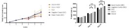
Influence of Olive Leaf Extract (OLE) Supplementation on Feed Consumption and Body Weight of Broilers Exposed to vNDV. A) Broiler chicks’ daily feed intake supplemented with OLE and challenged with vNDV. B) Body weight of broiler chicks supplemented with OLE and challenged with vNDV. Feed consumption and body weight were significantly elevated (p<0.05) in OLE and OLE-2+vNDV compared to the negative control, as determined by one-way ANOVA followed by Tukey’s HSD test.
Clinical signs and gross lesions
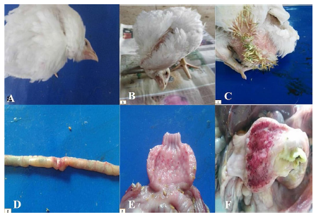
Clinical signs such as dyspnea, torticollis, greenish diarrhea, and opisthotonos were most severe in the NC group (Figure 2A-F). Birds in the OLE-1 and OLE-2 groups displayed milder symptoms. Behavioral scoring confirmed these findings: NC scored 5, OLE-1 scored 3, and OLE-2 scored 2 (Table 2). The blank control and OLE-only groups showed no signs of illness (score = 0). Birds from the blank control and OLE groups showed no prominent clinical signs and were given a 0 score, while the negative control exhibited the maximum signs and a 5 score, as shown in Table 2.
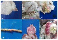
Clinical Signs and Gross Lesions Observed in Broilers Supplemented with Olive Leaf Extract (OLE) and Challenged with vNDV. (A) and (B) show torticollis and opisthotonos observed in the OLE-2 group. (C) shows severe greenish diarrhea in a bird from the Negative Control (NC) group. (D) shows a mild intestinal ulcer from the OLE-2 group. (E) and (F) display severe proventriculus hemorrhages and cecal tonsil congestion observed in the NC group. These lesions and clinical signs were significantly milder or absent in OLE-supplemented birds compared to NC.
Hemorrhages in the proventriculus, congestion of the cecal tonsils, and intestinal ulcers were observed postmortem in the negative control group following administration of a lethal dose of vNDV strain (Fig. 2). During necropsy, NC birds had severe proventricular hemorrhage, congested cecal tonsils, and intestinal ulceration (Figure 2E-F). These lesions were significantly less severe in OLE-treated birds, with only minimal intestinal ulceration observed for OLE-2 (Figure 2D).
Morbidity and mortality rates of experimental birds challenged with vNDV
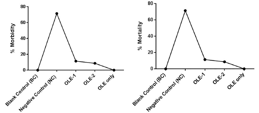
The survival curve was generated based on morbidity (display of disease signs) and mortality figures. The birds of OLE-2 showed more than an 80% survival rate compared to the positive control group, which showed a 30% survival rate (p<0.05). The birds in the OLE group showed the maximum survival rate (95%) (Fig 3). As shown in Figures 3A and 3B, both morbidity and mortality were most severe in the NC group, with a survival rate of only 30%. OLE-1 had an 80% survival rate, and OLE-2 was at 95%, demonstrating the protective action of OLE. Survival was also high in the OLE-only group, further supporting the immune-enhancing role of OLE.
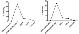
Morbidity and Mortality Rates in Broilers Experimentally Infected with vNDV and Supplemented with Olive Leaf Extract (OLE). Morbidity (A) and mortality (B) rates were considerably lower in OLE-treated groups compared to the negative control group. No significant difference was observed among the OLE-supplemented groups. Statistical significance was assessed using one-way ANOVA followed by Tukey’s HSD post-hoc test (p≤0.05; α=0.05, DF=4, 95% CI).
Antibody titer against vNDV
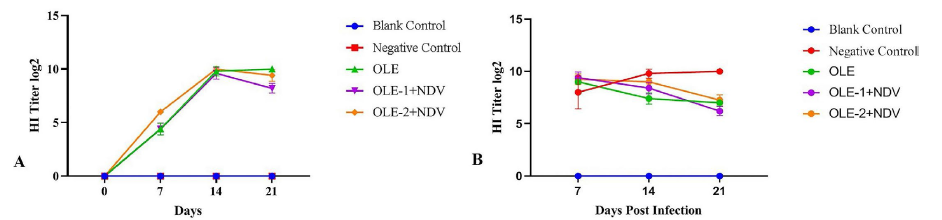
On day 14, the OLE-1 and OLE-2 groups showed higher antibody titers (p<0.05) than the NC group. On day 21, the antibody titer in NC was 317 ± 54, while the OLE-1 and OLE-2 groups had 488.2 ± 49.9 and 572 ± 72.5, respectively. The antibody titer was significantly higher (472±72.5) in the OLE group among the OLE-treated group on the 28th and 35th day of the experiment (Fig. 4). Figure 4 illustrates HI titers for all groups. At day 14, the OLE-1 and OLE-2 groups had significantly greater titers (488.2 ± 49.9 and 572 ± 72.5, respectively) than the NC group (317 ± 54, p<0.05). HI titers reached the peak at day 21 in all OLE-treated groups, and while the titers reduced after infection, they were still greater than those of non-supplemented birds.
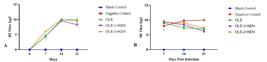
The Distribution of hemagglutination inhibition (HI) titers in birds supplemented with OLE via the oral route is presented. Panel (A) illustrates the HI titers across all groups at various time points before infection, while Panel (B) depicts the HI titers post-infection with the vNDV strain. In OLE-supplemented groups, the HI titer began to increase on day 7, peaking on day 21 and remaining significantly higher than the control groups throughout the experiment. Statistical differences in antibody titers among groups were evaluated using one-way ANOVA followed by Tukey’s HSD post-hoc test (p≤0.05). Following infection with vNDV, the log2 HI titer in OLE-supplemented groups began to decline from day 7 post-infection, stabilizing after day 14. In contrast, the unvaccinated negative control group showed a continuous increase in log2 HI titers, which persisted up to day 21 post-infection.
Histopathological Findings
Histopathological analysis showed severe tracheal mucosal disruption, intestinal hemorrhages, and glandular atrophy in NC birds. However, the OLE-treated groups demonstrated less severe lesions and better histopathological scores. Histopathologically, OLE-1 and OLE-3 birds showed no microscopic changes in visceral organs such as the trachea, intestine, and proventriculus. NC exhibited disrupted tracheal mucous epithelium, lymphoid infiltration, tubular gland atrophy in lamina propria of the intestine with necrotic epithelium and hemorrhages, proventriculus showed destruction of glands and infiltration of inflammatory cells (Fig. 5). Histological analysis showed extensive damage in the NC group, such as tracheal epithelial disruption, intestinal villi necrosis, and proventricular glandular degeneration (Figure 5B, 5F, 5J). OLE-treated birds had less severe changes. For example, OLE-2 birds had mostly intact tracheal lining (Figure 5D), intact intestinal architecture (Figure 5H), and normal proventricular glands (Figure 5L). The blank control group showed normal tissue in all organs.

Histopathological Examination of Organs from Broilers Supplemented with OLE and Infected with vNDV (H&E Staining, 10× Magnification). Trachea: (A) Normal trachea from Blank Control showing intact mucosa and epithelial lining. (B) Trachea from Negative Control showing mucosal erosion and lymphocytic infiltration. (C) Trachea from the OLE-1 group showing mild epithelial disruption. (D) Trachea from the OLE-2 group showing nearly normal histoarchitecture. Intestine: (E) Intestine from Blank Control showing regular villi and intact epithelium. (F) Intestine from Negative Control with necrotic villi, hemorrhage, and inflammation. (G) Intestine from the OLE-1 group showing mild mucosal damage. (H) Intestine from the OLE-2 group showing restored villi structure. Proventriculus: (I) Proventriculus from Blank Control with intact glandular structure. (J) Proventriculus from the NC group with severe glandular degeneration and infiltration. (K) Proventriculus from OLE-1 showing moderate damage. (L) Proventriculus from OLE-2 showing minimal lesions and intact architecture. (Scale bar=100 µm).
Hematology during the experimental trial
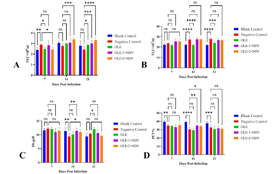
Hemoglobin (Hb), packed cell volume (PCV), total leukocytic count (TLC), and total erythrocytic count (TEC) were checked post-NDV-infection. Hematological parameters indicated that total leukocyte counts in the blank control were significantly (p<0.05) lower than in OLE-treated groups, i.e. the negative control group showed significantly (p<0.05) lower TLC (2.437±0.258), Hb (10.343±0.781), and PCV (32.571±3.155) at the 21st day post-infection (DPI) as compared to the OLE-treated groups. At 21st DPI, total leukocytic count and hemoglobin concentration were significantly (p<0.05) higher in the negative control group (27.681±1.333) as compared to OLE-treated groups (Fig. 6). As can be seen in Figure 6A-D, the total erythrocyte count, concentration of hemoglobin, and packed cell volume in OLE-treated birds were more elevated than in the NC group (p<0.05). The least total leukocytes were recorded in NC (2.437±0.258), whereas birds in OLE-1 and OLE-2 had their values within physiological range. All these hematologic changes indicate fewer systemic inflammations and improved oxygen-carrying capacities.

Hematological Parameters in Experimental Groups Supplemented with OLE and Infected with vNDV. A) Total erythrocyte count, B) total leukocyte count, C) hemoglobin concentration, and D) packed cell volume. OLE-supplemented groups showed improved hematological parameters compared to the negative control. Differences in total erythrocyte count, leukocyte count, hemoglobin concentration, and packed cell volume were analyzed using one-way ANOVA followed by Tukey’s HSD post-hoc test. Values with p≤0.05 were considered statistically significant.
Generally, the findings point to OLE supplementation as enhancing immune protection, reducing NDV-induced damage, and enhancing general performance in broiler chickens exposed to viral challenge.
DISCUSSION
The current study aimed to ascertain the immunomodulatory and growth-promoting effectiveness of olive leaf extract (OLE) in vNDV-infected broilers. It demonstrates that the supplementation of OLE, especially at 20 mL/L (OLE-2), had a marked capacity of increasing body weight gain, survival rates, hematological parameters, and humoral immune status, as well as reducing the severity of clinical signs and tissue pathology induced by NDV infection. Body weight increase and feed intake of the OLE-supplemented groups, especially the OLE-2, were greater due to the phytochemical content of OLE. Some phytochemicals present in OLE, like oleuropein, hydroxytyrosol, and tyrosol, have been shown to boost the activity of digestive enzymes, gut morphology, and nutrient absorption. This is consistent with Varmaghany et al. (2013), who documented improved growth performance in broilers fed with 1-2% olive leaf powder under normal and cold stress. Olive leaf polyphenols can resist oxidative stress and guard the intestinal system, improving feed conversion ratio and growth. At the immune response level, our results indicated that OLE-treated birds exhibited significantly elevated hemagglutination inhibition (HI) titers compared with the NC group. This indicates a heightened humoral immune response, potentially due to the immunostimulatory action of olive phenolics. Oleuropein has been found to increase macrophage activity and the production of nitric oxide, leading to the improved presentation of antigens and subsequent activation of B-cells (Kyriazis et al., 2016). Furthermore, the group that was given the higher dose of OLE-2 had the greatest antibody titers, in keeping with a dose-related immunopotentiating effect. The rise in hematological parameters-i.e., the increased erythrocyte count, hemoglobin concentration, and packed cell volume-is also in keeping with the systemic action of OLE. These hematological parameters are important indicators of physiological health and oxygen delivery. The decrease in the overall leukocyte counts seen in the OLE-treated groups versus NC is indicative of lowered systemic inflammation, which is likely due to the antiviral and anti-inflammatory nature of oleuropein. This is in line with Elsaaed et al. (2014), who showed that olive leaf supplementation in broiler chickens resulted in enhanced blood profiles and lower leukocyte counts under infectious stress. Histopathological and gross lesion analysis of the OLE-1 and OLE-2 groups showed fewer damages to the proventriculus, intestine, and trachea. Observation confirms the established antiviral potential of olive leaf constituents, which can inhibit viral replication and maintain epithelial integrity. Evidence at the mechanism level has been presented by studies carried out by Sabry (2014) and Omar (2010), where there is shown to be interference of olive leaf polyphenols with virus envelopes, as well as an effect of lowering pro-inflammatory cytokine production. Clinical scoring results indicated an impressive distinction between treated and untreated controls. While NC birds showed high respiratory and neurological clinical signs, OLE-1 and OLE-2 groups showed mild or no clinical signs, illustrating high protective efficacy. The high survival of OLE-2 (95%) compared to NC (30%) is a clear indication of OLE as a natural defense booster during outbreaks of NDV. Such a discovery not only highlights the antiviral property of OLE, but also justifies its utilization as a harmless substitute for antibiotics and synthetic growth promoters in broiler chicken rearing. Supplementation with natural bioactive materials such as OLE into feed is consonant with consumer needs for antibiotic-free poultry feed and benefits average flock productivity and well-being. Some limitations should be pointed out: although we noticed beneficial effects at the pathological as well as physiological levels, the study did not explore the molecular mechanism of OLE-induced immunomodulation. Future research needs to determine the levels of cytokines, oxidative stress markers, and signaling pathways to further elucidate the protective effect of OLE. Thus, our findings verify that water supplementation with OLE considerably improves immune processes, physical well-being, and mortality inhibition against disease in vNDV-infected broilers. Dose-response relationships of this study verify the viability of maximum health and production benefits from optimal use of OLE in broiler nutrition. Further studies are needed to investigate its utility for varied breeds, pathogens, and stresses.
CONCLUSION
The current study suggested that OLE supplementation of drinking water enhances broiler performance, immune responses, and ND-associated mortality reduction. The OLE-2 group (20 mL/L) had the best performance, with a survival rate of 95% and significantly elevated antibody titers. Future studies should aim to optimize the dosage and determine the molecular mechanisms of OLE’s immunomodulatory action.
ACKNOWLEDGEMENTS
The authors extend their appreciation to the ongoing Research Funding Program (ORF-2025-552) King Saud University, Riyadh, Saudi Arabia.
REFERENCES
-
Abd-El Ghany FT, Morsy SH, Hassan H, et al. Evaluation of olive leaves and pomace extracts in growing rabbit diets on productive performance, nutrient digestibility, carcass characteristics, antioxidant status and economic efficiency. International Journal of Veterinary Science 2023;12(1):37-44. https://doi.org/10.47278/journal.ijvs/2022.155
» https://doi.org/10.47278/journal.ijvs/2022.155 -
Afiouni A, Toghyani M, Landy N. Performance, morphological responses of the small intestine, and humoral immunity of broilers fed oak acorn (Quercus brantii) as a substitution for antibiotic growth promoters. Iranian Journal of Applied Animal Science 2023;1;13(2):321-31. https://doi.org/20.1001.1.2251628.2023.13.2.13.1
» https://doi.org/20.1001.1.2251628.2023.13.2.13.1 -
Bonsu F, Kagya-Agyemang J, Kwenin W, et al. Medicinal response of broiler chickens to diets containing neem (Azadirachta indica) leaf meal, haematology and meat sensory analysis. World Applied Sciences Journal 2012;19(6):800-5. https://doi.org/10.3923/ijps.2011.730.733
» https://doi.org/10.3923/ijps.2011.730.733 -
Ding Y, Xin C, Zhang C-W, et al. Natural molecules from Chinese herbs protecting against Parkinson's disease via anti-oxidative stress. Frontiers in Aging Neuroscience 2018;10:246. https://doi.org/10.3389/fnagi.2018.00246
» https://doi.org/10.3389/fnagi.2018.00246 -
Dono N. Turmeric (Curcuma longa Linn.) supplementation as an alternative to antibiotics in poultry diets. Indonesian Bulletin of Animal and Veterinary Sciences 2013;23(1):41-9. https://doi.org/10.14334/wartazoa.v23i1.958
» https://doi.org/10.14334/wartazoa.v23i1.958 -
Eevuri TR, Putturu R. Use of certain herbal preparations in broiler feeds - a review. Veterinary World 2013;6(3). https://doi.org/10.5455/vetworld.2013.172-179
» https://doi.org/10.5455/vetworld.2013.172-179 -
Elsaaed EM, Mahgoub K, Hassan ER, et al. Immune-stimulant effects of olive leaves in chickens infected with Escherichia coli. Global Veterinária 2014;13:649-55. https://doi.org/10.14202/vetworld.2021.2727-2734
» https://doi.org/10.14202/vetworld.2021.2727-2734 -
Farooq U, Mustafa R, Khalid MF, et al. Supplementation of herbal seeds to improve the growth performance and digestion in Japanese quail (Coturnix coturnix Japonica). Agrobiological Records 2022;10:19-25. https://doi.org/10.47278/journal.abr/2022.021
» https://doi.org/10.47278/journal.abr/2022.021 -
Hussain K, Abbas A, Rehman A, et al. Evaluating linum usitatissimum seeds extract as potential alternative biochemical and therapeutic agent against induced coccidiosis in broiler chicken. Kafkas Universitesi Veteriner Fakultesi Dergisi Journal 2024;30(6):803. https://doi.org/10.9775/kvfd.2024.32618
» https://doi.org/10.9775/kvfd.2024.32618 -
Kyriazis ID, Koutsoni OS, Aligiannis N, et al. The leishmanicidal activity of oleuropein is selectively regulated through inflammation-and oxidative stress-related genes. Parasites & Vectors 2016;9:1-16. https://doi.org/10.1186/s13071-016-1701-4
» https://doi.org/10.1186/s13071-016-1701-4 -
Landy N, Kheiri F. Effects of hydrolyzed cottonseed protein on growth performances, carcass traits, immunity, microbial and morphological responses of the small intestine and total antioxidant capacity of serum, and small intestine in broiler chickens. Iranian Journal of Applied Animal Science 2023;13(1):121-132. https://doi.org/20.1001.1.2251628.2023.13.1.13.9
» https://doi.org/20.1001.1.2251628.2023.13.1.13.9 -
Leila A, Lamjed B, Roudaina B, et al. Isolation of an antiviral compound from Tunisian olive twig cultivars. Microbial Pathogenesis 2019;128:245-9. https://doi.org/10.1016/j.micpath.2019.01.012
» https://doi.org/10.1016/j.micpath.2019.01.012 -
Mehala C, Moorthy M. Production performance of broilers fed with Aloe vera and Curcuma longa (Turmeric). International Journal of Poulttry Science 2008;7(9):852-6. https://doi.org/10.3923/ijps.2008.852.856
» https://doi.org/10.3923/ijps.2008.852.856 -
Mubashir A, Ghani A, Mubashar A. Common medicinal plants effective in peptic ulcer treatment: a nutritional review. International Journal of Agriculture and Biosciences 2022/11(2):70-4. https://doi.org/10.47278/journal.ijab/2022.010
» https://doi.org/10.47278/journal.ijab/2022.010 -
Omar GA, Mohammed LY. Physiochemical standardization and phytochemical screening of Urtica dioica L. leaves growing in Zakho, Kurdistan region, Iraq. Science Journal of University of Zakho 2023;11(3):306-16. https://doi.org/10.25271/sjuoz.2023.11.3.1069
» https://doi.org/10.25271/sjuoz.2023.11.3.1069 -
Omar SH. Oleuropein in olive and its pharmacological effects. Scientia Pharmaceutica 2010;78(2):133-54. https://doi.org/10.3797/scipharm.0912-18
» https://doi.org/10.3797/scipharm.0912-18 -
Palatty PL, Haniadka R, Valder B, et al. Ginger in the prevention of nausea and vomiting: a review. Critical Reviews in Food Science and Nutrition 2013;53(7):659-69. https://doi.org/10.1080/10408398.2011.553751
» https://doi.org/10.1080/10408398.2011.553751 -
Rasheed M, Zaman MA, Zafar A, et al. Prophylactic effects of methylene blue, coconut and olive oils supplements on hemato-biochemical and histo-pathological parameters against p-phenylenediamine toxicity in male albino rats. Pakistan Veterinary Journal 2024;44(3). https://doi.org/10.29261/pakvetj/2024.193
» https://doi.org/10.29261/pakvetj/2024.193 -
Rehan M, Aslam A, Khan M, et al. Potential economic impact of Newcastle disease virus isolated from wild birds on commercial poultry industry of Pakistan: a review. Hosts Viruses 2019;6(1):1-15. https://doi.org/10.17582/journal.hv/2019/6.1.1.15
» https://doi.org/10.17582/journal.hv/2019/6.1.1.15 -
Sabry OM. Beneficial health effects of olive leaves extracts. Journal of Natural Sciences Research 2014;4(19):1-9. https://doi.org/10.3390/antiox11081496
» https://doi.org/10.3390/antiox11081496 -
Sahin S, Bilgin M. Olive tree (Olea europaea L.) leaf as a waste by-product of table olive and olive oil industry: a review. Journal of the Science of Food and Agriculture 2018;98(4):1271-9. https://doi.org/10.1002/jsfa.8619
» https://doi.org/10.1002/jsfa.8619 -
Schrägle R, Müller W. The influence of selected tannin-containing plant species on the tenacity of pathogenic bacteria in an in-vitro rumen system. Journal of Veterinary Medicine, Series B 1990;37(1-10):181-6. https://doi.org/10.1111/j.1439-0450.1990.tb01044.x
» https://doi.org/10.1111/j.1439-0450.1990.tb01044.x -
Sonora M, Moreno P, Echeverría N, et al. An evolutionary insight into Newcastle disease viruses isolated in Antarctica. Archives of Virology 2015;160:1893-900. https://doi.org/10.1007/s00705-015-2434-y
» https://doi.org/10.1007/s00705-015-2434-y -
Uddin MM, Hasan A, Hossain I, et al. Molecular detection and epidemiological distribution of poultry respiratory viral pathogens in commercial chicken flocks in Bangladesh. Poultry Science 2025;104(2):104679. https://doi.org/10.1016/j.psj.2024.104679
» https://doi.org/10.1016/j.psj.2024.104679 -
Varmaghany S, Rahimi S, Torshizi MK, et al. Effect of olive leaves on ascites incidence, hematological parameters and growth performance in broilers reared under standard and cold temperature conditions. Animal Feed Science and Technology 2013;185(1-2):60-9. https://doi.org/10.1016/j.anifeedsci.2013.07.002
» https://doi.org/10.1016/j.anifeedsci.2013.07.002 -
Zhang Z, Zhang X, Chen H. TCID50 assay: a simple method to determine baculovirus titer. Methods and Molecular Biology 20242829:267-70. https://doi.org/10.1007/978-1-0716-3961-0_20
» https://doi.org/10.1007/978-1-0716-3961-0_20 -
Zuluaga G. Potential of bitter medicinal plants: a review of flavor physiology. Pharmaceuticals 2024;17(6):722. https://doi.org/10.3390/ph17060722
» https://doi.org/10.3390/ph17060722
-
FUNDING
Research Funding Program (ORF-2025-552) King Saud University, Riyadh, Saudi Arabia.
-
DATA AVAILABILITY STATEMENT
Data will be available upon request.
-
DISCLAIMER/PUBLISHER’S NOTE
The published papers’ statements, opinions, and data are those of the individual author(s) and contributor(s). The editor(s) disclaim responsibility for any injury to people or property resulting from any ideas, methods, instructions, or products referred to in the content.
Data will be available upon request.