 Dietary Supplementation of Arginine Promote Testicular Development in Muscovy Ducks Based on Transcriptome Analysis
ABSTRACT
The poor reproductive performance of male ducks hinders the development of the Muscovy duck industry. Adding arginine to the feed plays a crucial role in the testicular development and improvement of reproductive performance in Muscovy ducks, but there is limited research on the biochemical metabolism, gene expression changes, and other mechanisms involved. In this study, 60 male Muscovy ducks were randomly divided into two groups. One group was fed a basal diet (control group), and the other was fed a basal diet supplemented with 0.4% arginine (experimental group). The results showed that the reproductive cells, reproductive hormone levels, and antioxidant levels in the test group of Muscovy ducks were significantly higher than those in the control group. Through transcriptome sequencing analysis, this study identified a total of 1268 differentially expressed genes, including 740 genes that were significantly upregulated and 528 genes that were significantly downregulated. The expression of genes such as KIF13A, AFF4, DSCC1, and related pathways had a significant impact on the testicular development of Muscovy ducks. These findings provide an important basis for using arginine supplementation in feed to enhance the reproductive performance of Muscovy ducks.
Keywords:
Muscovy ducks; arginine; testicular tissue; transcriptome sequencing; differentially expressed genes
INTRODUCTION
Muscovy duck, also known as the French duck or American goose, is native to South America and is a high-quality, large, lean meat duck. Muscovy ducks exhibit strong disease resistance, tolerance to coarse feed, rapid growth and development, and offer high economic benefits in contemporary specialized poultry farming, endowing them with unique value and status. However, their poor reproductive performance severely hampers the rapid development of the Muscovy duck industry. The poor reproductive performance of Muscovy ducks is mainly reflected in late sexual maturity, low egg production rate, strong nest-seeking behavior, and low fertilization and hatching rates of eggs. For male Muscovy ducks, the development and performance of the testes play a crucial role in their reproductive success.
The testicles are vital organs in the male duck’s reproductive system. They are responsible for producing sperm through spermatogenesis and testosterone through steroidogenesis (Siu & Cheng, 2004). Spermatogenesis is a complex cell division process that relies on multiple factors and occurs in the seminiferous tubules of the testes. The formation of sperm in male animals is regulated by the hypothalamic-pituitary-gonadal axis. The hypothalamus secretes gonadotropin-releasing hormone (GnRH), which stimulates the pituitary gland to release luteinizing hormone (LH). Luteinizing hormone (LH) can stimulate the proliferation of interstitial cells in the testes, induce the secretion of testosterone, and affect the development of male reproductive organs, as well as the development of the germinal epithelium and spermatogenesis. The process of sperm formation is also regulated by many genes, and the fibroblast growth factor (FGF) gene family plays an important role in regulating the growth and development of several reproductive organs, including the testes (Wagener et al., 2003; Hiramatsu et al., 2010).
FOXO1 is one of the transcription factors in the FOX family, and has been shown to play an important role in mammalian reproduction. Yu et al. (2019) found that FOXO1 is localized in the Sertoli cells of chicken testes, which is consistent with its localization in mammals. They also discovered that FOXO1 is an important marker for supporting cells upstream of SOX9 expression, and a potential regulatory factor in the differentiation and normal function of the embryonic chicken testis (Yu et al., 2019). Bai et al. (2020) selected 24 pre- and post-sexually differentiated Putian ducks for transcriptome sequencing. They found that nine genes, including TTN, are related to testicular differentiation and development. After sexual differentiation, genes such as DMRT3 are highly expressed specifically in male ducks (Bai et al., 2020).
Arginine is an essential amino acid for poultry growth. It is not only an important raw material for protein synthesis in the body, but also a crucial precursor for the synthesis of bioactive substances such as nitric oxide, polyamines, ornithine, creatine, and agmatine (Wu & Morris, 1998). Relevant studies have shown that arginine has a significant impact on the production performance, reproductive performance, immune response, antioxidant performance, and growth and development of chickens (Duan et al., 2013). In terms of reproductive performance, provious study has shown that arginine is involved in the male reproductive process and plays a role in sperm formation. A deficiency in arginine can lead to a decrease in sperm count and quality (Miroueh, 1970). Turk et al. (2019) found that arginine has a protective effect against cyclosporine A-induced testicular toxicity (Turk et al., 2019). It reduces the impairment of testicular spermatogenic function and decreases apoptosis of germ cells. The aforementioned research indicates that arginine is essential for the reproductive performance of poultry. However, poultry do not have a urea cycle and lack the enzyme carbamoyl phosphate synthetase, which is a key enzyme required for the synthesis of arginine precursors. Therefore, poultry cannot synthesize arginine independently, making it feasible and necessary to add arginine to poultry feed to enhance their reproductive capacity. However, there is still insufficient research on the molecular mechanisms of gene regulation related to the development of testes and the improvement of reproductive performance in Muscovy ducks through the supplementation of arginine in their feed.
This study investigated the effects of adding 0.4% arginine to the feed of male Muscovy ducks on the morphology of testicular development and the regulation of hormone secretion. Transcriptome sequencing (RNA-seq) was also used to identify differentially expressed genes and related pathways in the testes of the experimental and control groups. This study provides a theoretical basis for the effects on testicular development and regulation of adding arginine to the feed of Muscovy ducks, and offers practical insights for enhancing reproductive capacity through arginine supplementation in the feed.
MATERIALS AND METHODS
Breeding and Sample Collection of Muscovy Ducks
The Muscovy ducks used in this experiment were sourced from Hewang Poultry Company in Lanxi City, Zhejiang Province. Sixty male breeding ducks at 49 weeks of age were randomly divided into two groups, with three replicates per group, and ten ducks per replicate. Group A served as the control group and was fed a basal diet, while Group B served as the experimental group and was fed a basal diet supplemented with a 0.4% concentration of L-arginine. The composition and nutrient levels of the basal diet are shown in Table 1. After 75 days of feeding, five male ducks were randomly selected from each replicate group for blood collection via the wing vein, resulting in a total of 30 blood samples. The blood samples were allowed to clot for 3-4 hours and then centrifuged at 3,000 rpm for 5 minutes. The serum was collected, transferred to 1.5 mL EP tubes, and stored at -20°C for further analysis of reproductive hormones and antioxidant capacity. In addition, one male duck was randomly selected from each replicate group for slaughter, resulting in a total of three ducks per group. After slaughter, the left and right testes were collected. The left testis was fixed in 4% paraformaldehyde for histological observation, while the right testis was collected in a cryotube and immediately placed in liquid nitrogen. Subsequently, it was transferred to a -80°C freezer for transcriptome sequencing.
Testicular tissue section
This study involved preparing paraffin sections of the testes of Muscovy ducks, which included the following four steps. Firstly, the testicular tissue samples were cut into small pieces of 2 cubic centimeters and fixed in Bouin’s solution for 12 hours. Secondly, the fixed tissues were trimmed and placed in a dehydration chamber for dehydration. After dehydration, the tissues were transferred to an embedding machine for paraffin embedding. The paraffin-embedded tissues were then sliced using a paraffin microtome, with a thickness of 6 micrometers. Finally, the paraffin sections were dewaxed and washed with distilled water. After the completion of sectioning, the sections were stained with hematoxylin and eosin. Hematoxylin stains the cell nuclei, while eosin stains the cytoplasm. After staining, the samples are mounted on slides and observed under a microscope to examine the morphological changes in the testicular tissue of both the experimental and control groups.
Serum biochemical index detection
The serum reproductive hormone indicators tested in this study include testosterone (T), estradiol (E2), follicle-stimulating hormone (FSH), and luteinizing hormone (LH), which were determined using enzyme-linked immunosorbent assay kits provided by Beijing Huaying Biotechnology Research Institute. The serum antioxidant capacity indicators include superoxide dismutase (SOD), malondialdehyde (MDA), glutathione peroxidase (GSH-Px), and total antioxidant capacity (T-AOC), which were analyzed using a fully automatic biochemical analyzer. The kits were provided by Beijing Huaying Biotechnology Research Institute.
RNA extraction and transcriptome sequencing
In this study, RNA was extracted using an Omega kit. The specific steps were as follows: an appropriate amount of testicular tissue was placed in a 1.5 mL centrifuge tube, and 500 mL of lysis solution and two sterilized steel beads were added. The tissue was homogenized using an automated sample homogenizer, and then centrifuged at the highest speed at room temperature for 5 minutes. The supernatant was transferred to a centrifugal column and centrifuged for 3 minutes. The filtrate was transferred to a 1.5 mL centrifuge tube, and twice the volume of 70% ethanol was added. The mixture was vortexed, and then the liquid was transferred to a HiBind RNA mini column and centrifuged for 3 minutes. The waste liquid was discarded, buffer I was added, and centrifuged for 2 minutes. The waste liquid was then discarded again, buffer II was added, and centrifuged (this step was repeated twice). After obtaining the dried mini column, elution solution was added, and the mixture was centrifuged for 2 minutes to extract the RNA. The Nanodrop 2000 was used to determine the concentration, purity, and integrity of the RNA. The Agilent Bioanalyzer 2100 was used to measure the OD value and RIN value of the samples. After RNA extraction, the PrimeScript RT reagent Kit with gDNA Eraser was used for library construction. The constructed library was quantified using qPCR to ensure the accurate concentration of the library. After passing the library quality control, paired-end sequencing with 150 base pairs was performed using the Illumina HiSeq platform.
Sequencing Data Alignment and Assembly
The raw data obtained from sequencing was subjected to quality control and filtering using Fastx_toolkit software. This process involved removing reads containing adapters and eliminating low-quality reads to obtain high-quality sequencing data (clean reads). The clean reads were then mapped to the duck reference genome (CAU_duck1.0) using HISAT2 software (Kim et al., 2019). This study utilized StringTie software for transcriptome assembly and splicing based on the existing reference genome, comparing it with the existing transcripts to identify unannotated new transcripts (Pertea et al., 2015).
Differential Expression Analysis
Differential expression of testicular tissue between the experimental group and the control group was analyzed using the Deseq2 software (Love et al., 2014). FPKM (Fragments Per Kilobase of transcript per Million fragments mapped) was used as a measure of gene expression level. Genes with a fold change (FC) ≥ 1 and a p-value < 0.05 were selected as the differentially expressed gene set. Genes with higher expression levels in the experimental group compared to the control group were classified as upregulated genes, while genes with lower expression levels were classified as downregulated genes.
Differential expression gene enrichment analysis
Gene Ontology (GO) analysis is a commonly used method in large-scale gene function enrichment studies. It consists of three main branches: biological process, molecular function, and cellular component. KEGG (Kyoto Encyclopedia of Genes and Genomes) stores a vast amount of data on genomics, biological pathways, signaling pathways, diseases, drugs, and chemicals. KEGG analysis helps interpret the function of genes by integrating gene and expression information comprehensively. The functional annotation of differentially expressed genes is conducted using the GOseq R package, which is based on the hypergeometric distribution. Additionally, KEGG pathway analysis is carried out using the KOBAS online platform.
Validation of Differentially Expressed Genes by qRT-PCR
To validate the sequencing results, qRT-PCR was carried out on 3 randomly selected DEGs, following the steps of the PrimeScript® RT regent Kit with gDNA Eraser to synthesize cDNA using identical samples with RNA-seq. Primers were designed using Primer3 (https://primer3.ut.ee/). Expression analysis of the obtained cDNA samples was performed using the SYBR Green method in a fluorescence 96-wells plate in an ABI 7500 Real-time PCR machine, using the following program: one cycle at 95 °C for 30 s and 40 cycles each at 95 °C for 5 s and 60 °C for 60 s. The β-actin (ACTB) gene was used as an internal control. The results were analyzed using the 2−ΔΔCt method.
Statistical analysis
The hypothesis testing between the experimental group and the control group in this study used non-parametric tests (wilcox.test), with p-values less than 0.05 and 0.01 indicating significant and highly significant results, respectively.
RESULTS
Observation of Testicular Tissue Morphology
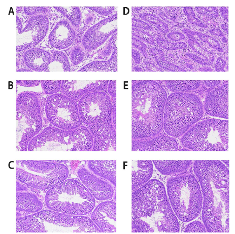
The paraffin sections of the testicular tissue of the Muscovy ducks are shown in Figure 1 (200X) and Figure 2 (400X). In the control group (Figure 1A, 1B, 1C) and (Figure 2A, 2B, 2C), there are fewer layers of germinal epithelium, fewer spermatocytes, a larger luminal area, and fewer mature sperm. In the arginine group (Figure 1D, 1E, 1F) and (Figure 2D, 2E, 2F), there are more layers of germinal epithelium, more spermatocytes, a fuller luminal area, and more mature sperm.

Histological sections of testes from the control group and the arginine group in male ducks (200X magnification). A, B, and C represent the control group, while D, E, and F represent the arginine group.

Histological sections of testes from the control group and the arginine group in male ducks (400X magnification). A, B, and C represent the control group, while D, E, and F represent the arginine group.
Serum biochemical index levels
The serum biochemical indicators data of the control group and the arginine group are shown in Supplementary Table 1. The results of the variance analysis between the two groups are presented in Table 2 and Table 3. In terms of serum reproductive hormones, the testosterone (T) and estradiol (E2) levels in the arginine group were significantly higher than those in the control group (p<0.05). The levels of follicle-stimulating hormone (FSH) and luteinizing hormone (LH) showed no significant differences between the experimental group and the control group. In terms of serum antioxidant capacity indicators, the levels of superoxide dismutase (SOD), glutathione peroxidase (GSH-Px), and total antioxidant capacity (T-AOC) in the arginine group were significantly higher than those in the control group (p<0.05). The malondialdehyde (MDA) level was significantly lower in the arginine group than in the control group (p<0.05).
Differentially expressed genes
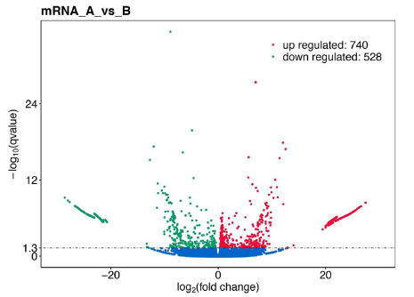
Using the Illumina sequencing platform, this experiment conducted high-throughput sequencing on six cDNA libraries of testicular samples. After quality control, each sample obtained an average of 5.38107 million clean reads (Table 4). DESeq2 software was used in this study to identify differentially expressed genes between the experimental and control groups using a screening criterion of p<0.05 and |log2FoldChange| > 1. A total of 1,268 differentially expressed genes were detected, with 740 genes being significantly upregulated and 528 genes significantly downregulated (Figure 3 and Supplementary Table 2). The top 20 genes with the most significant differences are shown in Table 5.

Volcano plot of differentially expressed genes in testicular tissue of experimental group and control group of Muscovy ducks.
GO annotation and enrichment analysis of differentially expressed genes
GO functional annotation was performed on differentially expressed genes, and a total of 3981 significantly enriched GO terms were identified in the experimental group compared to the control group (Supplementary Table 3 and Figure 4A). In terms of biological processes, these genes were mainly annotated for cellular process (GO:0009987), single-organism process (GO:0009987), metabolic process (GO:0009987), and organic substance metabolic process (GO:0009987). In terms of cellular components, the genes were mainly annotated for cell (GO:0005623), cell part (GO:0005623), intracellular (GO:0005623), and intracellular part (GO:0005623). In terms of molecular functions, the genes were mainly annotated for binding (GO:0005488), catalytic activity (GO:0003824), heterocyclic compound binding (GO:1901363), and organic cyclic compound binding (GO:0097159).
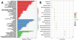
A and B are GO enrichment analysis and KEGGs enrichment analysis of the differential genes in the testicular tissue, respectively.
KEGG enrichment analysis was utilized to identify the primary biochemical metabolic pathways and signaling pathways associated with differentially expressed genes. The results showed that differentially expressed genes were significantly enriched in 212 pathways (Figure 4B, Supplementary Table 4), and the top 20 pathways with the most significant differences are shown in Table 6, including Oocyte meiosis (ko04114), mRNA surveillance pathway (ko03015), AMPK signaling pathway (ko04152), Folate biosynthesis (ko00790), Hippo signaling pathway (ko04391), Glucagon signaling pathway (ko04922), Fanconi anemia pathway (ko03460), Meiosis-yeast (ko04113), Phototransduction (ko04745), Insulin signaling pathway (ko04910), and Autophagy (ko04138).
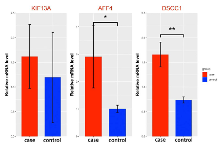
To technically validate the RNA-Seq results, three differentially expressed genes were selected for real-time PCR analysis. The results showed that the expression levels of the AFF4 gene and DSCC1 gene were significantly higher in the experimental group than in the control group, while the expression level of KIF13A in the experimental group was also higher than in the control group (but did not reach statistical significance) (Figure 5, Supplementary Table 4). RT-PCR results confirmed that the differentially expressed genes identified in this study had a significant impact on the testicular development of Muscovy ducks.
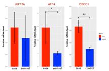
RT-PCR results of differentially expressed genes in the experimental group and control group.
DISCUSSION
Normal testicular tissue structure and function can maintain the reproductive ability of animals. In this study, paraffin sections were prepared, and it was observed that the experimental group exhibited an increased number of layers of germinal epithelium, more germ cells, a fuller lumen, and more mature sperm. These findings suggest that arginine can effectively enhance testes development in Muscovy ducks, promoting sperm production and maturation. Hormonal regulation is essential in the reproductive physiology of animals. Follicle-stimulating hormone and luteinizing hormone, secreted by the pituitary gland of male ducks, along with testosterone and estradiol secreted by the testes, are crucial reproductive hormones classified as steroid hormones. A study has shown that adding arginine to the feed can significantly increase the levels of testosterone, estradiol, follicle-stimulating hormone, and luteinizing hormone in Muscovy ducks. A study on HyLine Brown laying hens showed that adding arginine to low crude protein (LCP) feed can linearly upregulate (p<0.05) the gonadotropin-releasing hormone 1 (GnRH1) and gonadotropin inhibitory hormone in the hypothalamus. The pituitary growth hormone, GnRH receptor, and follicle-stimulating hormone (FSHβ) were also increased (p<0.05). Serum insulin-like growth factor 1 (IGF-1) and nitric oxide (NO) levels were found to increase significantly (p<0.05) with higher levels of arginine supplementation (Uyanga et al., 2022). Reproduction in poultry is regulated by a set of neuroendocrine mechanisms known as the hypothalamic-pituitary-gonadal (HPG) axis, which involves the hypothalamus, anterior pituitary gland, and the gonads. The stimulatory action of gonadotropin-releasing hormone (GnRH) enables the synthesis and release of gonadotropins, which is regulated by its inhibitory counterpart, gonadotropin-inhibitory hormone (GnIH) (Bedecarrats, 2015). These peptides coordinate the release of luteinizing hormone (LH) and follicle-stimulating hormone (FSH) from the anterior pituitary into the peripheral circulation. Therefore, we speculate that supplementing arginine to the basic daily diet of poultry modulated the gene expression of reproductive hormones in the hypothalamic-pituitary-gonadal axis of laying hens via actions that may be related to nitric oxide (NO) and insulin-like growth factor-1 (IGF-1) activity.
This study found that arginine has certain antioxidant and anti-inflammatory effects. Similar results were also obtained in the study by Akhigbe et al. (Akhigbe et al., 2023). The antioxidant and anti-inflammatory activities of arginine have been linked to its ability to suppress the generation of malondialdehyde (MDA), a marker of lipid peroxidation, and up-regulate the concentration of reduced glutathione (GSH) and the activities of enzymatic antioxidants (Saka et al., 2021). The intake of arginine has been shown to enhance its concentrations in the plasma and tissues and to promote the biosynthesis of nitric oxide, which is an important vasodilator (Nagase et al., 1997; Saka et al., 2021).
Through Gene Ontology (GO) functional annotation of differentially expressed genes, we identified enrichment in biological processes such as cellular component organization or biogenesis, cellular processes, cellular component organization, and cellular protein metabolic processes. The development of testes and sperm production in the testes are a complex process. Spermatogenesis can be divided into three major stages: proliferation and maintenance of the spermatogonial population, reduction of genetic information from diploid to haploid state through the meiosis process, and differentiation and maturation of the haploid sperm cell. All of these processes are cellular processes, and these stages are highly regulated at both the transcriptional and post-transcriptional levels, resulting in an accurate gene expression pattern depending on both space and time. There is evidence to suggest that arginine is a signaling molecule in some important pathways, such as the mTOR pathway (Takahara et al., 2020). mTOR is considered the key regulator of important cellular processes, including protein synthesis, proliferation, and metabolism (Starikova et al., 2023). Azhar et al. (2023) has revealed an essential role of methylarginine in coordinating spermatogonial development (Azhar et al., 2023).
Differentially expressed genes are mainly enriched in processes such as neuron projection, intracellular ribonucleoprotein complex, and meiotic cohesin complex in the Cellular Component. Neuronal projection pathways may influence the reciprocal regulation between the nervous system and the reproductive system during testicular development, sperm production, and maturation. Proteins and DNA complexes in neuronal projection pathways, such as CyclinH protein and CDK7, play important roles during meiosis and are associated with protein kinase activity and chromosome segregation in sperm production (Xu et al., 2021). Ribonucleoprotein complexes contain RNA and proteins closely related to cellular biological processes. During spermatogenesis, ribonucleoprotein complexes are involved in the regulation of RNA, affecting the formation and maturation of sperm (Griffin et al., 2022). The meiotic cohesin complex plays a key role in spermatocyte meiosis by maintaining chromosome connections and regulating chromosome separation during sperm formation to ensure the normal transmission of genetic information (Kim et al., 2013).
Differentially expressed genes in Molecular Function are mainly enriched in processes such as protein serine/threonine kinase activity and RNA binding. Previous studies have shown that the activity of enzymes such as protein serine/threonine kinase is closely related to testicular development and sperm capacitation, and some of them are mediated by NO (Tosti & Menezo, 2021). NO emerged as one the messengers involved in regulating protein phosphorylation levels, in particular on serine, threonine and tyrosine residues (Herrero et al., 1999; Thundathil et al., 2003; Rahman et al., 2014). We speculate that adding arginine to the diet of Muscovy ducks may have a positive effect on testicular development and sperm capacitation.
The differentially expressed genes in this study are mainly enriched in pathways such as oocyte meiosis, mRNA surveillance pathway, AMPK signaling pathway, folate biosynthesis, Hippo signaling pathway, and meiosis. This indicates that these signaling pathways are related to the testicular development and spermatogenic ability of Muscovy ducks. The AMPK is a type of heterotrimeric Ser/Thr kinase that acts as a sensitive energy sensor and regulator of cellular energy metabolism in Sertoli cells, making it an important regulatory factor. Furthermore, the AMPK signaling pathway maintains the stability of the blood-testes barrier, providing lactate for the energy metabolism of germ cells and supporting spermatogenesis (Galardo et al., 2007; Galardo et al., 2010; Crisostomo et al., 2018; Petricca et al., 2019). Additionally, studies have shown that the Hippo signaling pathway is involved in spermatogenesis, and its disruption is associated with azoospermia (He et al., 2009). The Hippo signaling pathway also plays a crucial role in cell differentiation, with related studies showing that it regulates the supporting cells of the salmon testis during puberty (Kjaerner-Semb et al., 2022).
This study identified certain genes that may influence the development of the testes in Muscovy ducks through functional screening of differentially expressed genes. Relevant studies have shown that the protein kinases KIF13A and HIPK4 are specifically enriched in the testes. Given the importance of phosphorylation in cell signaling and differentiation, it is not surprising that protein kinases are involved in spermatogenesis (Crapster et al., 2020). mTOR is an important cell signaling protein involved in regulating processes such as cell growth, metabolism, and proliferation. It plays a crucial role in the maintenance and differentiation of spermatogonial stem cells (SSCs), the maintenance and reconstitution of the blood-testis barrier, and sperm production. The RPTOR identified in this study is one of the important protein complexes that regulate the mTOR signaling pathway. Abnormal expression of the RPTOR gene may affect the normal function of the mTOR signaling pathway, thereby influencing the development of the testes in ducks (Moreira et al., 2019; Serra et al., 2019). Paukszto et al. (2023) found that the ODF2L gene is expressed differentially in testicular tissue during sperm maturation in birds. This expression pattern suggests that the gene may play a role in the development of post-testicular motility, which is a critical component of fertility in birds (Paukszto et al., 2023). Additionally, in a study on the genetic regulation of the ODF2L gene in pig tissues, researchers found that the cis-eQTL effects of ODF2L on chromosome 4 in blood and testis were significant (Tang et al., 2024). The above results indicate that ODF2L plays an important role in the genetic regulation of testicular development. Moreover, research indicates that the AFF4 gene has a significant influence on the development of the testes. The gene is responsible for determining testis development on the Y chromosome (Stiglec et al., 2007). DSCC1 has been found to be upregulated in testicular tissues, indicating a potential role in testicular function.
CONCLUSION
Taken together, this study indicated that adding arginine to the feed can increase the levels of testosterone, estradiol, follicle-stimulating hormone, and luteinizing hormone in the serum of Muscovy ducks. It also increases the levels of superoxide dismutase, glutathione peroxidase, and total antioxidant capacity. Adding arginine to the feed has a significant impact on the testicular development of Muscovy ducks and can activate the expression of genes such as KIF13A, HIPK4, RPTOR, ODF2L, AFF4, DSCC1, and related pathways. These findings provide an important basis for using arginine supplementation in feed to improve the reproductive performance of Muscovy ducks.
ACKNOWLEDGEMENTS
We thank the Foundation for the National Natural Science Foundation of China for its financial support of our research.
REFERENCES
-
AbAkhigbe R, Afolabi O, Ajayi A. L-Arginine abrogates maternal and pre-pubertal codeine exposure-induced impaired spermatogenesis and sperm quality by modulating the levels of mRNA encoding spermatogenic genes. Frontiers in Endocrinology 2023;14:1180085. https://doi.org/10.3389/fendo.2023.1180085
» https://doi.org/10.3389/fendo.2023.1180085 -
Azhar M, Xu C, Jiang X, et al. The arginine methyltransferase Prmt1 coordinates the germline arginine methylome essential for spermatogonial homeostasis and male fertility. Nucleic Acids Researcg 2023;51(19):10428-50. https://doi.org/10.1093/nar/gkad769
» https://doi.org/10.1093/nar/gkad769 -
Bai D, Chen Y, Hu Y, et al. Transcriptome analysis of genes related to gonad differentiation and development in Muscovy ducks. BMC Genomics 2020;21(1):438. https://doi.org/10.1186/s12864-020-06852-z
» https://doi.org/10.1186/s12864-020-06852-z -
Bedecarrats G. Control of the reproductive axis: Balancing act between stimulatory and inhibitory inputs. Poultry Science 2015;94(4):810-5. https://doi.org/10.3382/ps/peu042
» https://doi.org/10.3382/ps/peu042 -
Crapster JA, Rack P,G Hellmann ZJ, et al. HIPK4 is essential for murine spermiogenesis. Elife 2020;9:e50209. https://doi.org/10.7554/eLife.50209
» https://doi.org/10.7554/eLife.50209 -
Crisostomo L, Alves M, Gorga A, et al. Molecular mechanisms and signaling pathways involved in the nutritional support of spermatogenesis by sertoli cells. Methods in Molecular Biology 2018;1748:129-55. https://doi.org/10.1007/978-1-4939-7698-0_11
» https://doi.org/10.1007/978-1-4939-7698-0_11 -
Duan X, Li F, Mao S, et al. Effects of dietary L-arginine on laying performance and anti-oxidant capacity of broiler breeder hens, eggs, and offspring during the late laying period. Poultry Science 2013;94(12):2938-43. https://doi.org/10.1093/ps/pev283
» https://doi.org/10.1093/ps/pev283 -
Galardo M, Riera M, Pellizzari E, et al. Adenosine regulates Sertoli cell function by activating AMPK. Molecular and Cellular Endocrinology 2010;330(1-2):49-58. https://doi.org/10.1016/j.mce.2010.08.007
» https://doi.org/10.1016/j.mce.2010.08.007 -
Galardo M, Riera M, Pellizzari E, et al. The AMP-activated protein kinase activator, 5-aminoimidazole-4-carboxamide-1-b-D-ribonucleoside, regulates lactate production in rat Sertoli cells. Journal of Molecular Endocrinology 2007;39(4):279-88. https://doi.org/10.1677/JME-07-0054
» https://doi.org/10.1677/JME-07-0054 -
Griffin K, Walters B, Li H, et al. Widespread association of the Argonaute protein AGO2 with meiotic chromatin suggests a distinct nuclear function in mammalian male reproduction. Genome Research 2022;32(9):1655-68. https://doi.org/10.1101/gr.276578.122
» https://doi.org/10.1101/gr.276578.122 -
He Z, Kokkinaki M, Dym M. Signaling molecules and pathways regulating the fate of spermatogonial stem cells. Microscopy Research and Technique 2009;72(8):586-95. https://doi.org/10.1002/jemt.20698
» https://doi.org/10.1002/jemt.20698 -
Herrero M, de Lamirande E, Gagnon C. Nitric oxide regulates human sperm capacitation and protein-tyrosine phosphorylation in vitro. Biology of Reproduction 1999;61(3):575-81. https://doi.org/10.1095/biolreprod61.3.575
» https://doi.org/10.1095/biolreprod61.3.575 -
Hiramatsu R, Harikae K, Tsunekawa N, et al. FGF signaling directs a center-to-pole expansion of tubulogenesis in mouse testis differentiation. Development 2010;137(2):303-12. https://doi.org/10.1242/dev.040519
» https://doi.org/10.1242/dev.040519 -
Kim D, Paggi J, Park C, et al. Graph-based genome alignment and genotyping with HISAT2 and HISAT-genotype. Nature Biotechnology 2019;37(8):907-15. https://doi.org/10.1038/s41587-019-0201-4
» https://doi.org/10.1038/s41587-019-0201-4 -
Kim S, Spike C, Greenstein D. Control of oocyte growth and meiotic maturation in Caenorhabditis elegans Advances in Experimental Medicine and Biology 2013;757:277-320. https://doi.org/10.1007/978-1-4614-4015-4_10
» https://doi.org/10.1007/978-1-4614-4015-4_10 -
Kjaerner-Semb E, Ayllon F, Kleppe L, et al. Vgll3 and the Hippo pathway are regulated in Sertoli cells upon entry and during puberty in Atlantic salmon testis. Scientific Reports 2018;8(1):1-11. https://doi.org/10.1038/s41598-018-20308-1
» https://doi.org/10.1038/s41598-018-20308-1 -
Love M, Huber W, Anders S. Moderated estimation of fold change and dispersion for RNA-seq data with DESeq2. Genome Biol 2014;15(12):550. https://doi.org/10.1186/s13059-014-0550-8
» https://doi.org/10.1186/s13059-014-0550-8 -
Miroueh A. Effect of arginine on oligospermia. Fertility and Sterility 1970;21(3):217-219. https://doi.org/10.1016/s0015-0282(16)37384-8
» https://doi.org/10.1016/s0015-0282(16)37384-8 -
Moreira B, Oliveira P, Alves M. Molecular mechanisms controlled by mtor in male reproductive system. International Journal of Molecular Sciences 2019;20(7). https://doi.org/10.3390/ijms20071633
» https://doi.org/10.3390/ijms20071633 -
Nagase S, Takemura K, Ueda A, et al. A novel nonenzymatic pathway for the generation of nitric oxide by the reaction of hydrogen peroxide and D- or L-arginine. Biochemical and Biophysical Research Communications 1997;233(1):150-3. https://doi.org/10.1006/bbrc.1997.6428
» https://doi.org/10.1006/bbrc.1997.6428 -
Paukszto L, Wisniewska J, Liszewska E, et al. Specific expression of alternatively spliced genes in the turkey (Meleagris gallopavo) reproductive tract revealed their function in spermatogenesis and post-testicular sperm maturation. Poultry Science 2023;102(4):102484. https://doi.org/10.1016/j.psj.2023.102484
» https://doi.org/10.1016/j.psj.2023.102484 -
Pertea M, Pertea G, Antonescu C, et al. StringTie enables improved reconstruction of a transcriptome from RNA-seq reads. Nature Biotechnology 2015;33(3):290-5. https://doi.org/10.1038/nbt.3122
» https://doi.org/10.1038/nbt.3122 -
Petricca S, Flati V, Celenza G, et al. Tebuconazole and econazole act synergistically in mediating mitochondrial stress, energy imbalance, and sequential activation of autophagy and apoptosis in mouse sertoli TM4 Cells: possible role of AMPK/ULK1 Axis. Toxicological Sciences 2019;169(1):209-23. https://doi.org/10.1093/toxsci/kfz031
» https://doi.org/10.1093/toxsci/kfz031 -
Rahman M, Kwon WS Lee J, et al. Sodium nitroprusside suppresses male fertility in vitro. Andrology 2014;2(6):899-909. https://doi.org/10.1111/j.2047-2927.2014.00273.x
» https://doi.org/10.1111/j.2047-2927.2014.00273.x -
Saka W, Akhigbe R, Abidoye A, et al. Suppression of uric acid generation and blockade of glutathione dysregulation by L-arginine ameliorates dichlorvos-induced oxidative hepatorenal damage in rats. Biomedicine & Pharmacotherapy 2021;138:111443. https://doi.org/10.1016/j.biopha.2021.111443
» https://doi.org/10.1016/j.biopha.2021.111443 -
Serra N, Velte E, Niedenberger B, et al. The mTORC1 component RPTOR is required for maintenance of the foundational spermatogonial stem cell pool in micedagger. Biology and Reproduction 2019;100(2):429-39. https://doi.org/10.1093/biolre/ioy198
» https://doi.org/10.1093/biolre/ioy198 -
Siu M, Cheng C. Dynamic cross-talk between cells and the extracellular matrix in the testis. Bioessays 2004;26(9):978-92. https://doi.org/10.1002/bies.20099
» https://doi.org/10.1002/bies.20099 -
Starikova E, Rubinstein A, Mammedova J, et al. Regulated arginine metabolism in immunopathogenesis of a wide range of diseases: is there a way to pass between scylla and charybdis? Current Issues in Molecular Biology 2023;45(4):3525-51. https://doi.org/10.3390/cimb45040231
» https://doi.org/10.3390/cimb45040231 -
Stiglec R, Kohn M, James F, et al. Frequency of cancer genes on the chicken z chromosome and its human homologues: implications for sex chromosome evolution. Comparative and Functional Genomics. 2007;2007(1):43070. https://doi.org/10.1155/2007/43070
» https://doi.org/10.1155/2007/43070 -
Takahara T, Amemiya Y, Sugiyama R, et al. Amino acid-dependent control of mTORC1 signaling: a variety of regulatory modes. Journal of Biomedical Science 2020;27(1):87. https://doi.org/10.1186/s12929-020-00679-2
» https://doi.org/10.1186/s12929-020-00679-2 -
Teng J, Gao Y, Yin H, et al. A compendium of genetic regulatory effects across pig tissues. Nature Genetics 2024;56(1):112-23. https://doi.org/10.1038/s41588-023-01585-7
» https://doi.org/10.1038/s41588-023-01585-7 -
Thundathil J, de Lamirande E, Gagnon C. Nitric oxide regulates the phosphorylation of the threonine-glutamine-tyrosine motif in proteins of human spermatozoa during capacitation. Biology of Reproduction 2003;68(4):1291-8. https://doi.org/10.1095/biolreprod.102.008276
» https://doi.org/10.1095/biolreprod.102.008276 -
Tosti E, Menezo Y. Gamete activation: basic knowledge and clinical applications. Human Reproduction Update 2016;22(4):420-39. https://doi.org/10.1093/humupd/dmw014
» https://doi.org/10.1093/humupd/dmw014 -
Turk G, Atessahin A, Sonmez M, et al. Lycopene protects against cyclosporine A-induced testicular toxicity in rats. Theriogenology 2007;67(4):778-785. https://doi.org/10.1016/j.theriogenology.2006.10.013
» https://doi.org/10.1016/j.theriogenology.2006.10.013 -
Uyanga V, Xin Q, Sun M, et al. Research Note: Effects of dietary L-arginine on the production performance and gene expression of reproductive hormones in laying hens fed low crude protein diets. Poultry Science 2022;101(5):101816. https://doi.org/10.1016/j.psj.2022.101816
» https://doi.org/10.1016/j.psj.2022.101816 -
Wagener A, Blottner S, Goritz F, et al. Differential changes in expression of a and b FGF, IGF-1 and -2, and TGF-alpha during seasonal growth and involution of roe deer testis. Growth Factors 2003;21(2):95-102. https://doi.org/10.1080/08977190310001621023
» https://doi.org/10.1080/08977190310001621023 -
Wu G, Morris S. Arginine metabolism: nitric oxide and beyond. Biochemical Journal 1998;336(1):1-17. https://doi.org/10.1042/bj3360001
» https://doi.org/10.1042/bj3360001 -
Xu C, Li Y, Dai H, et al. Cyclin-dependent kinase 7 is essential for spermatogenesis by regulating retinoic acid signaling pathways and the STAT3 molecular pathway. IUBMB Life 2021;73(12):1446-59. https://doi.org/10.1002/iub.2574
» https://doi.org/10.1002/iub.2574 -
Yu X, Yuan Y, Qiao L, et al. The Sertoli cell marker FOXD1 regulates testis development and function in the chicken. Reproduction, Fertility and Development 2019;31(5):867-74. https://doi.org/10.1071/RD18214
» https://doi.org/10.1071/RD18214
-
FUNDING
This work was supported by the National Natural Science Foundation of China (32202662).
-
DATA AVAILABILITY STATEMENT
The data of this paper can be obtained by contacting the corresponding author.
-
DISCLAIMER/PUBLISHER’S NOTE
The published papers’ statements, opinions, and data are those of the individual author(s) and contributor(s). The editor(s) disclaim responsibility for any injury to people or property resulting from any ideas, methods, instructions, or products referred to in the content.
The data of this paper can be obtained by contacting the corresponding author.