
 Comparative evaluation of Real-Time and Insulated Isothermal PCR for detection of Mycoplasma gallisepticum
ABSTRACT
Mycoplasma gallisepticum is a common cause of chronic respiratory disease in commercial poultry, affecting both egg and meat production. The infection is often complicated by secondary bacterial and viral pathogens, adding complexity to the diagnosis. Administration of antibiotics to mitigate clinical disease outcomes and reduce production losses poses threats of increased antimicrobial resistance. PCR-based diagnostic techniques are of paramount significance for early and sensitive detection of M. gallisepticum, in-time management for disease therapeutics. So far, a number of molecular techniques have been devised for quick, efficient and cost-effective detection, including insulated isothermal PCR (iiPCR). The current study was performed to validate iiPCR in comparison with real-time PCR (qPCR) and conventional PCR (con-PCR). Analytical sensitivity was evaluated by preparing 10-fold diluted concentrations of M. gallisepticum F (live vaccine), and three field isolates in M. gallisepticum broth (100-107 CFU/ml). Diagnostic performance of iiPCR was assessed using 95 field samples. The analytical and diagnostic performance of the assays was evaluated and compared with the gold standard that is qPCR. In the present study, the detection limit of iiPCR was found to be comparable with that of qPCR. Statistical analysis and comparison of reliability of different PCR-based techniques for detection of M. gallisepticum provided almost perfect agreement between all techniques. It was found that iiPCR can be a good, efficient and relatively cost-effective alternative to qPCR. It can be employed for initial detection of infectious organisms, leading to effective management strategies.
Keywords:
Mycoplasma gallisepticum; insulated isothermal PCR; real time PCR; analytical performance; diagnostic performance
INTRODUCTION
Among the many challenges encountered by mankind, food security and infectious diseases stand as some of its most formidable existential threats. One health is a unifying approach prioritizing human, animal and environmental health by mitigating threats at the interface of these sectors. Poultry in Pakistan have experienced various outbreaks that directly or indirectly affects human health and food safety. White meat is an affordable source of protein, rich in nutrients such as essential vitamins and minerals, while offering fewer calories and less cholesterol as compared to red meat (Jaturasitha et al., 2008). Owing to the per capita consumption of white meat, poultry industry demands high production levels, disease management and quality assurance. However, in developing countries, poultry industries faces big challenges to guarantee the health and hygiene of flocks.
Respiratory diseases in poultry poses a serious health concern due to its high prevalence, substantial mortality and morbidity, expensive treatment, and adverse impacts on bird performance. Furthermore, some of these infections have zoonotic potential and exhibit major risk to public health. Major bacterial pathogens include Escherichia coli, Staphylococcus, Pasteurella multocida, and Mycoplasma gallisepticum (Yehia et al., 2023).
The genus Mycoplasma is comprised of species and subspecies with genome sizes ranging from 500kb to 1500kb. Mycoplasmas have developed various genetic systems, encoding variable surface proteins as mechanisms of immune evasion. M. gallisepticum is a cell wall deficient bacterium enclosed in triple layered plasma membrane. It exhibits independent replication, but lacks biosynthetic pathways (Razin, 1998; Halium et al., 2019; Lui et al., 2024). The genome of M. gallisepticum strain Rlow is 996kb in size with 742 coding DNA sequences (CDSs), showing 91% coding density. Strain A5969 possess 1076.9kb genome, with 766 CDS (Papazisi et al., 2003; Leigh & Evans, 2024).
M. gallisepticum infections in poultry cause high morbidity when complicated by secondary pathogens. In the absence of secondary infections, M. gallisepticum infections can be mild or asymptomatic and may be overlooked. In addition, when the organisms remain located in the upper respiratory tract of birds, initial signs of respiratory infections cannot be differentiated from other bacterial and viral respiratory infections (Farooq et al., 2020). Detection of M. gallisepticum is most successful during the acute phase, when a high number of organisms are present in the trachea (Gaunson et al., 2006). Early detection of M. gallisepticum infections and differentiation from other respiratory pathogens could aid in designing and implementing therapeutic or preventive approaches to maintain disease-free flocks. Losses attributed to mycoplasmosis, mainly M. gallisepticum infections, are due to decreases in egg production and egg quality, poor hatchability (high rate of embryonic mortality and culling of day-old birds), poor feed efficiency, increase in mortality and carcass condemnations, as well as medication costs (OIE, 2019). In an infected farm, infection can be transmitted horizontally via respiratory droplets, and vertical transmission can occur in breeding flocks from infected parental flocks (Levisohn & Kleven, 2000; Bradbury, 2005; Nascimento et al., 2005; Machado et al., 2017). To reduce M. gallisepticum outbreaks and to maintain biosecurity, routine monitoring of poultry farms is recommended (Umar et al., 2017).
The main approaches to diagnose avian mycoplasma infections are based on isolation of organisms, detection of immune response, and molecular detection of the organism’s nucleic acid by polymerase chain reaction (PCR) (Raviv & Kleven, 2009). Some studies comparing the sensitivity and specificity of different diagnostic tests - including serum plate agglutination (SPA), Haemagglutination inhibition (HI), polymerase chain reaction (PCR) and real time PCR - have been published, indicating the significance of different diagnostic tests when undertaking field diagnosis (Hanif & Najeeb, 2007; Rauf et al., 2013; Asif et al., 2015; Haque et al., 2015). Studies from Bangladesh and Egypt also reported bacterial isolation and molecular detection (Basit et al., 2021; Qorra et al., 2023). In addition, gene-targeted sequence analysis is now being used to discriminate the field and vaccinal strains of M. gallisepticum (Muhammad et al., 2021; Qorra et al., 2023)
For detection of four different avian mycoplasma, a real time Taq-man PCR assay was developed. The species-specific assay was targeted against the 16S-23S intergenic spacer region of M. synoviae and M. meleadridis, the upstream region to the 16S rDNA of M. iowea, and the conserved region of the mgc2 gene of M. gallisepticum (Raviv & Kleven, 2009). Multiplex real time PCR assay offers concurrent detection of 2 or more pathogens at the same time. The assay has been used for simultaneous detection of M. gallisepticum and M. synoviae by Fraga et al. (2013) to detect mycoplasma infection among Brazilian commercial poultry. A similar study from Idowu et al. (2024) reported detection of M. gallisepticum and M. synoviae when targeting 16S rRNA and vlhA by qPCR followed by sequence analysis.
The World Organization for Animal Health (OIE) recommends isolation, serology and polymerase chain reaction (PCR) for the diagnosis of M. gallisepticum infections in poultry. PCR provided a sensitive and rapid method of M. gallisepticum detection, since culturing the organism requires specialized media reagents and is time consuming (Nascimento et al., 1991; Kleven, 2008). Isolation of M. gallisepticum may also be compromised in case of complicated infections due to the presence of competing microorganisms, overgrowth of non-pathogenic mycoplasma spp., and low infectious dose of the organisms under investigation (Ley, 2008; Kleven, 2008). Real-time PCR proved to be more sensitive, with a higher detection limit than conventional PCR (con PCR). However, it needs a specialized laboratory setup and trained manpower to conduct the test. To facilitate in-time and rapid detection of M. gallisepticum, a sensitive, specific, and user friendly molecular diagnostic assay is in order. In developing countries, poultry farmers would benefit from early and cheap detection of pathogens, so that economic losses due to disease outbreaks, medication costs, and production losses could be minimized by adopting appropriate and timely measures.
The use of insulated isothermal PCR (iiPCR) for detection of various human and animal pathogens has been reported in different previous studies. The assay is based on the Rayleigh-Bernard convention (Krishnan et al., 2002), which operates on the principle of heat transfer due to movement of fluids. The POCKITTM Nucleic Acid analyzer by (POCKITTM; GeneReach, Taichung, Taiwan) claimed to provide a rapid and sensitive procedure for the diagnosis of different pathogens (Tsai et al., 2012). The application of iiPCR using POCKITTM Nucleic Acid analyzer could provide sensitive and rapid detection of M. gallisepticum for poultry in Pakistan. For this purpose, the analytical and diagnostic performance criteria of iiPCR (POCKIT Central Nucleic Acid Analyzer and POCKIT Micro Plus Nucleic Acid Analyzer) was evaluated, and a comparison between different PCR-based diagnostic techniques was made. The aim of the present study was to validate the use of iiPCR as a reliable technique for the detection of M. gallisepticum from field samples and to compare con-PCR, real time PCR, and iiPCR for the detection of M. gallisepticum.
MATERIALS AND METHODS
The study was conducted in the Microbiology Laboratory of Allama Iqbal Open University (AIOU), Islamabad, National Reference Laboratory for poultry Disease (NRLPD) (ISO/IEC 17025-2017) National Agriculture Research Centre, Pakistan and Averroes Laboratories (ISO 9001-2015), Islamabad, Pakistan.
Tracheal swabs received from M. gallisepticum-infected farms were processed according to established laboratory protocols for detection and isolation of infectious agents.
Initially, each sample was subjected to PCR using the 16S rRNA primer (OIE 2019), followed by isolation of M. gallisepticum on Mycoplasma gallisepticum agar (Farooq et al., 2020). Using selected laboratory isolates of M. gallisepticum, the analytical performance of iiPCR was evaluated. The diagnostic performance of the assay was assessed by comparing molecular detection of clinical samples already tested by con-PCR at NRLPD with real-time PCR and iiPCR.
Real-time / Quantitative Polymerase chain reaction (Q-PCR)
To evaluate the analytical and diagnostic performance of iiPCR, real-time PCR (qPCR) was performed as a standard assay. M. gallisepticum F (live vaccine) strain was used as a positive standard along with three laboratory isolates including Pak MG1 (ARL-1963) (Farooq et al., 2021; Farooq et al., 2024), Pak MG2 (ARL-2020), and Pak MG3 (ARL-2668). The laboratory isolates of M. gallisepticum included in the study were characterized on the basis of the gene-targeted sequence analysis described by Farooq (2021). Each sample was optimised at the concentration range of 100-107 CFU/ml by using 10-fold dilutions of M. gallisepticum broth. DNA extraction was done by using Invitrogen-Pure LinkTM Genomic DNA Kit-K182002. In addition, 95 clinical samples already tested by con-PCR were subjected to qPCR. Reactions with a threshold cycle (Ct) number value 35 or fewer were considered positive.
qPCR reaction was performed using Invitrogen Super ScriptTM One step QRT-PCR kit, following the manufacturer’s protocol. The qPCR was performed in a thermal-cycler (ABI 7500 Real time PCR Plus) using the temperature parameters mentioned by the manufacturer.
Validation of Insulated Isothermal Polymerase Chain Reaction (iiPCR)
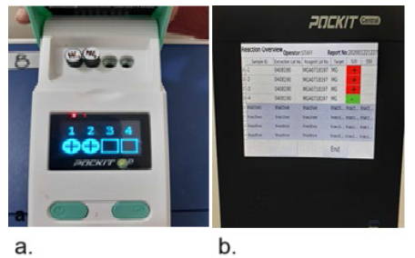
For validation of iiPCR, POCKIT Central Nucleic Acid Analyzer and POCKIT Micro Plus Nucleic Acid Analyzer (both with automatic nucleic acid extraction procedures) were appraised for the detection of M. gallisepticum. The analytical performance criteria of the assay comprising analytical sensitivity (ASe) and analytical specificity (ASp) were estimated. Diagnostic performance of assay was evaluated by comparison with M. gallisepticum qPCR using clinical samples.
Insulated Isothermal PCR (iiPCR) using Micro Plus Nucleic Acid Analyzer (Field deployable POCKIT TM device)
Test samples were transferred to the lysis buffer vial. Vials with lysis buffer and sample were capped and mixed by vigorous shaking for 30 times. Drop-n-Go cassette provided with the kit was unscrewed to load the sample. A single sample was loaded to each cassette. 60 µl of sample (pre-treated with lysis buffer) was transferred to the sample well with the help of a V-dropper. Fibre disc was gently pressed, and the sample was incubated for 5-10 secs. Two drops of wash buffer were added to the sample well and incubated for 10 mins to air dry the fibre disc. The fibre disc was transferred from sample well to elution buffer vial and mixed 30 times. Nucleic acid extract of sample was used to reconstitute the premix tube. The constituted premix was transferred to the respective R-tube, which was then was capped tightly. R-tubes for each sample were placed in the POCKITTM device and the “RUN” button was pressed. The Micro Plus Nucleic Acid Analyzer was run on a default program set by the manufacturer, with a total run time of 45 min.
Insulated Isothermal PCR (iiPCR) using POCKIT Central Nucleic Acid Analyzer
For each sample, 1 transfer cartridge and 1 extraction cartridge were prepared. The transfer cartridge was labelled with the sample ID, and premix vial was added to the well no. 3 of the respective cartridge. The extraction cartridge was labelled with sample ID, and 200µl sample was added to the first well of cartridge. Both cartridges were loaded to the POCKIT Central Nucleic Acid Analyzer, and the program was initiated. The sequence information of the iiPCR primers and probes and the components of the assay were proprietary to GeneReach Taiwan.
Analytical performance of iiPCR
Analytical Specificity (ASp) of iiPCR using POCKIT Central Nucleic Acid Analyzer and POCKIT Micro Plus Nucleic Acid Analyzer was assessed by testing live vaccine of M. gallisepticum F strain as reference, laboratory isolates Pak MG1 (ARL-1963), Pak MG2 (ARL-2020), and Pak MG3 (ARL-2668), along with reference antigens of other poultry pathogens M. synoviae (MS), New Castle disease virus (NDV), Avian influenza virus (AIV H9N2) and Salmonella pullorum (SP). Thea Analytical Sensitivity (ASe) and limit of detection of iiPCR by Micro Plus Nucleic Acid Analyzer and POCKIT Central Nucleic Acid Analyzer were assessed by using a range of 10-fold diluted concentrations in M. gallisepticum broth (100-107 CFU/ml) of M. gallisepticum F (live vaccine), Pak MG1 (ARL-1963), Pak MG2 (ARL-2020), and Pak MG3 (ARL-2668). Each sample/dilution was tested by iiPCR and qPCR, as well as con-PCR in triplicates.
Diagnostic performance of iiPCR
The diagnostic performance of iiPCR to detect M. gallisepticum in clinical samples was determined by comparing it with qPCR as reference. In total, 95 clinical samples, randomly selected from con-PCR positive and negative samples, were used in the study. Test samples were used for the detection of M. gallisepticum by both qPCR and iiPCR (POCKITTM Micro Plus Nucleic Acid Analyzer and POCKIT Central Nucleic Acid Analyzer) by following the manufacturer’s protocol.
Statistical Analysis
Percentage agreement between different PCR-based techniques was calculated, and statistical analysis was performed to assess reliability using Cohen’s kappa (Carossino et al., 2016).
RESULTS
Quantitative PCR of M. gallisepticum F strain and three field isolates - i.e. Pak MG1 (ARL-1963), Pak MG2 (ARL-2020), and Pak MG3 (ARL-2668) - was carried out (Table 1).
Among the 95 clinical samples subjected to qPCR, 70 were already declared positive and 25 were declared negative by con-PCR. However, qPCR revealed 72 positive and 23 negative samples with Ct ranging from 18-35. Samples showing Ct values greater than 35 were considered negative, which resulted in 23 negative samples.
Analytical sensitivity (Ase) of iiPCR for detection of M. gallisepticum
Analytical sensitivity (Ase) or limit of detection of iiPCR was established by using concentrations of M. gallisepticum F strain, Pak MG1 (ARL-1963), Pak MG2 (ARL-2020), and Pak MG3 (ARL-2668) corresponding to CFU ranging from 1×10⁷/ml to 100/ml. Minimum concentration giving 100% positive results by POCKIT Central Nucleic Acid Analyzer and POCKIT Micro Plus Nucleic Acid Analyzer was found to be 1x10³ CFU/ml as represented in Figure 1. Lack of uniformity in test results was found at 1x10² CFU/ml, with a similar trend being recorded by qPCR (Table 1). Comparison of conventional PCR (con-PCR) using 16S rRNA primers (OIE, 2019) and iiPCR in terms of sensitivity revealed that con-PCR achieved 100% detection with pure culture and at 10-1, 10-2, and 10-3 dilutions. At the 10-4 dilution, detection dropped to 33.3-66.7%, and no detection was observed at 10-5 (Table 2).
Contingency table for the comparison of qPCR and conventional PCR assay for the detection of Mycoplasma gallisepticum

Analytical specificity (Asp) of iiPCR for detection of M. gallisepticum
iiPCR was found to be very specific for detection of M. gallisepticum, as no cross reactivity was recorded with other respiratory pathogens on the POCKIT Central Nucleic Acid Analyzer and POCKIT Micro Plus Nucleic Acid Analyzer. Results were negative for M. synoviae (MS), New Castle disease virus (NDV), Avian influenza virus (AIV H9N2) and Salmonella pullorum (SP), which were used as negative control. On the other hand, M. gallisepticum F strain, Pak MG1 (ARL-1963), Pak MG2 (ARL-2020), and Pak MG3 (ARL-2668) were positive for M. gallisepticum iiPCR according to both analyzers. Analytical specificity of iiPCR using both analyzers was found to be 100%.
Diagnostic performance of iiPCR for detection of M. gallisepticum
A panel of clinical samples containing positive and negative samples already tested by con-PCR and by qPCR was used to assess the diagnostic performance of iiPCR using the POCKIT Central Nucleic Acid Analyzer and POCKIT Micro Plus Nucleic Acid Analyzer to detect M. gallisepticum. For this purpose, DNA extraction of the designated samples was carried out and qPCR was performed. Out of total 95 tested samples by qPCR, 72 were positive, with Ct ranging from 18-35, and 23 were negative. Results of iiPCR using the POCKIT Central Nucleic Acid Analyzer showed 71 positive and 22 negative sample, with one false positive and one false negative sample (Table 3). The same samples tested by iiPCR Micro Plus Nucleic Acid Analyzer showed 70 positive and 21 negative samples for M. gallisepticum, with one false positive and 02 false negative results (Table 4). Statistical analysis revealed almost perfect reliability of both techniques for the detection of M. gallisepticum from clinical samples with percentage agreement of 95% -97% (Table 5).
Contingency table for the comparison of qPCR and iiPCR using POCKIT Central Nucleic Acid Analyzer for the detection of Mycoplasma gallisepticum
Contingency table for the comparison of qPCR and iiPCR using Micro Plus Nucleic Acid Analyzer for detection of Mycoplasma gallisepticum
Comparison of reliability of different PCR-based diagnostic techniques for the detection of Mycoplasma gallisepticum
DISCUSSION
Diagnostic assays are validated based on analytical and diagnostic performance after completing their development, optimization and standardization. Modern day isothermal diagnostic techniques including insulated isothermal polymerase chain reaction PCR (iiPCR) has been demonstrated to be an useful tool for the detection of pathogens. In the present study, iiPCR was assessed for its analytical and diagnostic sensitivity, as well as its specificity for testing for the presence of M. gallisepticum causing respiratory infections in birds. Due to production losses and therapeutic burdens, early detection of infectious agents is of the foremost importance to curb transmission across flocks and farms.
PCR has been used for molecular detection and typing of infectious agents in many forms, including random amplification polymorphic DNA (RAPD) analysis or arbitrarily primed-PCR (AP-PCR). For routine diagnosis of M. gallisepticum, con-PCR has been the common practice (García et al., 2005; Fraga et al, 2013; Emam et al., 2024). iiPCR was developed based on the principle of convective heat transfer. Heat convection is a phenomenon of heat transfer based on motion of fluid. After initial development of iiPCR based on Rayleigh-Benard convection by Krishnan et al. (2002), the technique has undergone several modifications towards user-friendliness for the last couple of decades (Qiu et al., 2019; Song et al., 2022). In its present form, it is now available as an all-in-one version with minimum run time of 45 mins for 4 to 8 samples as a field portable device, i.e. Micro Plus Nucleic Acid Analyzer and the laboratory diagnostic device POCKIT Central Nucleic Acid Analyzer.
In the current study, M. gallisepticum-specific iiPCR assay exhibited first-rate analytical sensitivity and specificity. The analytical specificity of the assay was found to be 100%, with no cross reactions observed with the reference antigens of various pathogens. Analytical sensitivity and limit of detection of M. gallisepticum iiPCR was found to be 1×10³ CFU/ml, which is comparable to M. gallisepticum qPCR and far better than the con-PCR assay conducted for routine diagnosis.
To assess reliability of iiPCR for intended results, a comparative study was conducted with qPCR as a standard molecular method for the detection of M. gallisepticum. Using qPCR as the reference standard technique, the diagnostic performance of con-PCR and iiPCR was evaluated and compared. Comparison of reliability of different PCR based diagnostic techniques revealed 97.89% (Cohen’s Kappa value, 0.94) agreement between qPCR and con-PCR, as well as between qPCR and iiPCR POCKIT Central Analyser. qPCR and POCKIT Micro Plus Nucleic Acid Analyser had 95.7% (Cohen’s Kappa value, 0.88) agreement, in consistence with previous reports (Carossino et al., 2016). Statistical analysis to assess reliability of iiPCR in comparison to qPCR showed the compatibility of both techniques for use in the diagnosis of M. gallisepticum. The samples showing false positive results were processed tissue samples, and since iiPCR assay is based on direct detection from sample, tissue exudates can have been the hinderance for appropriate detection. The current study is consistent with the findings of Kuo et al. (2017), who reported 97.8% agreement of iiPCR with real time PCR for detection of M. synoviae infection in poultry by using the filed deployable POCKITTM device for timely detection in a suspected farm. In iiPCR, steady circulation of energy by fluid density gradient leads to sequential development of the denaturation, annealing and extension temperatures carried out in conventional process. The whole process has been described by Chou et al., (2011) in developing capillary convective PCR (CCPCR). Natural convection drives reagents in capillary to circulate through different temperature zones, corresponding to the PCR cycle, leading to the amplification of the target DNA. To overcome the influence of environmental temperature variations, a thermally baffled device has been developed by Chang et al., (2012). The insulated isothermal device (iiPCR) was further used by Tsai et al (2012) to successfully demonstrate the diagnosis of white spot syndrome virus (WSSV). Analysis of amplified products was carried out by agarose gel electrophoresis. Post-amplification processing was then replaced by real time detection, using fluorescent dyes and optical detection system (Tsen et al., 2013).
Insulated isothermal PCR has found wide application in prompt detection of several bacterial and viral pathogens for effective and swift control of infections. The assay was successfully used for detection of Salmonella from chicken meat samples employing TaqMan probes and PCR primers targeting yrfH gene (Tsen et al., 2013). A field deployable device, POCKITTM Micro Plus Nucleic Acid Analyzer, has been used for detection of Canine distemper virus (CDV) (Wilkes et al., 2014), Equine influenza Virus (EIV) H3N8 (Balasuriya et al., 2014), equine arteritis virus (having an agreement limit of more than 90% with the standard assays) (Carossino et al., 2016), Foot-and-mouth disease (FMD) and Bluetongue virus in ruminants (Ambagala et al., 2017a Ambagala et al., 2017b), classical swine fever virus (CSFV) (Lung et al., 2016), M. synoviae (MS) (Kuo et al., 2017), malaria (Chua et al., 2016), rotavirus (Soltan et al., 2016), Feline leukaemia virus (FeLV) (Wilkes et al., 2018), Dengue virus (DENV) infection and its serotypes (Go et al., 2016; Wang and Gubler 2018; Tsai et al., 2019), Seneca Valley virus (SVV) (Zhang et al., 2019), and Staphylococcus aureus from food samples (Yin et al., 2019). The current study further demonstrated the utility of iiPCR for rapid detection of M. gallisepticum for the first time, thus paving the way for its integration into national surveillance frameworks to control mycoplasma infections in avian populations.
CONCLUSION
Insulated isothermal PCR (iiPCR) using Micro Plus Nucleic Acid Analyzer and POCKIT Central Nucleic Acid Analyzer showed similar results and both were good alternatives to qPCR for the detection of M. gallisepticum. The result showed a high sensitivity of 90.9% and 100% of Micro Plus Nucleic Acid Analyzer and POCKIT Central Nucleic Acid Analyzer when considering qPCR as the gold standard. The detection limit of iiPCR was found to be comparable with qPCR. No non-specific detections were observed.
ACKNOWLEDGEMENTS
We would like to acknowledge Hina Fatimah (Late), Allama Iqbal Open University, who offered unwavering support during this project. The National Reference Lab for Poultry Diseases (NRLPD) has facilitated for the project.
REFERENCES
-
Ambagala A, Fisher M, Goolia M, et al. Field-deployable reverse transcription-insulated isothermal PCR (RT-II PCR) assay for rapid and sensitive detection of foot-and-mouth disease virus. Transboundary and Emerging Diseases 2017a;64(5):1610-23. https://doi.org/10.1111/tbed.12554
» https://doi.org/10.1111/tbed.12554 -
Ambagala A, Pahari S, Fisher M, Lee, et al. A rapid field-deployable reverse transcription-insulated isothermal polymerase chain reaction assay for sensitive and specific detection of bluetongue virus. Transboundary and Emerging Diseases 2017 b;64(2):476-86. https://doi.org/10.1111/tbed.12388
» https://doi.org/10.1111/tbed.12388 - Asif M, Khalid S, Sarfaraz I, et al. Comparative efficacy of rapid serum agglutination and haeagglutination inhibition tests in the seroprevalence study of mycoplasma gallisepticum in poultry birds. Science International 2015;27(5):4317-9.
-
Balasuriya UB, Lee PYA, Tiwari A, et al. Rapid detection of equine influenza virus H3N8 subtype by insulated isothermal RT-PCR (iiRT-PCR) assay using the POCKIT(tm) nucleic acid analyzer. Journal of Virological Methods 2014;207:66-72. https://doi.org/10.1016/j.jviromet.2014.06.016
» https://doi.org/10.1016/j.jviromet.2014.06.016 -
Basit SI, Mamun MA, Rahman M, et al. Isolation and molecular detection of Mycoplasma gallisepticum in commercial layer chickens in Sylhet, Bangladesh. World's Veterinary Journal 2021;11(4):614-20. https://doi.org/10.54203/scil.2021.wvj78
» https://doi.org/10.54203/scil.2021.wvj78 -
Bradbury JM. Poultry mycoplasmas:sophisticated pathogens in simple guise. British Poultry Science 2005;46(2):125-36. https://doi.org/10.1080/00071660500066282
» https://doi.org/10.1080/00071660500066282 -
Carossino M, Wagner B, Loynachan AT, et al. Equine arteritis virus elicits a mucosal antibody response in the reproductive tract of persistently infected stallions. Clinical Vaccine and Immunology 2017;24(10):e00215-17. https://doi.org/10.1128/CVI.00215-17
» https://doi.org/10.1128/CVI.00215-17 -
Chang HFG, Tsai YL, Tsai CF, et al. A thermally baffled device for highly stabilized convective PCR. Journal of Biotechnology 2012;7(5):662-6. https://doi.org/10.1002/biot.201100453
» https://doi.org/10.1002/biot.201100453 -
Chou WP, Chen, PH, Miao JM, et al. Rapid DNA amplification in a capillary tube by natural convection with a single isothermal heater. Biotechniques 2011;50(1):52-7. https://doi.org/10.2144/000113589
» https://doi.org/10.2144/000113589 -
Chua KH, Lee PC, Chai HC. Development of insulated isothermal PCR for rapid on-site malaria detection. Malaria Journal 2016;15(1):134. https://doi.org/10.1186/s12936-016-1183-z
» https://doi.org/10.1186/s12936-016-1183-z -
Emam M, Hashem YM, Ismael E, et al. Comparative study of various diagnostic methods for detection of Mycoplasma gallisepticum and Mycoplasma synoviae in Egyptian chicken flocks. Journal of World Poultry Research 2024;14(2):255-63. https://doi.org/10.36380/jwpr.2024.26
» https://doi.org/10.36380/jwpr.2024.26 - Farooq S, Biological characterization and comparative diagnostics of Mycoplasma gallisepticum Isolates [dissertation]. Islamabad (PAK): Quaid-i-Azam University; 2021. Available from: prr.hec.gov.pk/jspui/bitstream/123456789/17400/1/Saba Farooq Microbiology 2021 qau isb.pdf
-
Farooq S, Naeem K, Akbar J, et al. Development of serum plate agglutination antigen using mycoplasma gallisepticum field isolate. Journal of Agriculture and Veterinary Science 2024;3(3):439-45. https://doi.org/10.55627/agrivet.003.03.0875
» https://doi.org/10.55627/agrivet.003.03.0875 -
Farooq S, Shah AA, Siddique N, et al. Biological characterization of locally circulating Mycoplasma gallisepticum in poultry. Pakistan Veterinary Journal 2021;41(1):112-6. https://doi.org/10.29261/pakvetj/2020.079
» https://doi.org/10.29261/pakvetj/2020.079 -
Fraga AP, de Vargas T, Ikuta N, et al. A Multiplex real-time PCR for detection of Mycoplasma gallisepticum and Mycoplasma synoviae in clinical samples from Brazilian commercial poultry flocks. Brazilian Journal Microbiology 2013;44(2):505-10. https://doi.org/10.1590/S1517-83822013000200028
» https://doi.org/10.1590/S1517-83822013000200028 -
García M, Ikuta N, Levisohn S, et al. Evaluation and comparison of various PCR methods for detection of Mycoplasma gallisepticum infection in chickens. Avian Diseases. 2005;49(1):125-32. https://doi.org/10.1637/7261-0812204R1
» https://doi.org/10.1637/7261-0812204R1 -
Gaunson, JE, Philip, CJ, Whithear, KG, et al. The cellular immune response in the tracheal mucosa to Mycoplasma gallisepticum in vaccinated and unvaccinated chickens in the acute and chronic stages of disease. Vaccine 2006;24(14):2627-33. https://doi.org/10.1016/j.vaccine.2005.12.008
» https://doi.org/10.1016/j.vaccine.2005.12.008 -
Go YY, Rajapakse RJ, Kularatne SA, et al. A pan-dengue virus reverse transcription-insulated isothermal PCR assay intended for point-of-need diagnosis of dengue virus infection by use of the POCKIT nucleic acid analyzer. Journal of Clinical Microbiology 2016;54(6):1528-35. https://doi.org/10.1128/JCM.00225-16
» https://doi.org/10.1128/JCM.00225-16 -
Halium MMA, Salib FA, Marouf SA, et al. Isolation and molecular characterization of Mycoplasma spp. in sheep and goats in Egypt. Veterinary World 2019;12(5):664-70. https://doi.org/10.14202/vetworld.2019.664-670
» https://doi.org/10.14202/vetworld.2019.664-670 - Hanif A, Najeeb MI. Comparison of conventional bacterial isolation, rapid slide agglutination and polymerase chain reaction for detection of Mycoplasma gallisepticum in breeder flocks. Pakistan Journal of Life and Social Science 2007;5(1-2):1-5.
-
Haque ES, Rahman S, Khan M, et al. A simplified duplex real-time PCR incorporating TaqMan minor groove binder (MGB) probes and an exogenous internal positive control for the simultaneous detection of Mycoplasma gallisepticum and Mycoplasma synoviae cultures. The Journal of Veterinary Medicine 2015;60(5):268-73. https://doi.org/10.17221/8179-VETMED
» https://doi.org/10.17221/8179-VETMED -
Idowu PA, Mpofu TJ, Zishiri OT, et al. Molecular detection and genetic characterization of Mycoplasma gallisepticum and Mycoplasma synoviae in selected chicken breeds in South Africa. BMC Infectious Diseases 2024;24(1):562. https://doi.org/10.1186/s12879-024-09437-3
» https://doi.org/10.1186/s12879-024-09437-3 -
Jaturasitha S, Srikanchai T, Kreuzer M, et al. Differences in carcass and meat characteristics between chicken indigenous to northern Thailand (Black-boned and Thai native) and imported extensive breeds (Bresse and Rhode Island red). Poultry Science 2008;87(1):160-9. https://doi.org/10.3382/ps.2006-00398
» https://doi.org/10.3382/ps.2006-00398 -
Kleven SH. Control of avian mycoplasma infections in commercial poultry. Avian Diseases 2008;52(3):367-74. https://doi.org/10.1637/8323-041808
» https://doi.org/10.1637/8323-041808 -
Krishnan M, Ugaz VM, Burns MA. PCR in a Rayleigh-Benard convection cell. Science 2002;298(5594):793-4. https://doi.org/10.1126/science.298.5594.793
» https://doi.org/10.1126/science.298.5594.793 -
Kuo HC, Lo DY, Chen CL, et al. Rapid and sensitive detection of Mycoplasma synoviae by an insulated isothermal polymerase chain reaction-based assay on a field-deployable device. Poultry Science 2017;96(1):35-41. https://doi.org/10.3382/ps/pew228
» https://doi.org/10.3382/ps/pew228 -
Leigh SA, Evans JD.2024.Genome sequence of Mycoplasma gallisepticum strain A5969. Microbiology Resource Announcement 2024. 13:e00813-24.https://doi.org/10.1128/mra.00813-24
» https://doi.org/10.1128/mra.00813-24 - Levisohn S, Kleven, SH. Avian mycoplasmosis (Mycoplasma gallisepticum). Revue scientifique et technique / Office International des Épizooties 2000;19(2):425-42
- Ley DH. Mycoplasma gallisepticum infection. In: Saif YM, editor. Diseases of poultry. 12th ed. Ames: Blackwell Publishing; 2008. p.807-12.
-
Liu Y, Wang Y, Zheng, SJ. Immune evasion of Mycoplasma gallisepticum: an overview. International Journal of Molecular Sciences 2024;25(5):2824. https://doi.org/10.3390/ijms25052824
» https://doi.org/10.3390/ijms25052824 -
Lung O, Pasick J, Fisher M, et al. Insulated isothermal reverse transcriptase PCR (ii RT-PCR) for rapid and sensitive detection of classical swine fever virus. Transboundary and Emerging Diseases 2016;63(5):e395-402. https://doi.org/10.1111/tbed.12318
» https://doi.org/10.1111/tbed.12318 -
Machado LDS, Abreu DLDC, Lemos M, et al. Performance, serologic and tracheal responses of laying hens exposed to F strain of Mycoplasma gallisepticum. Arquivos do Instituto Biológico 2017;84:1-4. https://doi.org/10.1590/1808-1657000052016
» https://doi.org/10.1590/1808-1657000052016 -
Muhammad J, Rabbani M, Sheikh AA, et al. Molecular detection of Mycoplasma gallisepticum in different poultry breeds of Abbottabad and Rawalpindi, Pakistan. Brazilian Journal of Biology 2021;83:e246514. https://doi.org/10.1590/1519-6984.246514
» https://doi.org/10.1590/1519-6984.246514 -
Nascimento ER, Pereira VLA, Nascimento MGF, et al. Avian mycoplasmosis update. Brazilian Journal Poultry Science 2005;7(1):1-9. https://doi.org/10.1590/S1516-635X2005000100001
» https://doi.org/10.1590/S1516-635X2005000100001 - Nascimento ER, Yamamoto R, Herrick KR, et al. Polymerase chain reaction for detection of Mycoplasma gallisepticum. Avian Diseases 1991;35(1):62-9.
-
Papazisi L, Gorton TS, Kutish G, et al. The complete genome sequence of the avian pathogen Mycoplasma gallisepticum strain Rlow. Microbiology 2003;149(9):2307-16. https://doi.org/10.1099/mic.0.26427-0
» https://doi.org/10.1099/mic.0.26427-0 -
Qiu X, Shu JI, Baysal O. et al. Real-time capillary convective PCR based on horizontal thermal convection. Microfluid Nanofluid 2019;23;39 https://doi.org/10.1007/s10404-019-2207-0
» https://doi.org/10.1007/s10404-019-2207-0 - Qoraa AM, Salem HM, Shakal M. Phenotypic and molecular detection of mycoplasma gallisepticum in broiler and layer chickens in some Egyptian governorates. Journal of Advanced Veterinary Research 2023;13(5):799-805. 0000-0002-4453-7884
- Rauf M, Chaudhary ZI, Younus M, et al. Identification of Mycoplasma gallisepticum by polymerase chain reaction and conventional diagnostics from White Leghorn layer flocks. The Journal of Animal and Plant Science 2013;23(2):393-7.
-
Raviv Z, Kleven SH. The development of diagnostic real-time TaqMan PCRs for the four pathogenic avian mycoplasmas. Avian Diseases 2009;53(1):103-7. https://doi.org/10.1637/8469-091508-Reg.1
» https://doi.org/10.1637/8469-091508-Reg.1 -
Razin S, Yogev D, Naot Y. Molecular biology and pathogenicity of mycoplasmas. Microbiology and Molecular Biology Reviews: MMBR 1998;62(4):1094-56. https://doi.org/10.1128/MMBR.62.4.1094-1156.1998
» https://doi.org/10.1128/MMBR.62.4.1094-1156.1998 -
Soltan MA, Tsai YL, Lee PYA, et al. Comparison of electron microscopy, ELISA, real time RT-PCR and insulated isothermal RT-PCR for the detection of Rotavirus group A (RVA) in feces of different animal species. Journal of Virological Methods 2016;235:99-104. https://doi.org/10.1016/j.jviromet.2016.05.006
» https://doi.org/10.1016/j.jviromet.2016.05.006 -
Song R, Liu P, Yang Y, et al. Development of a duplex insulated isothermal pcr assay for rapid on-site detection and differentiation of genotypes 1 and 2 of African Swine Fever Virus. Frontiers in Cellular and Infection Microbiology 2022;12:948771. https://doi.org/10.3389/fcimb.2022.948771
» https://doi.org/10.3389/fcimb.2022.948771 -
Tsai JJ, Liu WL, Lin PC, et al. An RT-PCR panel for rapid serotyping of dengue virus serotypes 1 to 4 in human serum and mosquito on a field-deployable PCR system. PloS One 2019;14(3):e0214328. https://doi.org/10.1371/journal.pone.0214328 eCollection 2019
» https://doi.org/10.1371/journal.pone.0214328 -
Tsai YL, Wang HTT, Chang, HFG, et al. Development of TaqMan probe-based insulated isothermal PCR (iiPCR) for sensitive and specific on-site pathogen detection. PloS One 2012;7(9):e45278. https://doi.org/10.1371/journal.pone.0045278
» https://doi.org/10.1371/journal.pone.0045278 -
Tsen HY, Shih CM, Teng PH, et al. Detection of Salmonella in chicken meat by insulated isothermal PCR. Journal of Food Protection 2013;76(8):1322-9. https://doi.org/10.4315/0362-028X.JFP-12-553
» https://doi.org/10.4315/0362-028X.JFP-12-553 -
Umar S, Munir MT, Ur-Rehman Z, et al. Mycoplasmosis in poultry:update on diagnosis and preventive measures. Worlds Poultry Science Journal 2017;73(1):12-7. https://doi.org/10.1017/S0043933916000830
» https://doi.org/10.1017/S0043933916000830 -
Wang WK, Gubler DJ. Potential point-of-care testing for dengue virus in the field. Journal of Clinical Microbiology 2018;56(5):e203. https://doi.org/10.1128/JCM.00203-18
» https://doi.org/10.1128/JCM.00203-18 -
Wilkes RP, Anis E, Dunbar D, et al. Rapid and sensitive insulated isothermal PCR for point-of-need feline leukaemia virus detection. Journal of Feline Medicine and Surgery 2018;20(4):362-9. https://doi.org/10.1177/1098612X17712847
» https://doi.org/10.1177/1098612X17712847 -
Wilkes RP, Tsai YL, Lee PY, et al. Rapid and sensitive detection of canine distemper virus by one-tube reverse transcription-insulated isothermal polymerase chain reaction. BMC Veterinary Research 2014;10(1):213. https://doi.org/10.1186/s12917-014-0213-8
» https://doi.org/10.1186/s12917-014-0213-8 - World Organization for Animal Health. Avian mycoplasmosis (Mycoplasma gallisepticum, M. synoviae). In: WOAH. Manual of diagnostic tests and vaccines for terrestrial animals. Paris: OIE; 2019. v.2, p.844-59
-
Yehia N, Salem HM, Mahmmod Y, et al. Common viral and bacterial avian respiratory infections:an updated review. Poultry Science 2023;102(5):102553. https://doi.org/10.1016/j.psj.2023.102553
» https://doi.org/10.1016/j.psj.2023.102553 -
Yin YH, Lin YY, Lin C H, et al. Rapid and sensitive detection of Staphylococcus aureus in processed foods using a field-deployed device to perform an insulated isothermal polymerase chain reaction-based assay. Journal of Food Safety 2019;39(6):e12690. https://doi.org/10.1111/jfs.12690
» https://doi.org/10.1111/jfs.12690 -
Zhang J, Nfon C, Tsai CF, et al. Development and evaluation of a real-time RT-PCR and a field-deployable RT-insulated isothermal PCR for the detection of Seneca Valley virus. BMC Veterinary Research 2019;15(1):1-11. https://doi.org/10.1186/s12917-019-1927-4
» https://doi.org/10.1186/s12917-019-1927-4
-
FUNDING
No funding was received for the research.
-
DATA AVAILABILITY STATEMENT
All relevant data is available in manuscript tables.
-
DISCLAIMER/PUBLISHER’S NOTE
The published papers’ statements, opinions, and data are those of the individual author(s) and contributor(s). The editor(s) disclaim responsibility for any injury to people or property resulting from any ideas, methods, instructions, or products referred to in the content.
All relevant data is available in manuscript tables.

 b) POCKIT Central Nucleic acid Analyzer