 CHITOSAN/GELATIN AND Jatropha mollissima OVULES FOR THE TREATMENT OF FUNGAL VAGINITIS
Abstract
Fungal vaginitis is an inflammatory condition of the vaginal mucosa caused by fungal proliferation, characterized by symptoms such as pruritus, dysuria, and dyspareunia. Despite antifungal agents are widely used orally and topically, women report adverse effects including burning, hypersensitivity and irritation. In this context, this study proposes an alternative therapeutic approach using herbal medicine Jatropha mollissima, which exhibits pharmacological potential, ovules composed of chitosan, gelatin, and Jatropha mollissima ethanolic extract were developed, aiming to enhance antifungal treatment efficacy while minimizing side effects. Ovules were fabricated via lyophilization, using three different gelatin concentrations in a chitosan matrix. After determining the optimal concentration, ovules were formulated with 2% (w/v) chitosan in 1% (v/v) acetic acid, 15% (w/w) gelatin, and Jatropha mollissima extract at 1, 3, and 5% (w/w). Samples were characterized by optical microscopy, scanning electron microscopy, Fourier transform infrared spectroscopy, swelling degree, in vitro biodegradation, antimicrobial and antifungal activities, and cytotoxicity. The results confirmed porous structures, extract incorporation, complete degradation within 28 days, antimicrobial activity against E. coli and S. aureus, antifungal activity against C. albicans, and absence of cytotoxicity in L929 cells. This approach represents a significant advancement in the treatment of fungal vaginitis, offering therapeutic benefits with reduced side effects.
Keywords:
phytotherapy; ovules; vaginal drug delivery; Jatropha mollissima.
INTRODUCTION
Fungal vaginitis is an infectious and/or inflammatory process of the female lower genitourinary tract that develops due to infection by yeasts that inhabit the vaginal mucosa, with Candida albicans being the most common etiological agent. Pruritus, leucorrhea, whitish plaques, edema, and erythema in the vulva and vagina have been the most common clinical manifestations of this type of infection. The symptoms of fungal vaginitis are not pathognomonic, and clinical suspicion can be confirmed by tests that reveal the nature of the etiological agent.1
Candidiasis is an infection caused by the fungus Candida albicans in the genital region. Treatment involves oral and vaginal antifungal agents, including 2% fenticonazole nitrate cream, an imidazole derivative with broad-spectrum antifungal activity. However, many women report burning and skin irritation at the application site, hypersensitivity, itching, scaling, eczema, and erythema.2 These limitations highlight the need for new therapeutic strategies that reduce side effects while maintaining or enhancing antifungal efficacy.
Encapsulation of medicinal plant extracts in biodegradable carriers has emerged as an effective approach to modulate pharmacokinetics, enabling targeted and sustained release while minimizing adverse reactions. Among the materials studied for this purpose, gelatin, chitosan, and their derivatives stand out due to their biocompatibility and biodegradability.3,4 Chitosan is a non-toxic polysaccharide widely used as a drug delivery system for antibiotics, anti-inflammatories, antifungals, and other therapeutic agents.5,6 Gelatin, a natural polymer derived from collagen, provides structural support and mechanical stability, making it suitable for biomedical applications.7
Several biodegradable polymers have been explored for vaginal drug delivery systems, including alginate, hydroxypropyl methylcellulose (HPMC), polyvinyl alcohol (PVA), and various plant-derived gums, which have been used to formulate mucoadhesive gels, films, microspheres, and ovules for antifungal therapy.8 These systems aim to prolong drug residence time in the vaginal mucosa, improve local bioavailability, and reduce systemic exposure.9 Additionally, chitosan and gelatin themselves have been widely investigated in other biomedical applications, such as wound dressings, hemostatic materials, scaffolds for tissue engineering, and controlled drug-release platforms, owing to their biocompatibility, biodegradability, and bioadhesive characteristics.10 Previous studies11 have reported the antifungal effectiveness of chitosan-based formulations against Candida species and the capacity of gelatin to enhance structural stability and modulate degradation rates in composite matrices. Together, these findings reinforce the relevance of combining these polymers to develop vaginal ovules and highlight the potential advantages of incorporating plant extracts into such systems.12,13
Incorporating Jatropha mollissima extract into chitosan/gelatin ovules offers the possibility of combining the plant’s antifungal potential with the bioadhesive and healing properties of chitosan. This strategy may not only eliminate the fungal agent responsible for vaginitis but also enhance tissue recovery.14,15 The extract of Jatropha mollissima contains phenolic compounds, flavonoids, tannins, diterpenes, alkaloids, and lignans, which are associated with antimicrobial, anti-inflammatory, and antioxidant activities, supporting its use in vaginal infections.15-18
Jatropha mollissima is widely used in traditional medicine in Northeastern Brazil and has demonstrated antifungal activity against Candida spp., which reinforces its potential for incorporation into vaginal therapeutic formulations. Combining this medicinal extract with biodegradable ovules represents an innovative strategy that unites phytotherapy with controlled-release drug delivery systems. Additionally, the use of natural compounds such as Jatropha mollissima may reduce the risk of antifungal resistance, a growing concern in current clinical practice.19
Given this context, the present study aimed to develop chitosan/gelatin ovules loaded with Jatropha mollissima extract for the treatment of fungal vaginitis.
EXPERIMENTAL
Materials
The chitosan used had a molecular weight of approximately 270 kDa and a degree of deacetylation (DD) of 85-90%, as provided by the manufacturer. Chitosan with genetic heritage (Sisgen Registration AEBD12D) and the sap of Jatropha mollissima (Sisgen Registration AD5B98B) were produced and supplied by CERTBIO (Laboratory for Evaluation and Development of Biomaterials of the Northeast). Gelatin type A, phosphate buffer (pH 7.2; P3288-1VL), and lysozyme were purchased from Sigma-Aldrich. Glacial acetic acid (P.A.) and sodium hydroxide were obtained from Vetec. Distilled water and 0.9% saline solution were used in all experiments.
Methods
Preparation of chitosan and chitosan/gelatin solutions
A 2% (w/v) chitosan solution was prepared by dissolving the polymer powder in 1% (v/v) acetic acid under constant mechanical stirring (200 rpm) at 25 °C for 2 h. Chitosan/gelatin solutions were then prepared by incorporating gelatin at 5, 10, and 15% (w/w) relative to chitosan, under heating at 40 °C and stirring for 30 min until complete solubilization.
Preparation of the ethanolic extract of Jatropha mollissima (EEJM)
The ethanolic extract was obtained from 1 L of plant sap using a 1:1 (v/v) sap-to-solvent ratio. The mixture was protected from light and macerated at room temperature for 72 h. After extraction, the suspension was vacuum filtered, and the filtrate was concentrated in a SL-126 rotary evaporator at 50 °C until complete solvent removal. The concentrate was frozen at -80 °C for 48 h and lyophilized for 72 h to obtain the powdered extract.
Preparation of chitosan/gelatin/EEJM ovules
After defining 15% gelatin as the optimal concentration, EEJM was incorporated into the chitosan/gelatin solution at 1, 3, and 5% (w/w). The mixture was stirred at 200 rpm for 1 h at 24 °C. Then, 2 mL aliquots were dispensed into silicone egg-shaped molds and frozen at -80 °C for 48 h, followed by lyophilization for 72 h.
The dried ovules were neutralized in 1 mol L-1 NaOH for 10 min, rinsed repeatedly with distilled water until neutral pH (ca. 7.4), refrozen at -80 °C for 48 h, and lyophilized again for 72 h. During the neutralization step with 1 mol L-1 NaOH, a slight partial solubilization of gelatin was observed, which is inherent to its sensitivity to alkaline environments. This effect was minimized by limiting the exposure time to 10 min and performing immediate washing with distilled water. The samples were subsequently refrozen and lyophilized to restore structural integrity. Samples were coded according to Table 1.
Characterizations
Physical-chemical analysis
Morphological analysis was performed using a Hirox KH-1300 microscope at 350× magnification. Samples were positioned on glass slides, and micrographs were captured in reflected light mode to observe surface features and cavity distribution.
Surface morphology, pore size, and pore distribution were evaluated using a Shimadzu SuperScan SS500 Scanning Electron Microscope (SEM). Samples were mounted on aluminum stubs with carbon tape and gold-sputter-coated with a thin gold layer (ca. 10 nm) prior to analysis. Imaging was performed at 500× magnification under an acceleration voltage of 10-15 kV.
Fourier transform infrared spectroscopy (FTIR) spectra were obtained using a PerkinElmer Spectrum 400 in the 4000 650 cm-1 range, with a resolution of 4 cm-1 and 32 scans per sample. Lyophilized powdered samples were analyzed using an attenuated total reflectance (ATR) accessory equipped with a diamond crystal, enabling the identification of characteristic functional groups of chitosan, gelatin, and EEJM and the detection of possible intermolecular interactions.
The swelling capacity of the systems was evaluated by measuring their ability to absorb liquid and expand. Samples were weighed (Wi) and immersed separately in physiological saline solution (0.9% NaCl) and lactic acid solution (pH 4.5). Immersion was conducted at 37 °C for 1 h, a period previously established in preliminary tests as sufficient for the samples to reach swelling equilibrium without compromising their structural integrity. After immersion, the samples were surface-dried with absorbent paper and weighed again (Wf). The swelling degree (SD) was calculated as:
Biodegradation was assessed over 28 days following ASTM F1635-11.20 Samples were incubated at 37 °C in phosphate buffer solution (pH 7.2), distilled water, lactic acid solution (pH 4.5), and phosphate buffer with lysozyme (1.5 µg mL-1) to simulate enzymatic degradation. Mass loss was determined at 7, 14, 21, and 28 days using:
where W0 is initial mass and Wt is mass at time t.
Each measurement was performed in quintuplicate.
Biological assays
Antimicrobial and antifungal activity was evaluated using the disc diffusion method on Mueller-Hinton agar inoculated with Escherichia coli, Staphylococcus aureus, and Candida albicans. Ovule fragments (6 mm diameter) were placed on each plate and incubated at 37 °C for 24-72 h. Inhibition zones were measured at 24 h intervals. Tests were performed in triplicate.
The cytotoxic activity of the samples was evaluated using the agar diffusion method according to ISO 10993-5:2009.21 L929 mouse fibroblast cells (Banco de Células do Rio de Janeiro, BCRJ) were cultured in RPMI (Roswell Park Memorial Institute) 1640 medium until reaching maximum confluence. Cells were then trypsinized with 0.25% trypsin/EDTA (ethylenediaminetetraacetic acid) and adjusted to a density of 1.0-3.0 × 10⁵ cells mL-1. Aliquots of 4 mL were dispensed into 6-well plates and incubated for 48 h at 37 °C and 5% CO2, forming monolayers with > 80% confluence.
A solidifying overlay medium was prepared consisting of 1.8% agar containing 0.01% neutral red dye, mixed 1:1 with Eagle’s minimum essential medium (MEM, 2×). After complete homogenization, 1 mL of the agar-medium mixture was added to each well. Plates were allowed to rest for 10 min at room temperature to ensure solidification.
Test samples (1 × 1 cm2 squares), prepared in duplicate, were placed at the center of each well. Positive controls consisted of latex sheets, and negative controls consisted of non-toxic filter paper, both also tested in duplicate. Plates were incubated for 24 h at 37 °C with 5% CO2.
Following incubation, qualitative cytotoxicity was assessed by observing the presence of decolorization zones around the samples under a Nikon Eclipse TS100 inverted microscope. The diameter of the discoloration halos was measured visually, and the degree of cytotoxicity was classified according to ISO 10993-521 criteria: Grade 0, non-cytotoxic; Grade 1, slight; Grade 2, moderate; Grade 3, severe cytotoxicity. Cytotoxicity analyses were conducted at the CERTBIO, accredited according to ABNT ISO/IEC 17025:2005 (CRL 0799)22 for Chemical and Biological Testing. All procedures followed validated protocols and certified good laboratory practices.
RESULTS AND DISCUSSION
Characterization and physicochemical analysis of eggs without Jatropha mollissima
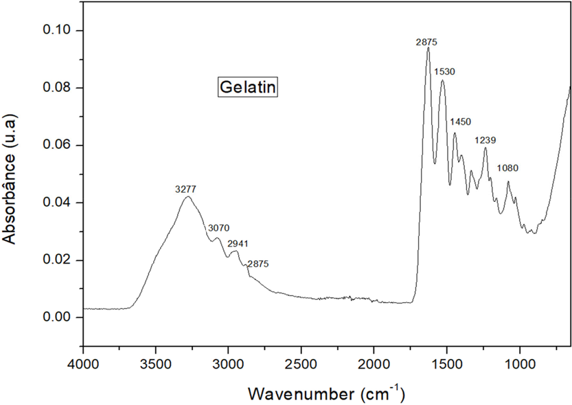
Figure 1 illustrates the FTIR spectrum obtained for the gelatin sample, and Table 2 provides the corresponding main absorption bands identified.

In this spectrum, the band at 3277 cm-1 is attributed to the stretching vibration of the OH groups, at 2941 cm-1 referring to the asymmetric stretching vibration CH2, at 1627 cm-1 the stretching vibration of the C=O group is noted, while at 1530 cm-1 the bending vibration NH and at 1450 cm-1 the symmetric stretching of -COOH groups are noted, confirming the protein molecule.23Figure 2 shows the FTIR spectra of the chitosan samples without and with different concentrations of gelatin and Table 3 provides the corresponding main absorption bands identified.
Main absorption bands (FTIR) of chitosan samples with different gelatin concentrations (Q, QG5, QG10, QG15)

In the chitosan (Q) spectrum, the band around 3250 cm-1 corresponds to the axial stretching of OH, superimposed on the NH stretching band. At 2885 cm-1, it is attributed to the asymmetric stretching of the C-H group. At 1646 cm-1, it is associated with the C=O axial deformation of the primary amide, and at 1581 cm-1, it refers to the vibrational deformation of the NH3+ group. The band at 1379 cm-1 can be attributed to the -CN axial deformation of amino groups. The intense band at 1016 cm-1 is associated with the C-O-C stretching of the pyranoside rings. The spectrum shows the absorptions relative to chitosan. For chitosan with different gelatin concentrations, no changes were observed.24Figure 3 illustrates the swelling profiles of the samples with different gelatin concentrations.

Degree of swelling of the samples with different gelatin concentrations after 1 h of immersion in phosphate buffer solution (pH 7.2)
It is noteworthy that the increased gelatin percentage contributes to the increased degree of swelling, which is related to the changes observed in the morphology and chemical profile of the material. The gelatin concentration influences the growth of ice crystals during freezing, the initial stage of lyophilization. Water molecules are separated by macromolecules, preventing them from concentrating and arranging themselves, altering the porosity of the structure. The swelling test measures the ability of the biomaterial to absorb liquids and expand without compromising its structure, influenced by the chitosan composition, which ensures structural stability, and gelatin, which increases its swelling capacity.25,26
Biodegradation of chitosan, a process that generally involves the enzymatic cleavage of glycosidic bonds between glucosamine units, is influenced by its molecular weight and degree of deacetylation. Additionally, factors such as the type of buffer solution, the pH of the medium, and the type and concentration of the enzymes also affect the degradation rate.27
Evaluating the biodegradation results of the samples immersed in phosphate buffer solution and lysozyme/phosphate buffer presented in Table 4 with periods of 7, 14, 21 and 28 days, it was noted in all samples that biodegradation was more pronounced under the action of lysozyme, evidencing the efficiency of this enzyme in the biodegradation of chitosan.
Percentage mass loss of samples (Q, QG5, QG10 and QG15) at intervals of 7, 14, 21 and 28 days subjected to the biodegradation test
Comparing the samples, it is clear that the eggs with the highest percentage of gelatin also showed greater degradation, indicating that the incorporation of gelatin accelerates the degradation of the structure. Regarding the degradation byproducts, the oligomers (N-glucosamine) exhibit bioactivity as probiotics that inhibit the growth of harmful bacteria, strengthening the immune system.28
Based on the results obtained, it was decided to continue working with sample QG15 to incorporate EEJM, because despite similar behavior in the FTIR spectra, in relation to swelling and biodegradation, the values of this sample were higher, indicating that this composition is more suitable for the proposed application. In addition, the combination of high swelling capacity and complete biodegradation within 28 days in physiologically relevant media suggests that this composition presents a favorable profile for use as a vaginal delivery system, supporting its potential application in the local treatment of fungal vaginitis.
Physical-chemical analysis of ovules with Jatropha mollissima
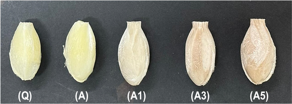
Figure 4 shows the ovules obtained with different compositions and concentrations of EEJM. Sample Q, containing only chitosan, presents a clear and uniform color, indicating that chitosan alone maintains characteristics reflecting an efficient freeze-drying process.

Sample A combines chitosan with gelatin. A slight change in color is observed, which remains clear but slightly less bright, and the texture is still similar to that of sample Q. This is related to the addition of gelatin, which modifies the structure, suggesting that its inclusion does not significantly impact the formation of a homogeneous material during the freeze-drying process.
With the inclusion of EEJM, starting at 1% in sample A1, a change in color is observed, becoming more opaque and brownish, as well as a slightly rough texture, indicating that even at low concentrations, changes in the final structure of the material occur due to the interaction of EEJM with the chitosan/gelatin matrix. As the EEJM concentration increases, as seen in samples A3 at 3% and A5 at 5%, the changes become evident, with the color becoming darker and the material becoming rougher.
Similar studies reported by Gomes et al.,28 also observed color changes to brownish tones in the membranes due to the incorporation of J. mollissima. Figure 5 illustrates optical microscopy images of ovules without and with different concentrations of EEJM. Sample A presents a relatively homogeneous structure with well-defined fibers and a uniform appearance, indicating a cohesive structure.

The EEJM content caused a slight change in the color of the ovules, and insoluble particles could be identified in the structures. All samples presented a porous structure, characteristic of the freeze-drying method.25,26
In sample A1, a slight change in structural uniformity was observed; the fibers began to show darker and slightly irregular regions, where the dark spots may indicate insoluble particles in the matrix. As the EEJM concentration increased in sample A3, the structural changes became more evident, with the fibers becoming less defined and more dark areas visible, indicating greater dispersion in the matrix. In sample A5, the changes demonstrated less distinct fibers and an increase in insoluble particles, suggesting the incorporation of EEJM into the matrix in a dispersed form.
In the SEM analysis (Figure 6) of samples A, A1, A3, and A5, variations in structure were observed compared to control sample A.

Sample A presented a fibrous structure, with overlapping layers and well-defined morphology. The fibers were visibly organized, indicating good polymer matrix formation and a porous structure resulting from the freeze-drying process. In sample A1, the structure maintained characteristics similar to sample A, but there was a slight increase in fiber density and compaction. The layers were still present, but there was a slight reduction in porosity, attributed to the interaction between the EEJM and the matrix components. These changes indicate that even small concentrations of EEJM alter the structure, possibly affecting its mechanical and absorption properties.
In sample A3, an increase in fiber disorganization was noted, with a denser and more compact structure, increased particle size, and fewer areas of visible porosity. In this sense, sample A5 exhibited greater disorganization and compaction compared to the previous samples, with a more pronounced reduction in porosity and the formation of agglomerates. In summary, the addition of EEJM, especially in higher concentrations, tends to compact the structure of the material, reducing its porosity.29,30
These results were corroborated by the study of Souza et al.,5 who also observed that the incorporation of gelatin and Jatropha mollissima into the chitosan matrix leads to a reduction in pore size in the resulting scaffolds.
Partial gelatin solubilization during NaOH neutralization is expected, as gelatin is susceptible to alkaline hydrolysis. However, the controlled exposure time used in this study prevented significant structural loss. The final ovules maintained their morphology and porosity, indicating that this effect did not compromise the final properties of the material.
Figure 7 illustrates the FTIR spectra and Table 5 lists the absorption bands identified in the samples with Jatropha mollissima extract. Sample A without extract has characteristic bands at 3400 3200 cm-1, corresponding to the O-H stretching resulting from the polymeric association of chitosan; at 2870 cm-1, corresponding to the C-H axial deformation of aliphatic compounds; at 1630 and 1580 cm-1, the C=O vibrations of N-substituted amides and symmetrical angular deformation in the NH2 plane; in the 1420 cm-1 region, angular deformation in CH2 adjacent to the carbonyl; and in the 1030 cm-1 region, vibrations are observed related to the C-O-C grouping of the saccharide structure.29
Key FTIR absorption bands identified in samples with Jatropha mollissima extract (A, A1, A3, A5)
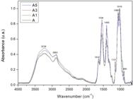
When comparing samples A, A1, A3, and A5, the band around 3300 cm-1 and the bands at 2883 and 2895 cm-1 show intensification with the incorporation of EEJM, being more evident for composition A5, due to its higher concentration. The observed intensifications suggest a possible secondary binding of EEJM to the matrix, indicating its incorporation.2
Figure 8 illustrates the degree of swelling (SD) of samples A, A1, A3 and A5. It was observed that sample A presented an SD of around 1200%, while for the samples incorporated with EEJM, there was a reduction in SD, which was more pronounced in sample A5.

Degree of swelling of samples obtained with and without Jatropha mollissima extract after 1 h of immersion (ác: acid)
Sample A presented a high SD, indicative of a highly porous and hydrophilic structure, suggesting significant liquid absorption capacity, essential for biomaterial applications. In sample A1, a slight decrease in SD was observed compared to sample A. This was related to the incorporation of EEJM, which, when interacting with the chitosan and gelatin matrix, reduced its porosity, making it less permeable to liquid. Nevertheless, the degree of swelling indicated that the addition of a small amount of EEJM does not severely compromise swelling capacity of the structure.
As the EEJM concentration increases, as observed in samples A3 and A5, the reduction in SD becomes more evident, related to the higher concentration and its interference with the structure, possibly due to the pore-blocking effect or the introduction of hydrophobic components, which limit liquid absorption.
Using lactic acid as the immersion medium, the results show a similar pattern, but with slightly higher SD values, resulting from the differences in interactions between the medium and the samples. The differences between the immersion media demonstrate that they have a relevant impact on swelling.31-33
The results of the biodegradation evaluation of samples immersed in phosphate buffer solution, lysozyme/phosphate buffer, and lactic acid are presented in Table 6 for the periods of 7, 14, 21, and 28 days. It was observed that in all samples, biodegradation was more pronounced under the action of acid, followed by lysozyme. The data demonstrate how the incorporation of EEJM influences its biodegradation in different media.
Percentage mass loss of samples (A, A1, A3 and A5) at intervals of 7, 14, 21 and 28 days subjected to the biodegradation test
Sample A showed a mass loss in phosphate buffer solution, reaching 93.23% after 28 days. In the lysozyme/phosphate buffer medium, it showed even more accelerated degradation, with complete mass loss in 28 days, indicating that the presence of enzymes accelerates the breaking of polymer bonds. In an acidic medium, degradation was faster, resulting in a complete mass loss in 21 days, indicating that the structure is susceptible to acidic environments.
In sample A1, the biodegradation rate in phosphate buffer solution was similar to that of sample A, with a mass loss of 89.41% after 28 days. However, in the lysozyme/phosphate buffer medium, degradation was slightly slower, with a 98.15% loss in 28 days, while in the acidic medium, it showed complete mass loss in 28 days. In sample A3, a slight reduction in the degradation rate was observed in phosphate buffer solution, with 92.12% mass loss after 28 days. In lysozyme/phosphate buffer medium, sample A3 also showed slightly less degradation than sample A, with 97.10% mass loss in 28 days. However, in acidic medium, mass loss was rapid, completing within 28 days, similar to samples A and A1.
Sample A5 showed the lowest degradation rate of all: in phosphate buffer solution, the mass loss was 88.01% after 28 days; in lysozyme/phosphate buffer medium, the mass loss was 96.03% in 28 days. In acidic medium, sample A5 also showed complete degradation within 28 days, although slightly slower than the other samples. These results indicate that the incorporation of EEJM may offer some additional resistance to degradation.34-36 Overall, the degradation behavior observed in phosphate buffer solution, enzymatic, and acidic media indicates that the developed ovules are able to maintain their structure for sufficient time to allow local drug release at the vaginal site, while still undergoing complete resorption within clinically acceptable periods. This temporal profile is consistent with the requirements for bioresorbable intravaginal formulations, reinforcing the potential applicability of these systems in the treatment of fungal vaginitis.
Biological assays
The evaluation of antimicrobial activity was performed using the Mueller-Hinton agar diffusion method, using solutions A1, A3, and A5 against three types of microorganisms: Escherichia coli, Staphylococcus aureus, and the fungus Candida albicans, as shown in Figure 9.

Antimicrobial activity of samples A1, A3, and A5. P: positive control (cephalexin for bacteria and ketoconazole for fungi), N: negative control
The positive control used was cephalexin for the bacteria (E. coli and S. aureus) and ketoconazole for the fungus (C. albicans). The inhibition halos measured for the positive controls were: E. coli: 18 mm; S. aureus: 17 mm. Figure 9 and Table 7 show the efficiency of the compositions, evidenced by the formation of inhibition halos resulting from the release of chitosan compounds and the plant extract.
The values correspond to the diameter of the inhibition zones (mm), corrected by subtracting the diameter of the disc (6 mm), according to standard ISO 10993-5 (2009).21 For E. coli, a common and representative pathogen of Gram-negative bacteria, increasing the EEJM concentration from 1% (A1) to 3% (A3) results in an increase in the inhibition zone, from 5.0 to 7.3 mm. However, increasing the concentration to 5% (A5) does not increase the inhibition zone beyond that observed for A3, suggesting that antimicrobial efficacy may have reached a plateau at the 3% concentration.
For S. aureus, a Gram-positive bacterium, the response to increasing the EEJM concentration is even more pronounced. The inhibition zone increases from 5.3 mm in A1 to 7.7 mm in A3 and reaches 9.7 mm in A5. This result suggests that S. aureus is particularly sensitive to EEJM, and that increasing the concentration improves its antimicrobial efficacy within the tested range. Regarding C. albicans, a pathogenic fungus, the inhibition pattern is similar to that observed for E. coli. Increasing the EEJM concentration from 1% (A1) to 3% (A3) results in an increase in the inhibition zone from 5.6 to 7.1 mm, but further increasing the concentration to 5% (A5) results in only a slight increase to 7.3 mm, again indicating a possible limit in efficacy with increasing concentration.
The results indicate that EEJM exhibits dose-dependent antimicrobial action, with variations in response among Gram-negative and Gram-positive bacteria and fungi. This suggests that, when formulating chitosan ovules for antimicrobial applications, the EEJM concentration can be optimized depending on the target pathogen.1
Figure 10 shows the light microscopy images of the results obtained from the agar diffusion cytotoxicity test using L929 cells.

Optical microscopy images for the cytotoxicity assay by the agar diffusion method of the samples: positive control (Cont.P), negative control (Cont.N), A, A1, A3 and A5
The positive control showed a high number of dead or damaged cells, reaching a cytotoxicity level of 4 (severe). The negative control showed no signs of toxicity, with few or no dead cells, validating the experimental conditions.
Samples A, A1, A3, and A5 showed results similar to the negative control, with little or no cell death evident in the images, and are considered biocompatible. Although the use of chitosan scaffolds is widely described in the literature37,38 as non-cytotoxic, their combination with EEJM has not been extensively researched. Taken together, the antimicrobial activity against E. coli, S. aureus and C. albicans, along with the absence of relevant cytotoxicity toward L929 fibroblasts, demonstrate that the chitosan/gelatin ovules loaded with Jatropha mollissima extract are both bioactive and biocompatible. These findings support their potential application as intravaginal formulations for the local treatment of fungal vaginitis and justify further preclinical investigation of this delivery system.
In summary, the developed chitosan/gelatin ovules incorporating Jatropha mollissima extract exhibited suitable porosity, tunable swelling capacity, controlled biodegradation in simulated vaginal environments, broad antimicrobial activity, and in vitro biocompatibility. This combination of properties indicates that these systems are promising candidates for alternative intravaginal therapies for fungal vaginitis, particularly in contexts where reduced side effects and the use of phytotherapeutic agents are desirable.
CONCLUSIONS
The results demonstrated that chitosan/gelatin ovules incorporated with Jatropha mollissima extract present suitable physicochemical, structural, biodegradable, biocompatible, and antimicrobial properties for potential application in the treatment of fungal vaginitis. The formulation containing 5% EEJM exhibited the best performance, especially against Candida albicans, indicating its promise as a phytotherapeutic vaginal delivery system.
ACKNOWLEDGMENTS
The authors express their gratitude to CNPq (Brazil) and CAPES (Brazil) for their financial support. We also extend our thanks to the Federal University of Campina Grande (UFCG, PB, Brazil) and the Northeast Biomaterials Evaluation and Development Laboratory (CERTBIO) for their invaluable support in the execution of this work.
DATA AVAILABILITY STATEMENT
All data used and analyzed in this study are fully presented within the body of the manuscript.
REFERENCES
-
1 Diniz, C. M. G. P.; Tissiani, J. N. A.; Sousa, W. J. B.; Barbosa, R. C.; Buriti, J. S.; Barreto, M. E. V.; Macêdo, M. D. M.; Fook, M. V. L.; Research, Society and Development 2022, 11, e60111032269. [Crossref]
» Crossref -
2 Cardoso, M. J. B.; Barbosa, R. C.; Ries, A.; Fidelis, T.; Fook, M. V. L.; Revista Eletrônica de Materiais e Processos 2012, 7, 1. [Link] accessed in January 2026
» Link -
3 Heras-Mozos, R.; Gavara, R.; Hernández-Muñoz, P.; Carbohydr. Polym. 2022, 283, 119137. [Crossref]
» Crossref -
4 Xue, B.; Yu, Y.; Peng, G.; Sun, M.; Lv, P.; Li, X.; Molecules 2022, 27, 3079. [Crossref]
» Crossref -
5 de Souza, M. F.; da Silva, H. N.; Rodrigues, J. F. B.; Macêdo, M. D. M.; de Sousa, W. J. B.; Barbosa, R. C.; Fook, M. V. L.; Polymers 2023, 15, 603. [Crossref]
» Crossref -
6 Yu, S.; Xu, X.; Feng, J.; Liu, M.; Hu, K.; Int. J. Pharm. 2019, 560, 282. [Crossref]
» Crossref -
7 Battogtokh, G.; Joo, Y.; Abuzar, S. M.; Park, H.; Hwang, S.-J.; Molecules 2022, 27, 1041. [Crossref]
» Crossref - 8 Nayak, A. K.; Hasnain, M. S.; Pal, D.; Natural Polymers for Pharmaceutical Applications: Plant-Derived Polymers, 1st ed.; CRC Press: Boca Raton, 2019.
-
9 Baloglu, E.; Senyigit, Z. A.; Karavana, S. Y.; Bernkop-Schnürch, A.; J. Pharm. Pharm. Sci. 2009, 12, 312. [Link] accessed in January 2026
» Link -
10 Yarahmadi, A.; Dousti, B.; Karami-Khorramabadi, M.; Afkhami, H.; Front. Bioeng. Biotechnol. 2024, 12, 1397668. [Crossref]
» Crossref -
11 Perinelli, D. R.; Campana, R.; Skouras, A.; Bonacucina, G.; Cespi, M.; Mastrotto, F.; Baffone, W.; Casettari, L.; Pharmaceutics 2018, 10, 23. [Crossref]
» Crossref -
12 Tejada, G.; Lamas, M. C.; Sortino, M.; Alvarez, V. A.; Leonardi, D.; AAPS PharmSciTech 2018, 19, 3712. [Crossref]
» Crossref -
13 Chanaj-Kaczmarek, J.; Rosiak, N.; Szymanowska, D.; Rajewski, M.; Wender-Ozegowska, E.; Cielecka-Piontek, J.; Pharmaceutics 2022, 14, 740. [Crossref]
» Crossref -
14 Hamedi, H.; Moradi, S.; Hudson, S. M.; Tonelli, A. E.; King, M. W.; Carbohydr. Polym. 2022, 282, 119100. [Crossref]
» Crossref -
15 de Queiroz Neto, R. F.; de Araújo Júnior, H. N.; Freitas, C. I. A.; Costa, K. M. F. M.; Abrantes, M. R.; de Almeida, J. G. L.; Torres, T. M.; Moura, G. H. F.; Batista, J. S.; Semina: Cienc. Agrar. 2019, 40, 2613. [Crossref]
» Crossref -
16 Braquehais, I. D.; Vasconcelos, F. R.; Ribeiro, A. R. C.; da Silva, A. R. A.; Franca, M. G. A.; de Lima, D. R.; de Paiva, C. F.; Guedes, M. I. F.; Magalhães, F. E. A.; Rev. Bras. Plant. Med. 2016, 18, 582. [Crossref]
» Crossref -
17 de Queiroz Neto, R. F.: Pinhão-Bravo (Jatropha mollissima pohl baill.): Caracterização Fitoquímica e Atividades Farmacológicas do Látex e dos seus Extratos; Dissertação de Mestrado, Universidade Federal Rural do Semi-Árido, Mossoró, Rio Grande do Norte, Brasil, 2018. [Crossref]
» Crossref -
18 Araújo, J. R. S.; Morais, J. G. S.; Santos, C. M.; Rocha, K. C. A.; Fagundes, A. C. A. R.; Silva Filho, F. A.; Martins, F. A.; de Almeida, P. M.; J. Toxicol. Environ. Health, Part A 2021, 84, 743. [Crossref]
» Crossref -
19 Bakshi, P. S.; Selvakumar, D.; Kadirvelu, K.; Kumar, N. S.; Int. J. Biol. Macromol. 2020, 150, 1072. [Crossref]
» Crossref - 20 ASTM F1635: Standard Test Method for in vitro Degradation Testing of Hydrolytically Degradable Polymer Resins and Fabricated Forms for Surgical Implants, Pennsylvania, 2011.
- 21 ISO 10993-5: Biological Evaluation of Medical Devices Part 5: Tests for in vitro Cytotoxicity; ISO: Geneva, 2009.
- 22 ABNT NBR ISO/IEC 17025: Requisitos Gerais para a Competência de Laboratórios de Ensaio e Calibração, Brasil, 2005.
-
23 Diniz-Neto, H.; Silva, S. L.; Cordeiro, L. V.; Silva, D. F.; Oliveira, R. F.; Athayde-Filho, P. F.; Oliveira-Filho, A. A.; Guerra, F. Q. S.; Lima, E. O.; Braz. J. Biol. 2024, 84, e255080. [Crossref]
» Crossref -
24 Ndlovu, S. P.; Ngece, K.; Alven, S.; Aderibigbe, B. A.; Polymers 2021, 13, 2959. [Crossref]
» Crossref -
25 Agarwal, T.; Narayan, R.; Maji, S.; Behera, S.; Kulanthaivel, S.; Maiti, T. K.; Banerjee, I.; Pal, K.; Giri, S.; Int. J. Biol. Macromol. 2016, 93, 1499. [Crossref]
» Crossref -
26 Negut, I.; Dorcioman, G.; Grumezescu, V.; Polymers 2020, 12, 2010. [Crossref]
» Crossref -
27 Shagdarova, B.; Konovalova, M.; Zhuikova, Y.; Lunkov, A.; Zhuikov, V.; Khaydapova, D.; Il’ina, A.; Svirshchevskaya, E.; Varlamov, V.; Materials 2021, 15, 15. [Crossref]
» Crossref -
28 Gomes, V. V.; de Azevedo, A. C. S.; de Lima, P. H. C.; Tavares, A. A.; Fialho, D. A.; de Sousa, W. J. B.; Barbosa, R. C.; Silva, S. M. L.; Fook, M. V. L.; Quim. Nova 2024, 47, e-20230114. [Crossref]
» Crossref -
29 Chang, B.; Ahuja, N.; Ma, C.; Liu, X.; Materials Science and Engineering: R: Reports 2017, 111, 1. [Crossref]
» Crossref -
30 Zhang, J.; Chen, Y.; Xu, J.; Wang, J.; Li, C.; Wang, L.; Regener. Biomater. 2018, 5, 213. [Crossref]
» Crossref -
31 Batista, J. G. S.: Desenvolvimento de Matrizes Poliméricas Biodegradáveis à Base de Quitosana e Possíveis Blendas como Sistemas de Liberação Controlada de Fármacos; Dissertação de Mestrado, Universidade de São Paulo, São Paulo, Brasil, 2015. [Link] accessed in January 2026
» Link -
32 Bona, J. C.: Preparação e Caracterização de Filmes Biodegradáveis a partir de Blendas de Amido com Polietileno; Dissertação de Mestrado, Universidade Federal de Santa Catarina, Florianópolis, Brasil, 2007. [Link] accessed in January 2026
» Link -
33 Sun, L.; Li, M.; Ma, C.; Li, P.; Fuel 2017, 206, 27. [Crossref]
» Crossref -
34 Bektas, C. K.; Hasirci, V.; J. Tissue Eng. Regener. Med. 2018, 12, e1899. [Crossref]
» Crossref -
35 Tondera, C.; Hauser, S.; Krüger-Genge, A.; Jung, F.; Neffe, A. T.; Lendlein, A.; Klopfleisch, R.; Steinbach, J.; Neuber, C.; Pietzsch, J.; Theranostics 2016, 6, 2114. [Crossref]
» Crossref -
36 Ullm, S.; Krüger, A.; Tondera, C.; Gebauer, T. P.; Neffe, A. T.; Lendlein, A.; Jung, F.; Pietzsch, J.; Biomaterials 2014, 35, 9755. [Crossref]
» Crossref -
37 Mohonta, S. K.; Maria, K. H.; Rahman, S.; Das, H.; Hoque, S. M.; Int. Nano Lett. 2021, 11, 381. [Crossref]
» Crossref -
38 Xu, J.; Fang, H.; Zheng, S.; Li, L.; Jiao, Z.; Wang, H.; Nie, Y.; Liu, T.; Song, K.; Int. J. Biol. Macromol. 2021, 187, 840. [Crossref]
» Crossref
Edited by
-
Executive Editor handled this article:
Júlio S. Rebouças