
 Atypical clinical scrapie in a Dorper ram mimicking spinal cord injury: measuring vacuolar changes in the central nervous system
ABSTRACT:
Scrapie is a disease in sheep that manifests with neurological signs and involves prion deposition in the central nervous system. This study aimed to describe an atypical case of scrapie in a sheep and to measure vacuolar changes in the central nervous system. A 3-year-old Dorper ram presented with kyphosis, difficulty standing still, fasciculations, proprioceptive deficits in the pelvic limbs, and a positive scratch test. The lymphoid tissue biopsy from the third eyelid and rectal mucosa, and the enzyme-linked immunosorbent assay (ELISA) were positive for the scrapie isoform of the prion protein (PrPsc). Immunohistochemical and western immunoblot analyses were positive, and the ram genotype was naturally susceptible (ARQ/ARQ). A necropsy revealed no macroscopic changes. After fixation of the brain in 10% buffered formalin, eight serial transverse sections of the brain and one longitudinal section of the cerebellum were obtained. Histological measurement of the number of vacuoles was performed for each region. The vacuoles were counted with a 40x objective, counting three to four randomly selected regions and obtaining the average. The degree of vacuolization was evaluated descriptively on a scale of 0 to 3. The second classification was performed based on the number of affected regions. Histopathological examination revealed multiple well-delimited, round vacuoles of varying sizes in the perikarya of neurons and neuroparenchyma. The areas with the highest average of vacuoles in neuroparenchyma were in the thalamus (average 8), medulla oblongata at the level of the obex (average 6.25) and colliculi (5.5). In neurons, the highest average was observed in the thalamus (average 1), the rostral colliculi (average 0.5), and the medulla oblongata at the level of the obex (average 0.25). This study describes a case of scrapie in a sheep with atypical clinical signs, highlighting the need to consider scrapie when any neurological signs appear. Vacuolizations were most pronounced in the thalamus, rostral colliculi and medulla oblongata at the level of the obex, with degrees of severity ranging from moderate to severe. The distribution of vacuolization, predominantly in the neuropil, reinforces the neuropathological characteristics associated with scrapie. The ARQ/ARQ genotype, linked to susceptibility, was identified, and diagnostic analyses confirmed PrPsc deposition in the brain. These findings highlight the importance of genetic screening and early diagnosis to help control scrapie in sheep populations.
INDEX TERMS:
Nervous system; transmissible spongiform encephalopathy; prion diseases; small ruminants; sheep
RESUMO:
Scrapie é uma doença de ovinos que apresenta sinais neurológicos e envolve a deposição de príons no sistema nervoso central. Este estudo teve como objetivo descrever um caso atípico de scrapie em um carneiro e realizar a mensuração de alterações vacuolares no sistema nervoso central. Um carneiro Dorper de três anos apresentou cifose, dificuldade de manter-se em estação, fasciculações, déficits proprioceptivos dos membros pélvicos e com teste de coçar positivo. A biópsia do tecido linfoide da terceira pálpebra e da mucosa retal, o ensaio imunoenzimático (ELISA) foi positivo para a isoforma de scrapie da proteína príon (PrPsc). As análises imuno-histoquímica e western immunoblot foram positivas e o genótipo do carneiro foi considerado naturalmente suscetível (ARQ/ARQ). Na necropsia não houve alterações macroscópicas. Após a fixação do cérebro em formalina tamponada a 10%, foram obtidas oito secções transversais seriadas do cérebro e uma secção longitudinal do cerebelo. Foi realizada a mensuração histológica do número de vacúolos para cada região. Os vacúolos foram contados com uma objetiva de 40x, contando de três a quatro regiões selecionadas aleatoriamente e obtendo a média. O grau de vacuolização foi avaliado descritivamente em uma escala de 0 a 3. A segunda classificação foi realizada com base no número de regiões afetadas. O exame histopatológico revelou múltiplos vacúolos redondos, bem delimitados, de tamanhos variados nos pericários dos neurônios e neuroparênquima. As áreas com maior média de vacúolos no neuroparênquima foram no tálamo (média 8), medula oblonga no nível do óbex (média 6,25) e coliculos (5,5). Nos neurônios, a maior média foi observada no tálamo (média 1), colículos rostrais (média 0,5) e medula oblonga no nível do obex (média 0,25). Este relato descreve um caso de scrapie em um carneiro com sinais clínicos atípicos, destacando a necessidade de considerar scrapie quando sinais neurológicos aparecem. As vacuolizações foram mais pronunciadas no tálamo, colículos rostrais e medula oblongata no nível do óbex, com graus de gravidade variando de moderado a grave. A distribuição da vacuolização, predominantemente no neuropilo, reforça as características neuropatológicas associadas ao scrapie. O genótipo ARQ/ARQ, ligado à suscetibilidade, foi identificado, e análises diagnósticas confirmaram a deposição de PrPsc no cérebro. Essas descobertas destacam a importância da triagem genética e do diagnóstico precoce para auxiliar a controlar o scrapie em populações de ovinos.
TERMOS DE INDEXAÇÃO:
Sistema nervoso; encefalopatias espongiformes transmissíveis; doenças priônicas; genotipagem de pequenos ruminantes; ovinos
Introduction
Scrapie is an infectious disease of sheep and goats characterized by paresis, pruritus, and locomotor disorders (Martins et al. 2012). It belongs to a group of diseases known as transmissible spongiform encephalopathies (Ladogana et al. 2005). Unaltered cellular prion protein (PrPc) modulates nervous and immune system functions such as cell proliferation, differentiation, and sensitivity to apoptosis (Prusiner 1998). When PrPc changes its conformation, it accumulates inside neurons, called the scrapie isoform of prion protein (PrPsc), and disseminates through the central nervous system (Prusiner 1998, Linden et al. 2008). By identifying which cells and molecules are involved in the transepithelial transport of the prion, it is possible to establish processes that influence susceptibility to the disease, from which intervention methods can be developed (Donaldson et al. 2012). Variations in brain changes may be observed, such as involvement of gray matter or gray and white matter, intracellular or extracellular accumulations, and greater or lesser involvement of glial cells. These accumulations give rise to distinct manifestations of the neuropathological phenotype (González et al. 2014). These lesion patterns are altered due to host PrPc gene polymorphism or prion strain (Beck et al. 2010, González et al. 2010).
Macroscopically, no changes are observed. However, certain signs can help in the suspicion and diagnosis of the disease, such as poor body condition and extensive wool loss areas (Martins et al. 2012). In the histopathological evaluation, the lesions are characterized by round, well-defined vacuoles of different diameters in the perikaryon of neurons and distributed throughout the neuropil, mainly in the region of the medulla at the height of the obex. However, they can also occur in the midbrain, pons, medulla oblongata, lateral and ventral horns of the medulla, and the dorsal motor nucleus of the vagus nerve. In the cerebellum and cortex, mild spongiform changes can be observed (Martins et al. 2012, Greenlee 2019). Histological measurement is important to assess the severity and distribution of vacuolization caused by the prion. Furthermore, it is suggested that some strains may present different vacuolization profiles in the central nervous system (Ryder et al. 2001, Dustan et al. 2008).
This study aimed to report the clinicopathological aspects with measures of vacuolar changes in the central nervous system, and genotypic characteristics of an isolated scrapie case in a ram with atypical clinical signs in Brazil.
Materials and Methods
Ethical approval. The study was submitted and approved by the Animal Ethics Committee of “Universidade do Estado de Santa Catarina” (CEUA-CAV-UDESC) and obtained a favorable opinion under the protocol number 4671221122.
A 3-year-old Dorper sheep developed neurological signs, and it was sent to the veterinary hospital, where it underwent a physical examination, a full blood count, an X-ray, and a myelographic exam. A lymphoid tissue biopsy from the third eyelid and rectal mucosa was performed and referred for the scrapie isoform of the prion protein (PrPsc) screening using enzyme-linked immunosorbent assay (ELISA).
The ram was euthanized because of an unfavorable prognosis. All organs were collected and fixed in 10% buffered formalin for routine histopathological processing and hematoxylin and eosin (HE) staining. Fragments of the spinal cord, obex, tonsil, mesenteric lymph node, ileum, and spleen were sent refrigerated to the Canadian Food Inspection Agency, located in Canada, for immunohistochemistry and Western immunoblot examination.
For immunohistochemistry (IHC), the spinal cord tissues, tonsils, mesenteric lymph nodes, ileum and spleen were used (Andrade et al. 2015).
Prion protein (PRNP) genotyping was performed using genomic DNA extracted from frozen brains, and polymorphisms in codons 136, 154, and 171 of PRNP were examined using polymerase chain reaction (PCR) to classify sheep according to the degree of susceptibility and resistance to scrapie according to the National Scrapie 2010 (USDA & APHIS 2019). DNA extraction was performed individually through the kit GeneEluteTM Blood Genomix DNA (Sigma-Aldrich®, St. Louis/MO, United States) according to the manufacturer’s instructions. The DNA concentrations were measured in a spectrophotometer Nano Drop 2000 (Thermo Scientific®, Wilmington/DE, United States).
PCR was conducted using primer pair 136 (5’ ATGAAGCATGTGGCAGGAGC-3’) and 171 (5’ GGTGACTGTGTGTTGCTTGACTG-3’) covering the regions of the three main codons described for prion resistance or susceptibility, 136, 154 and 171 (Andrade et al. 2011), amplifying a 245 base pair fragment. The reaction was conducted in a thermocycler ABI Veriti (Applied Biosystems®, Foster City/CA, United States) using the following conditions: 95 °C for 5 minutes, followed by 35 cycles of 95 °C for 30 seconds, 58 °C for 30 seconds and 72 °C for 30 seconds, with a final extension of 72 °C for 10 minutes. Amplified samples were purified manually using 2.5 µL of 3 M sodium acetate and 100 µL of 100% ethanol. The purified reactions were quantified again in a spectrophotometer Nano Drop 2000 (Thermo Scientific®, Wilmington/DE, United States).
The sequences of each sample were obtained through Sanger sequencing, using the kit BigDye Terminator 3.1 Cycle Sequencing in an ABI PRISM 3130 Genetic Analyzer (Applied Biosystems®, Foster City/CA, United States). Each sample was sequenced once in the forward and reverse directions. The sequences were evaluated through the BioEdit 7.2 Software (Hall 1999), edited and concatenated. Then, they were aligned using the ClustalW method (Thompson et al. 1994) in MEGA11 (Tamura et al. 2021). The sequences from this study were aligned with a complete reference sequence deposited on the BLAST and GenBank platforms, “Ovis aries PrP mRNA for prion protein ARQ complete cds” (GenBank access number AB621927), to identify the amino acids present in positions 136, 154, and 171 of the gene. For genotyping and classification of the susceptibility of these sheep to scrapie, the allele frequencies of codons 136, 154, and 171 were verified for subsequent tabulation and classification of the genotypes, as described by Roels et al. (2004).
Measurement of vacuolization in the central nervous system of sheep with scrapie. After fixation of the brain in 10% buffered formalin, eight serial transverse sections of the brain and one longitudinal section of the cerebellum were obtained.
The eight sections were as follows: 1) frontal lobe at the level of the knee of the corpus callosum - frontal cortex and basal nuclei. 2) Diencephalon through the interthalamic adhesion-parietal cortex, hippocampus, and thalamus. 3) Occipital cortex. 4) Cerebellum. 5) Midbrain - rostral colliculi. 6) Pons with cerebellar peduncles. 7) Medulla oblongata at the obex level. 8) Cervical spinal cord (Fig. 1-8). Subsequently, these sections were embedded in paraffin and stained with HE.

Transverse and longitudinal sections of formalin-fixed 10% sheep brains with scrapie. The points represent the microscopic areas evaluated for vacuole measurements. (1) Transverse section of the frontal lobe: frontal cortex in yellow (gray matter) and white matter in red. Black represents the basal nuclei. (2) Transverse section of the diencephalon: parietal lobe in yellow (gray matter) and white matter in red. Hippocampus: blue. Thalamus: black. (3) Transverse section of the occipital cortex: yellow (gray matter) and white matter in red. (4) Longitudinal section of the cerebellum: granular layer in yellow, molecular layer in red, Purkinje cells in blue, and white matter in black. (5) Transverse section of rostral colliculi: area 1 in red, area 2 in yellow, area 3 in blue, and area 4 in black. (6) Transverse section of the pons with cerebellar peduncles: area 1 in red, area 2 in yellow, and area 3 in blue. (7) Transverse section of the medulla oblongata at the obex level: area 1, red; area 2, yellow; area 3, blue; and area 4, black. (8) Transverse section of the spinal cord: red and yellow represent the dorsal horns; blue and black represent the ventral horns; and lilac, pink, orange, and green represent the white matter.
Analysis of vacuolizations. Histological measurement of the number of vacuoles was performed for each region. The vacuoles were counted using a Zeiss Axio Scope.A1 microscope (AxioCam ICc, AxioVision 4.8.1 software, November 2009 version) with a 40x objective, counting three to four randomly selected regions, and the average was obtained. When vacuolization was observed in the neuron, it was marked with a red X, and when observed in the neuropil, it was marked with a black X in HE staining. The degree of vacuolization was evaluated descriptively on a scale of 0 to 3. A grade of 0 was assigned when no vacuolization was present, grade 1 (mild) for 1-5 vacuoles, grade 2 (moderate) for 6-10 vacuoles, and grade 3 (severe) for more than 11 vacuoles in the 40x objective, based on the average count in each region. The averages of vacuolizations were tabulated for all regions evaluated.
The second classification was performed based on the number of affected regions. Vacuolization observed in regions 1 and 2 was categorized as type I, type II for three to five regions, and type III for more than five regions. This classification distinguishes between vacuolization in neurons and vacuolization in the neuropil. All assessments of vacuolization grade were adapted from Mould et al. (1967).
Results
A 3-year-old Dorper sheep developed clinical signs of muscle tremors, difficulty in locomotion, dog-sitting posture, and difficulty standing for 30 days. The sheep were acquired approximately 40 days prior and remained in a paddock with some bulls; however, no conflict was observed between them. Sheep ingested food and water normally, and their diet was based on feed composed of corn grain, soybean hulls, Tifton hay, and Jiggs pasture. The animals remained on a Tangola pasture during the day and were kept on a sheepfold at night.
Physical examination revealed a body condition of 2.5 (1-5), rectal temperature of 38.6 °C, respiratory rate of 96 movements per minute, 36 beats per minute, normokinetic pulse, and four ruminal movements every 5 min (rumen hypomotility). Kyphosis attracted the most attention in the neurological examination, in addition to preferential sternal recumbency and difficulty standing. The animal presented a dog sitting posture (Fig. 9) and fell in an attempt to remain in the standing position (Fig. 10). Proprioceptive deficits, reduction of deep pain, and absence of superficial pain, spasticity, and muscle fasciculations were observed in the pelvic limbs. The scratch test showed a positive pruritus reflex characterized by satisfactory lip licking. Radiographic and myelographic examination revealed no significant abnormalities. There were no changes in the blood count, total plasma protein, or plasma fibrinogen levels. The enzymatic activity of creatine phosphokinase was increased.

A 3-year-old male Dorper ram with an atypical case of scrapie. Dog sitting posture and kyphosis.

A 3-year-old male Dorper ram with an atypical case of scrapie. Dorsal recumbency after a fall in an attempt to remain in a standing position.
ELISA for anti-PrPsc of lymphoid tissue from the third eyelid and rectal mucosa was positive. A necropsy revealed no macroscopic changes. Immunohistochemical evaluation revealed that the spinal cord, medulla at the obex, pharyngeal tonsils, mesenteric lymph nodes, ileum, and spleen were positive for anti- PrPsc. Moreover, the medulla in the obex and spinal cord samples were considered characteristic of classic scrapie using western immunoblot analysis. The genotype was classified as naturally susceptible (ARQ/ARQ) to disease development and group 3 of light resistance.
Histopathological examination revealed multiple well-delimited, moderately round vacuoles of varying sizes in the perikarya of neurons and parenchyma, as shown in Table 1 (Fig. 11-16).
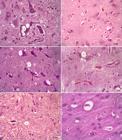
Atypical case of scrapie in a 3-year-old Dorper ram. (11) Medulla oblongata at the obex level, five vacuoles in parenchyma, one in neuron cytoplasm. (12) Frontal cortex, two vacuoles in parenchyma. (13) Thalamus, two vacuoles in the neuron cytoplasm and eight vacuoles in the parenchyma. (14) Rostral colliculus, two vacuoles in neuron cytoplasm and three vacuoles in parenchyma. (15) Pons, two vacuoles in parenchyma. (16) Parietal cortex, two vacuoles in the parenchyma. HE, bar = 20 µm.
Discussion
Purebred, white Dorpers and their crossbreeds are considered more predisposed to the development of scrapie (Andrade et al. 2015). The breed of the ram in this study is an essential epidemiological factor in scrapie cases, as well as age, because in sheep that develop neurological clinical signs are between two and five years of age (Moore et al. 2016). This diagnosis is the second in an animal of the Dorper breed in Santa Catarina; the first was reported by Andrade et al. (2015). The clinical course usually ranges from 10 days to months, depending on the amount of PrPsc deposition until the animal’s death (Cantile & Youssef 2016, Cassmann et al. 2019). In the evaluated case, the sheep was euthanized approximately 40 days after clinical presentation, so the time of progression cannot be estimated.
The clinical diagnosis of the ram was initially attributed to thoracolumbar spinal cord trauma between T3 and L3 due to the dog sitting posture. However, it was discarded when evaluated by radiography and myelography. Despite this, scrapie had not yet been considered owing to neurological clinical signs not specific to the disease, characterized by wool loss and head tremors (Andrade et al. 2015). The only neurological signs that could lead to suspicion of scrapie are limb weakness and sternal recumbency, as animals usually develop ataxia and fasciculations of the pelvic limbs (Moore et al. 2016). A positive scratch test and unfavorable diagnosis justified euthanasia, allowing complementary examinations and confirming the diagnosis of scrapie (Martins et al. 2012, Andrade et al. 2015). The “scratch” test consists of manually scratching the animal’s back, which will respond with a “nibble” reflex (Martins et al. 2012). Cases of atypical scrapie, stiff gait, ataxia, hypermetria (dysmetria), abnormal pelvic limb posture, generalized tremors, and lip tremors have been reported (Cassmann et al. 2021). In an experimental study, animals with greater accumulation of PrPsc in the cerebellum with the ARQ/ARR genotype developed severe dysmetria (Cassmann et al. 2021).
No lesions were observed macroscopically. However, some signs can aid in suspecting and diagnosing the disease, such as poor body condition and extensive areas of loss of hair due to scratching (Martins et al. 2012, Meling et al. 2012). Therefore, preclinical diagnosis with the use of lymphoid tissue biopsies from the third eyelid and rectal mucosa is essential to prevent the spread of the prion in suspected cases. However, it should be noted that it is important to perform biopsies of both the third eyelid and rectal mucosa, as described by Leal et al. (2012), as immunostaining may not always occur in both evaluated locations (Brasil 2009, Leal et al. 2012). The definitive diagnosis in this sheep was obtained through histopathology and immunohistochemistry of samples of the spinal cord, medulla, mesenteric lymph nodes, spleen, and tonsils (Andrade et al. 2015). ELISA is usually performed for screening when there is a clinical suspicion of scrapie (Esteves et al. 2021), as in this case.
The histological lesions caused by prions are characterized by vacuolization in both the parenchyma of the central nervous system and neurons without infiltration of inflammatory cells (Marín-Moreno et al. 2021). These vacuolizations are considered more intense in the region of the medulla oblongata at the level of the obex, although they are also observed in other regions (Martins et al. 2012, Greenlee 2019). This histological pattern is also observed when manipulating the neuropil to produce artifacts, myelin edema, spongiform myelopathies, neuronal alterations caused by viruses or toxins, metabolic disorders, and lysosomal storage diseases (Wohlsein et al. 2012). In this study, in addition to the observation of vacuoles, the clinical presentation and complementary tests were important to exclude the aforementioned alterations of artifacts or other conditions that present with neurological clinical presentations (Wohlsein et al. 2012, Prins et al. 2024). Vacuolar lesions were considered only for well-circumscribed vacuoles since manipulation of nervous tissue can cause artifacts, leading to errors in interpretation for the evaluator (Wohlsein et al. 2012).
Clinically healthy animals usually do not develop vacuolar changes even when they test positive in immunohistochemistry. Animals with significant vacuolization typically develop neurological signs (Wood et al. 1997). However, the presence of vacuolization in the central nervous system is not a requirement for the development of neurological clinical signs (Ryder et al. 2009). The subject of this study exhibited vacuolar alterations, along with clinical signs considered unusual for scrapie.
The development of the clinical presentation depends on the infectious load, the age at which the animal was exposed, and susceptibility will depend on the age of the animal and the incubation period (McIntyre et al. 2006). In the outbreak evaluated by these authors, it was found that the occurrence of scrapie in sheep with the VRQ/VRQ genotype began earlier when compared to VRQ/ARQ animals, followed by animals of the VRQ/ARR genotype.
Genotyping is important in sheep flocks for selecting genetically resistant animals, as in Europe, where selection programs are performed to eliminate sheep with high susceptibility (Ortiz-Pelaez et al. 2014). In Brazil, the ARQ/ARQ genotype, the same as that observed in the animal in this study, is the most common in sheep (Andrade et al. 2015) and is classified as moderate risk (R3) for the development of scrapie according to the United States Department of Agriculture (Sotomaior et al. 2008, Ianella et al. 2012). In Brazil, the occurrence of scrapie has been considered only recently, and genotyping, considered one of the methods for describing and accurately controlling the disease, is rarely performed (Andrade et al. 2011, Santos et al. 2012).
The present study was based on similar methodologies that also established scores for grading neurological lesions; thus, an adaptation of the grading and scores was performed (Mould et al. 1967, Salvesen et al. 2020). Thus, a higher score was observed in the thalamus, in the medulla oblongata at the level of the obex, and in the rostral colliculi. Similar data to those found in a study conducted on goats in the thalamic region showed higher scores, followed by the gray matter of the rostral colliculus, septal nuclei, and hippocampus. However, in the medulla oblongata at the level of the obex, these scores decreased considerably in goats compared to those obtained in the present study (Salvesen et al. 2020).
The spinal cord proved to be important for identifying vacuolar lesions. However, evaluation was not performed on all portions, such as the cervical and lumbar enlargements, only the initial portion near the medulla oblongata at the level of the obex. The assessment of these portions is considered important because the deposition of the prion protein often occurs in this region, and possibly the entry point of the prion is through parasympathetic motor neurons in the vagus nerve that innervate the gastrointestinal tract (Ryder et al. 2001).
Although the degree of vacuolization in the central nervous system of sheep is studied in cases of scrapie, the association of vacuolization in different regions of the brain with clinical changes in sheep has not yet been performed (Mould et al. 1967, Fraser & Dickinson 1973, Scott & Fraser 1984, Salvesen et al. 2020). Therefore, this study can be considered pioneering and serves as a basis for future studies.
Conclusions
This study describes a case of scrapie in a sheep with atypical clinical signs of the disease, indicating that when animals show neurological signs, such as kyphosis and proprioceptive deficits in the pelvic limbs, scrapie should always be considered a differential diagnosis. This genotype was classified as naturally susceptible (ARQ/ARQ) to disease development, demonstrating the importance of early diagnosis.
Histological analysis revealed significant vacuolization in different regions of the brain, supporting that vacuolar lesions, although not inflammatory, are characteristic of prion-related diseases. Vacuolizations were most pronounced in the thalamus, rostral colliculi and medulla oblongata at the level of the obex, with degrees of severity ranging from moderate to severe. The distribution of vacuolization, predominantly in the neuropil, reinforces the neuropathological characteristics associated with scrapie. Immunohistochemical and molecular analyses provided a definitive diagnosis, demonstrating PrPsc deposition and identifying the ARQ/ARQ genotype, which is associated with moderate susceptibility to scrapie. These findings highlight the importance of PRNP genotyping in the management of scrapie in sheep populations, as selection for genetic resistance may help control the disease, as seen in Europe. This study, therefore, contributes valuable insights into the clinical spectrum and genetic markers associated with scrapie, reinforcing the need for proactive monitoring and genetic screening to mitigate the spread of this fatal disease in sheep.
Acknowledgments
This work was supported by “Fundação de Amparo à Pesquisa e Inovação do Estado de Santa Catarina” (FAPESC, 027/2020 - No. 2021TR872) and by “Programa de Apoio à Pós-Graduação” (PROAP), maintained by the “Coordenação de Aperfeiçoamento de Pessoal de Nível Superior” (CAPES, Finance code 001).
References
-
Andrade CP, Almeida LL, Castro LA, Leal JS, Silva SC, Driemeier D. Single nucleotide polymorphisms at 15 codons of the prion protein gene from a Scrapie-affected herd of Suffolk sheep in Brazil. Braz J Vet Res 2011; https://doi.org/10.1590/S0100-736X2011001000010
» https://doi.org/10.1590/S0100-736X2011001000010 -
Andrade CP, Oliveira EC, Leal JS, Almeida LL, Castro LA, Silva SC, Driemeier D. Report of outbreaks of classical Scrapie in Dorper sheep and associated prion protein gene polymorphisms in affected flocks. Trop Anim Health Prod 2015; https://doi.org/10.1007/s11250-015-0849-9
» https://doi.org/10.1007/s11250-015-0849-9 -
Beck KE, Sallis RE, Lockey R, Simmons MM, Spiropoulos J. Ovine PrP genotype is linked with lesion profile and immunohistochemistry patterns after primary transmission of classical scrapie to wild-type mice. J Neuropathol Exp Neurol 2010; https://doi.org/10.1097/NEN.0b013e3181db2497
» https://doi.org/10.1097/NEN.0b013e3181db2497 - Brasil. Manual de Legislação: programas nacionais de saúde animal do Brasil. Brasília: Ministério da Agricultura, Pecuária e Abastecimento, Secretaria de Defesa Agropecuária, Departamento de Saúde Animal; 2009. 440p.
-
Cantile C, Youssef S. Nervous System, p.250-406. In: Maxie MG. Jubb, Kennedy and Palmer’s Pathology of Domestic Animals. 2016; https://doi.org/10.1016/B978-0-7020-5317-7.00004-7
» https://doi.org/10.1016/B978-0-7020-5317-7.00004-7 -
Cassmann ED, Mammadova N, Moore SJ, Benestad S, Greenlee JJ. Transmission of the atypical/Nor98 Scrapie agent to Suffolk sheep with VRQ/ARQ, ARQ/ARQ, and ARQ/ARR genotypes. PLoS One 2021; https://doi.org/10.1371/journal.pone.0246503
» https://doi.org/10.1371/journal.pone.0246503 -
Cassmann ED, Moore SJ, Smith JD, Greenlee JJ. Sheep with the homozygous lysine-171 prion protein genotype are resistant to classical scrapie after experimental oronasal inoculation. Vet Pathol 2019; https://doi.org/10.1177/0300985818817066
» https://doi.org/10.1177/0300985818817066 -
Donaldson DS, Kobayashi A, Ohno H, Yagita H, Williams IR, Mabbott NA. M cell-depletion blocks oral prion disease pathogenesis. Mucosal Immunol. 2012; https://doi.org/10.1038/mi.2011.68
» https://doi.org/10.1038/mi.2011.68 -
Dustan BH, Spencer YI, Casalone C, Brownlie J, Simmons MM. A histopathologic and immunohistochemical review of archived UK caprine scrapie cases. Vet Pathol 2008; https://doi.org/10.1354/vp.45-4-443
» https://doi.org/10.1354/vp.45-4-443 -
Esteves A, Vieira-Pinto M, Quintas H, Orge L, Gama A, Alves A, Seixas F, Pires I, Pinto ML, Mendonça AP, Lima C, Machado CN, Silva JC, Tavares P, Silva F, Bastos E, Pereira J, Gonçalves-Anjo N, Carvalho P, Sargo R, Matos A, Figueira L, Pires MA. Scrapie at abattoir: monitoring, control, and differential diagnosis of wasting conditions during meat inspection. Animals 2021; https://doi.org/10.3390/ani11113028
» https://doi.org/10.3390/ani11113028 -
Fraser H, Dickinson AG. Scrapie in mice: agent-strain differences in the distribution and intensity of grey matter vacuolation. J Comp Pathol 1973;https://doi.org/10.1016/0021-9975(73)90024-8
» https://doi.org/10.1016/0021-9975(73)90024-8 -
González L, Martin S, Hawkins SAC, Goldmann W, Jeffrey M, Sisó S. Pathogenesis of natural goat scrapie: modulation by host PRNP genotype and effect of co-existent conditions. Vet Res 2010; https://doi.org/10.1051/vetres/2010020
» https://doi.org/10.1051/vetres/2010020 -
González L, Pitarch JL, Martin S, Thurston L, Moore J, Acín C, Jeffrey M. Identical pathogenesis and neuropathological phenotype of scrapie in valine, arginine, glutamine/valine, arginine, glutamine sheep infected experimentally by the oral and conjunctival routes. J Comp Pathol 2014; https://doi.org/10.1016/j.jcpa.2013.06.006
» https://doi.org/10.1016/j.jcpa.2013.06.006 -
Greenlee JJ. Review: update on classical and atypical scrapie in sheep and goats. Vet Pathol 2019; https://doi.org/10.1177/0300985818794247
» https://doi.org/10.1177/0300985818794247 - Hall TA. BioEdit: a user-friendly biological sequence alignment editor and analysis program for windows 95/98/NT. Nucl Acids Symp Ser 1999;41:95-98.
-
Ianella P, McManus CM, Caetano AR, Paiva SR. PRNP haplotype and genotype frequencies in Brazilian sheep: issues for conservation and breeding programs. Res Vet Sci 2012; https://doi.org/10.1016/j.rvsc.2011.06.025
» https://doi.org/10.1016/j.rvsc.2011.06.025 -
Ladogana A, Puopolo M, Croes EA, Budka H, Jarius C, Collins S, Klug GM, Sutcliffe T, Giulivi A, Alperovitch A, Delasneire-Laupretre L, Brandel J-P, Poser S, Kretzchmar H, Rietveld I, Mitrova E, Cuesta JP, Martinez-Martin P, Glatzel M, Aguzzi A, Knight R, Ward H, Pocchiari M, van Duijn CM, Will RG, Zerr I. Mortality from Creutzfeldt-Jakob disease and related disorders in Europe, Australia, and Canada. Neurol 2005; https://doi.org/10.1212/01.WNL.0000160117.56690.B2
» https://doi.org/10.1212/01.WNL.0000160117.56690.B2 -
Leal JS, Correa GLF, Dalto AGC, Boos GS, Oliveira EC, Bandarra PM, Lopes RFF, Driemeier D. Use of third eyelid and rectal mucosa biopsies for diagnosis of sheep Scrapie on a farm in Southern Brazil. Braz J Vet Res 2012; https://doi.org/10.1590/S0100-736X2012001000007
» https://doi.org/10.1590/S0100-736X2012001000007 -
Linden R, Martins VR, Prado MAM, Cammarota M, Izquierdo I, Brentani RR. Physiology of the príon protein. Physiol Rev 2008; https://doi.org/10.1152/physrev.00007.2007
» https://doi.org/10.1152/physrev.00007.2007 -
Marín-Moreno A, Aguilar-Calvo P, Espinosa JC, Zamora-Ceballos M, Pitarch JL, González L, Fernández-Borges N, Orge L, Andréoletti O, Nonno R, Torres JM. Classical scrapie in small ruminants is caused by at least four different prion strains. Vet Res 2021; https://doi.org/10.1186/s13567-021-00929-7
» https://doi.org/10.1186/s13567-021-00929-7 -
Martins HM, Carvalho NM, Ribas NLKS, Driemeier D, Lemos RAA, Guimarães EB. Scrapie and differential diagnosis in sheep in Mato Grosso do Sul, Brazil. Braz J Vet Res 2012; https://doi.org/10.1590/S0100-736X2012001200004
» https://doi.org/10.1590/S0100-736X2012001200004 -
McIntyre KM, Gubbins S, Goldmann W, Stevenson E, Baylis M. The time-course of a scrapie outbreak. BMC Vet Res 2006; https://doi.org/10.1186/1746-6148-2-20
» https://doi.org/10.1186/1746-6148-2-20 -
Meling S, Bårdsen K, Ulvund MJ. Presence of an acute phase response in sheep with clinical classical scrapie. BMC Vet Res 2012; https://doi.org/10.1186/1746-6148-8-113
» https://doi.org/10.1186/1746-6148-8-113 -
Moore SJ, Smith JD, Greenlee MH, Nicholson EM, Richt JA, Greenlee JJ. Comparison of two US sheep Scrapie isolates supports identification as separate strains. Vet Pathol 2016; https://doi.org/10.1177/0300985816629712
» https://doi.org/10.1177/0300985816629712 -
Mould DL, Dawson AM, Slater JS, Zlotnik I. Relationships between chemical changes and histological damage in the brains of scrapie affected mice. J Comp Pathol 1967; https://doi.org/10.1016/0021-9975(67)90024-2
» https://doi.org/10.1016/0021-9975(67)90024-2 -
Ortiz-Pelaez A, Thompson CE, Dawson M. The impact of the National Scrapie Plan on the PRNP genotype distribution of the British national flock, 2002-2012. Vet Rec 2014; https://doi.org/10.1136/vr.102255
» https://doi.org/10.1136/vr.102255 -
Prins S, Hamer K, Cloquell A, Spiropoulos J, Sargison N, Vellema P. MRI changes observed in a case of atypical scrapie in a 7-year-old Herdwick ewe. J Vet Diagn Investig 2024; https://doi.org/10.1177/10406387241267849
» https://doi.org/10.1177/10406387241267849 -
Prusiner SB. Prions. Proc Natl Acad Sci 1998; https://doi.org/10.1073/pnas.95.23.13363
» https://doi.org/10.1073/pnas.95.23.13363 -
Roels S, Renard C, De Bosschere H, Geeroms R, Van Poucke MV, Peelman L, Vanopdenbosch E. Detection of polymorphisms in the prion protein gene in the Belgian sheep population: Some preliminary data. Vet Q 2004; https://doi.org/10.1080/01652176.2004.9695163
» https://doi.org/10.1080/01652176.2004.9695163 -
Ryder SJ, Dexter GE, Heasman L, Warner R, Moore SJ. Accumulation and dissemination of prion protein in experimental sheep scrapie in the natural host. BMC Vet Res 2009; https://doi.org/10.1186/1746-6148-5-9
» https://doi.org/10.1186/1746-6148-5-9 -
Ryder SJ, Spencer YI, Bellerby PJ, March SA. Immunohistochemical detection of PrP in the medulla oblongata of sheep: the spectrum of staining in normal and scrapie-affected sheep. Vet Rec 2001; https://doi.org/10.1136/vr.148.1.7
» https://doi.org/10.1136/vr.148.1.7 -
Salvesen Ø, Espenes A, Reiten MR, Vuong TT, Malachin G, Tran L, Andréoletti O, Olsaker I, Benestad SL, Tranulis MA, Ersdal C. Goats naturally devoid of PrPC are resistant to scrapie. Vet Res 2020; https://doi.org/10.1186/s13567-019-0731-2
» https://doi.org/10.1186/s13567-019-0731-2 -
Santos CR, Mori E, Leão DA, Maiorka PC. Genotyping of polymorphisms in the PRNP gene in Santa Ines sheep in the State of São Paulo, Brazil. Braz J Vet Res 2012; https://doi.org/10.1590/S0100-736X2012000300007
» https://doi.org/10.1590/S0100-736X2012000300007 -
Scott JR, Fraser H. Degenerative hippocampal pathology in mice infected with scrapie. Acta Neuropathol 1984; https://doi.org/10.1007/BF00689829
» https://doi.org/10.1007/BF00689829 -
Sotomaior CS, Sotomaior VS, Madeira HMF, Thomaz-Soccol V. Prion protein gene polymorphisms in sheep in the state of Paraná, Brazil. Anim Genet 2008; https://doi.org/10.1111/j.1365-2052.2008.01775.x
» https://doi.org/10.1111/j.1365-2052.2008.01775.x -
Tamura K, Stecher G, Kumar S. MEGA11: molecular evolutionary genetics analysis version 11. Mol Biol Evol 2021; https://doi.org/10.1093/molbev/msab120
» https://doi.org/10.1093/molbev/msab120 -
Thompson JD, Higgins DG, Gibson TJ. Clustal W: Improving the sensitivity of progressive multiple sequence alignment through sequence weighting, position-specific gap penalties and weight matrix choice. Nucleic Acids Res 1994; https://doi.org/10.1093/nar/22.22.4673
» https://doi.org/10.1093/nar/22.22.4673 - USDA, APHIS. Scrapie Program Standards Volume 1: National Scrapie Erradication Program. United States Department of Agriculture, Animal and Plant Health Inspection Service. 2019.
-
Wohlsein P, Deschl U, Baumgärtner W. Nonlesions, unusual cell types, and postmortem artifacts in the central nervous system of domestic animals. Vet Pathol 2012; https://doi.org/10.1177/0300985812450719
» https://doi.org/10.1177/0300985812450719 -
Wood JLN, McGill IS, Done SH, Bradley R. Neuropathology of scrapie: a study of the distribution patterns of brain lesions in 222 cases of natural scrapie in sheep, 1982-1991. Vet Rec 1997; https://doi.org/10.1136/vr.140.7.167
» https://doi.org/10.1136/vr.140.7.167
The authors confirm that the data supporting the findings of this study are available within the article. Derived data supporting the findings of this study are available from the corresponding author upon request.


