Metastatic gallbladder adenocarcinoma and peritonitis as the cause of death in one of the world’s oldest captive jaguars (Panthera onca) in southern Brazil
ABSTRACT:
A 25-year-old captive female jaguar (Panthera onca) presented with clinical signs of anorexia, adipsia, vomiting, and difficulty moving that progressed over a duration of three days. This eventually led to death. During necropsy, a significant amount of brown fluid was detected in the abdominal cavity. The gallbladder was markedly enlarged, diffusely distended with thickened walls, and had an irregular mucosa ranging from black to white. Bile spilled into the abdominal cavity through a rupture in the wall. Additionally, a focally extensive, 7 x 5 x 3 cm, white mass with adherent friable brown material was observed in the mucosa. A 2 x 2 x 1 cm, firm white nodular mass was present in the extrahepatic bile duct and caused obstruction of the bile passage. Histopathologic evaluation confirmed adenocarcinoma of the gallbladder and adjacent ducts. The neoplasm was arranged as acini and tubules and supported by abundant fibrovascular stroma. Metastases were observed in the liver, intestines, and lungs. Several other tumors were identified in this jaguar. These included papillary carcinomas of the ovaries, fibroleiomyomas in the uterus, leiomyomas in the stomach, and follicular adenomas in the thyroid gland. Chronic kidney disease and moderate dilated cardiomyopathy were also observed. Immunohistochemistry showed positive staining for pan-cytokeratin in the neoplastic cells of the gallbladder, ovary, and thyroid gland. Positive staining for vimentin was observed in the neoplastic cells of the uterus and stomach. The identification of multiple tumors in this aged jaguar highlights the need for routine examinations throughout life for early detection and management of tumors.
INDEX TERMS:
Adenocarcinoma; biliary disease; neoplasia; non-domestic feline
RESUMO:
Uma onça-pintada (Panthera onca) fêmea de 25 anos em cativeiro apresentou sinais clínicos de anorexia, adipsia, vômito e dificuldade de locomoção, que progrediram ao longo de três dias, levando a morte. Durante a necropsia, foi detectada uma quantidade significativa de fluido marrom na cavidade abdominal. A vesícula biliar estava significativamente aumentada, difusamente distendida com paredes espessadas e apresentava mucosa irregular, variando de preto a branco. Bile havia extravasado para a cavidade abdominal devido a uma ruptura na parede. Além disso, foi observada uma massa branca, focalmente extensa, medindo 7 x 5 x 3 cm, com material marrom friável aderido à mucosa. Uma massa nodular branca, firme, de 2 x 2 x 1 cm, estava presente no ducto biliar extra-hepático, causando obstrução da passagem da bile. A avaliação histopatológica confirmou adenocarcinoma de vesícula biliar na vesícula e nos ductos adjacentes. A neoplasia estava organizada em ácinos e túbulos, sustentada por estroma fibrovascular abundante. Metástases foram observadas no fígado, intestinos e pulmões. Vários outros tumores também foram identificados nessa onça-pintada, incluindo carcinomas papilíferos nos ovários, fibroleiomioma no útero, leiomioma no estômago e adenoma folicular na glândula tireoide. Doença renal crônica e cardiomiopatia dilatada moderada também foram constatadas. A imuno-histoquímica revelou marcação positiva para pancitoqueratina nas células neoplásicas da vesícula biliar, ovário e glândula tireoide. Marcação positiva para vimentina foi observada nas células neoplásicas do útero e estômago. A identificação de múltiplos tumores nessa onça-pintada idosa destaca a importância de exames de rotina ao longo da vida para a detecção precoce e manejo de neoplasias.
TERMOS DE INDEXAÇÃO:
Adenocarcinoma; doença biliar; neoplasia; felino não doméstico
Introduction
In the Americas, the jaguar (Panthera onca) ranges from Mexico to Argentina. It inhabits savannas, tropical forests, and semi-arid areas. The species is classified as near threatened due to hunting, habitat loss, and human-wildlife conflicts (IUCN 2022). In the wild, an estimated 15,000 to 20,000 individuals remain, with much smaller numbers in captivity. In Brazil, agricultural expansion remains a significant challenge for the species, but conservation and reintroduction programs aim to mitigate these impacts (Wultsch et al. 2016, Cuyckens et al. 2017, IUCN 2022).
The lifespan of captive wild animals has increased owing to improvements in nutrition, management, and veterinary care. This has led to an increase in the number of age-related diseases affecting these species (Longley 2011). Among large felids of the genus Panthera, individuals in captivity may reach or slightly exceed 20 years of age (Kloft et al. 2019). In contrast, the lifespan of jaguars in the wild is poorly described, generally ranging from 14 to 16 years (Hunter 2011, Harmsen et al. 2017).
Post mortem examinations that include histopathological and immunohistochemical analyses are important to diagnose and contextualize diseases (Effron et al. 1977, Chu et al. 2012). Neoplasms, renal diseases, and dental diseases are among the most common diseases affecting captive jaguars (Hope & Deem 2006, Junginger et al. 2015, Norton et al. 2018). In one study, felids in the Panthera genus showed a greater susceptibility for developing neoplasms than those of other non-domestic felid species (Moresco et al. 2020).
Previous studies have shown that neoplasms are amongst the most important causes of euthanasia or death in captive wild felids (19.8-58.3%), with mammary and hepatic neoplasms being the most common (Owston et al. 2008, Junginger et al. 2015, D’Aquino et al. 2022, Duque-Correa et al. 2022). In one study that evaluated neoplasms in large felids of the Panthera genus, the most commonly identified tumors were mammary carcinomas, thyroid adenomas, and multicentric lymphomas (Kloft et al. 2019). In comparison, the causes of death in jaguars in the wild are scarce and poorly documented, with most of the information from sporadic cases (Duque-Correa et al. 2022, Díaz-Vaquero et al, 2024).
The objective of this study was to report on what seems to be the first recorded metastatic gallbladder adenocarcinoma with subsequent bile peritonitis as the primary cause of death in one of the world’s oldest captive jaguars (Panthera onca), in conjunction with other non-fatal pathology.
Case Report
Ethical approval. The animal in this case report died naturally under human care and was submitted for postmortem examination at the “Laboratório de Patologia Animal” (LAPA), “Universidade do Estado de Santa Catarina” (UDESC). Therefore, approval by the Ethics Committee on the Use of Animals was not required.
A 25-year-old captive female jaguar (Panthera onca) (Fig. 1) was referred for necropsy to the “Laboratório de Patologia Animal” (Laboratory of Veterinary Pathology - LAPA), “Universidade do Estado de Santa Catarina” (UDESC) in Lages/SC, Brazil. Clinical signs included anorexia, adipsia, vomiting, and difficulty moving over a period of three days prior to death. Additionally, one month before death, the jaguar underwent mastectomy for the removal of a mammary tumor. The animal was rescued in 1997 in the state of Minas Gerais by a local zoobotanical park and, in the same year, as a kitten, was sent to a zoo in the state of Santa Catarina, where it lived until its death.

Carcass of a jaguar (Panthera onca) diagnosed with metastatic gallbladder adenocarcinoma with perforation and bile peritonitis.
At the necropsy, sections of the gallbladder, liver, lungs, heart, thyroid glands, adrenal glands, intestines, spleen, stomach, pancreas, kidneys, bladder, ovary, uterus, bone marrow and brain were collected and fixed in formalin before routine histopathological processing, and hematoxylin and eosin (HE) staining. Sections of the gallbladder were also stained with Masson’s trichrome stain.
Sections of the gallbladder, ovary, and thyroid glands were subjected to immunohistochemical labeling for pan-cytokeratin and vimentin, whereas sections of the uterus and stomach were labeled only for vimentin. Three-micron thick tissue sections were placed on charged glass slides and endogenous peroxidase was blocked with hydrogen peroxide p.a. (Synth, Diadema, São Paulo, Brazil) diluted to 10% in methanol p.a. (Synth, Diadema/SP, Brazil) at room temperature (20 °C approximately). Subsequently, antigen recovery was carried out in a water bath for 40 minutes at 100 °C using tris-EDTA buffer (pH 9) for pan-cytokeratin and citrate buffer (pH 6) for vimentin. The blocking of nonspecific binding was carried out using 5% skimmed milk powder (Molico®, Nestlé S.A., Araçatuba/SP, Brazil) in distilled water for 20 minutes. The primary antibodies consisted of pan-cytokeratin (1:100, AE1/AE3 clone cocktail, Biocare Medical, Concord/CA, USA) and vimentin (1:200, V9 clone, Biocare Medical, Concord, California, USA), diluted in phosphate-buffered saline (PBS). The sections were incubated in a humidity chamber for 1.5 hours at 37 °C.
The slides were incubated with a universal MACH 4 HRP polymer probe kit (Biocare Medical, Concord/CA, USA) for 15 min in the first stage (probe) and 30 minutes in the second stage (polymer). Then, staining with 3,3′-diaminobenzidine (Chromogen DAB, Dako, Glostrup, Denmark) was performed, followed by counterstaining with hematoxylin. The positive controls included tissue sections from a domestic canine with mammary carcinoma for pan-cytokeratin detection and fibrosarcoma for vimentin detection, and a universal negative control reagent (EasyPath, SP, Brazil) was used as the negative control.
Necropsy revealed the jaguar was in a thin body condition. The abdominal cavity contained abundant watery, opaque brown fluid (Fig. 2). The gallbladder was markedly enlarged (20 cm in diameter), filled with watery and black bile, and there was diffuse thickening of the wall. The mucosal surface was irregular with multifocal areas ranging from black to white. Within the wall of the gallbladder was a focal, 1 cm diameter, transmural rupture with hemorrhage at the edges and leakage of biliary content into the abdominal cavity. Additionally, on the mucosa, there was a broad-based, 7 cm x 5 cm x 3 cm white mass that extended deeply into the wall of the gallbladder. Friable brown material was adhered to its surface. In the extrahepatic bile duct, there was a firm, demarcated, broad-base white nodular mass (2 cm x 2 cm x 1 cm) that occluded bile passage (Fig. 3). Nodular masses similar to those in the gallbladder and bile duct were seen in the liver (0.5-3 cm in diameter), lungs (0.2 cm in diameter), and on the serosal surfaces of the small and large intestine (0.5-2 cm diameter).

Macroscopic, microscopic and immunohistochemistry findings observed in metastatic adenocarcinoma of the gallbladder. (3) Gallbladder: the gallbladder is expanded by a white mass with adherent brown material (white arrow) on the surface of the mass, and black and white discoloration of the mucosa (asterisk). Inset: white nodule in the extrahepatic bile duct (red circle). (4) Gallbladder adenocarcinoma with multiple acinar and tubular projections. HE, obj. 100x. Inset: higher magnification of gallbladder adenocarcinoma. HE, obj. 400x. (5) Gallbladder adenocarcinoma with strong immunostaining for pan-cytokeratin (Cocktail AE1/AE3). IHC, obj. 200x. Polymer linked to endogenous peroxidase (MACH 4 HRP), chromogen DAB 3,3’-diaminobenzidine.
Histopathological evaluation of the gallbladder and adjacent ducts revealed a non-delineated, non-encapsulated, malignant epithelial neoplasm composed of acini and/or tubules separated by abundant fibrovascular stroma compatible with gallbladder adenocarcinoma (Fig. 4). The cells were polyhedral to columnar, with round to oval nuclei, finely stippled chromatin, and a single prominent nucleolus. The cytoplasm was eosinophilic and distinct. Moderate anisocytosis to anisokaryosis was observed with zero mitotic figures per 2.37 mm². Fibrinoid necrosis accompanied by multifocal and accentuated neutrophil and macrophage infiltration was observed in the neoplasm. Immunohistochemistry (IHC) showed moderate-to-strong and diffuse positive staining for pan-cytokeratin in the cytoplasm of neoplastic cells (Fig. 5). Additionally, the gallbladder adenocarcinoma led to a focal rupture of the organ, resulting in the development of biliary peritonitis in the abdominal cavity. The parenchyma of the lung, liver and the intestinal submucosa and serosa were expanded by tumors that were morphologically similar to those in the gallbladder and bile ducts.
Additionally, but not related to the demise of the animal, were a number of incidental findings. This included several benign tumors and chronic renal and cardiac changes.
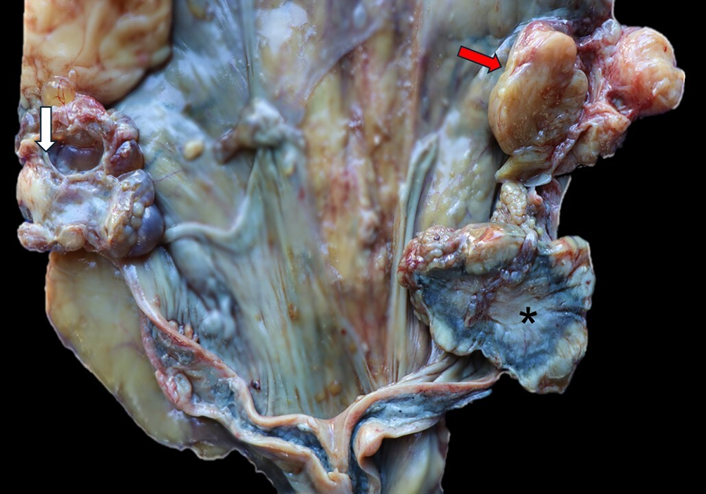
In the left ovary, an irregular nodule measuring 4 cm x 2 cm x 3 cm was observed; it was firm with solid, multilobulated areas that were slightly irregular and white. Multiple cysts (0.4-4 cm diameter) were also present; these had thin walls and a translucent content. In the right ovary, an irregular nodule measuring 8.5 cm x 5.5 cm x 5 cm was present, with macroscopic features similar to the left ovary (Fig. 6).

Papillary carcinoma in the ovaries and uterine fibroleiomyoma: left ovary (white arrow) - white nodule with multilobulated and irregular areas. Right ovary (red arrow) - white, multilobulated nodule. Left uterine body- white to black nodule (asterisk).
Histologically, by expanding the parenchyma in both ovaries, a non-encapsulated and poorly delineated malignant epithelial neoplastic, composed of papillae, sometimes arboriformly, projecting into cystic areas, and separated by abundant fibrovascular stroma compatible with papillary carcinoma. Immunohistochemistry revealed moderate-to-strong and diffuse positive staining for pan-cytokeratin in the cytoplasm of neoplastic cells. In addition, the cyst walls were lined with simple squamous epithelium containing moderate multifocal eosinophilic amorphous proteinaceous material and fibrin thrombi.
In the left uterine body was a 6.5 cm x 5 cm x 4 cm, white to black, firm, irregular, multilobulated nodule (Fig. 6). Histologically, the myometrium was expanded, well-demarcated, unencapsulated, mesenchymal neoplasm composed of multidirectional bundles separated by moderate fibrovascular stroma compatible with a uterine fibroleiomyoma. Masson’s trichrome staining revealed that approximately 50% of the neoplastic cells showed red-stained cytoplasm (muscle tissue), whereas approximately 50% of the neoplastic cells exhibited a blue-stained cytoplasm (fibrous connective tissue). Immunohistochemistry revealed strong positive staining for vimentin in the neoplastic cells.
In the stomach was a 1 cm x 1.5 cm x 1 cm, diffusely white, firm, rounded raised nodule (Fig. 7). Histologically, the muscular layer was expanded, well-defined, encapsulated, benign mesenchymal neoplasm composed of multidirectional bundles separated by moderate fibrovascular stroma that was compatible with leiomyoma. Masson’s trichrome staining revealed positive blue staining in the cytoplasm of neoplastic cells (fibrous connective tissue). Immunohistochemistry revealed strong positive staining for vimentin in the neoplastic cells.

The left thyroid gland was moderately enlarged and contained a 2 cm x 2.5 cm x 2 cm, soft and dark red nodule (Fig. 8). Histologically, a well-defined, partially encapsulated, benign epithelial neoplasm was present within the glandular parenchyma of the thyroid. It was composed of small follicular nests that contained scant colloid and were separated by moderate fibrovascular stroma (colloid was sometimes totally absent). The nodule was compatible with a follicular adenoma. Immunohistochemistry revealed moderate-to-strong and diffuse positive staining for pan-cytokeratin in the cytoplasm of neoplastic cells.

Follicular adenoma: red nodule in left thyroid gland. Insert: lateral view of the follicular adenoma in the left thyroid (red circle).
Both kidneys were moderately reduced in size and had a mild diffuse irregular surface, with multiple tubular cysts (0.3-0.5 cm in diameter). Additionally, the kidneys exhibited fibrous connective tissue proliferation in the interstitium, with discrete to moderate multifocal lymphoplasmacytic infiltration, multifocal moderate glomerulosclerosis, retraction of the glomerular tuft, and thickening of the Bowman’s capsule, compatible with chronic kidney disease.
Dilated cardiomyopathy associated with tricuspid and mitral valve endocardiosis was diagnosed in the heart. Macroscopically, the heart occupied five intercostal spaces; it was moderately globular and exhibited moderate eccentric hypertrophy of the left ventricle. The atrioventricular valves (tricuspid and mitral) were moderately thickened, shortened, and wrinkled.
Macroscopic, histopathological, histochemical, and immunohistochemical findings confirmed the diagnosis of gallbladder adenocarcinoma with perforation and bile peritonitis. The tumor was associated with metastasis to the liver and lungs and with intestinal carcinomatosis. Other tumors included papillary carcinoma in the ovaries, uterine fibroleiomyoma, gastric leiomyoma, and follicular adenoma in the thyroid gland. Chronic kidney disease and dilated cardiomyopathy associated with tricuspid and mitral valve endocardiosis were also seen in this 25-year-old captive jaguar (Panthera onca).
Discussion
The development of neoplastic and degenerative diseases in non-domestic felids is common in aged (adult, geriatric) animals in captive settings (Junginger et al. 2015, Norton et al. 2018, Kloft et al. 2019, D’Aquino et al. 2022). Previous studies have demonstrated that the age of non-domestic felids affected by some types of neoplasia ranges from seven to 20 years, with a higher incidence in animals over 12 years of age (Owston et al. 2008, Kloft et al. 2019). The current report described the presence of multiple tumors, including fatal gallbladder adenocarcinoma, in one of the oldest recorded jaguars in captivity. In a global database of jaguars managed by zoos, there is currently a jaguar in a European zoo that may be older; however, the available data does not directly confirm its exact age, possibly around 25 years. Additionally, the literature on diseases affecting jaguars mostly involves studies with captive animals.
As a comparison, biliary tract neoplasms have rarely been reported in non-domestic felids. In a study reporting gallbladder adenocarcinoma in two captive African lions, the animals were 17 and 18 years old (Sakay et al. 2003). Similar to previous studies, tumors were diagnosed in this jaguar at an advanced age. In comparison, biliary tumors in domestic cats have been reported in animals between 12 and 13 years of age (Broadbridge et al. 2021, Pandolfo et al. 2023).
In domestic animals, biliary neoplasms are often accompanied by nonspecific clinical signs, such as anorexia, apathy, prostration, weight loss, lethargy, vomiting, and jaundice (Diogenes et al. 2020, Pandolfo et al. 2023). In non-domestic felids, the clinical signs are more subtle, with severe anorexia and ascites being reported; however, in certain cases, the animals are asymptomatic (McClure et al. 1977, Sakay et al. 2003). In this report, the jaguar exhibited mild clinical signs with anorexia similar to those reported in other studies; however, it also had adipsia, vomiting, and difficulty in mobility.
Macroscopically, gallbladder adenocarcinoma lesions in two lions were characterized by a markedly thickened gray wall with areas of fibrosis in association with pronounced ascites (Sakay et al., 2003). In our case, the jaguar showed similar alterations. However, in the jaguar, obstruction of the extrahepatic bile duct due to tumor presence led to a marked accumulation of bile and subsequent gallbladder rupture and bile peritonitis, which has not previously been described in other gallbladder adenocarcinomas in domestic or non-domestic felids. In contrast, in domestic felids, the gallbladder typically contains a well-defined mass of gelatinous bile, and bile duct obstruction occurs secondary to mass effect that can occasionally block bile flow (Patnaik 1992, Diogenes et al. 2020, Pandolfo et al. 2023), but without gallbladder perforation.
Gallbladder adenocarcinoma is a malignant neoplasm that can be associated with metastatic dissemination. The most commonly affected sites are the lungs and lymph nodes (Meuten 2017). However, neoplastic infiltration also occurs in the liver, predominantly in the lobes adjacent to the gallbladder, and has been observed in the mesentery, omentum, and intestinal serosa (Patnaik 1992, Sakay et al. 2003, Pandolfo et al. 2023). In the present case, metastases were observed in the liver, lungs, and intestinal serosa.
In addition to the gallbladder and bile duct adenocarcinoma, several other tumors were present in this aged jaguar. This included tumors in the reproductive tract and thyroid gland. The most frequently reported reproductive tract neoplasms in non-domestic felids in several previous studies were mammary tumors (Owston et al. 2008, Kloft et al. 2019, Duque-Correa et al. 2022). In one study that evaluated the causes of mortality in jaguars of the genus Panthera onca, uterine hyperplasia, ovarian cysts, and mammary and uterine adenocarcinomas were the most common (Hope & Deem 2006). The jaguar in this case report had a mammary tumor but underwent mastectomy without histopathological examination.
Reproductive diseases are more commonly observed in females than males. In an experimental study where melengestrol acetate, a progestin medication, was administered in jaguars, the data suggest that exposure to the drug increased the risk of developing female reproductive diseases and should be seriously considered before being used as a contraceptive (Hope & Deem 2006). It has also been suggested that jaguars may have an increased incidence of mammary gland carcinomas even in the absence of melengestrol acetate administration (Munson & Moresco 2007). Another study highlighted the use of contraceptive medications in non-domestic female felids, which they also suggested may increase the rate of reproductive neoplasms (Junginger et al. 2015). The jaguar in the current report had both an ovarian papillary carcinoma and a uterine fibroleiomyoma. In this context, it is important to note that uterine leiomyomas are more common than fibroleiomyomas in non-domestic felids (Walzer et al. 2003, Budiono 2012). In one study that described diseases in captive wild felids in a German zoo, reproductive neoplasms were the second most common type of tumor and were diagnosed in 24% of the animals. Leiomyomas were the most commonly detected tumor type and were seen in four tigers, three leopards, and one lion (Junginger et al. 2015).
Endocrine tumors have also been reported in several non-domestic felid species, and a follicular adenoma was seen in this jaguar. In a study of neoplastic lesions in four leopards, three tigers, and three pumas, with an average age of 15 years, thyroid gland adenomas were the most common neoplasms, followed by parathyroid adenomas and thyroid carcinomas (Pope et al. 2017). The cellular morphology frequently observed is a follicular pattern with a discrete amount of colloid. However, a spindle cell morphology can be observed in some neoplastic areas (Pope et al. 2017). Another study on neoplasms in Panthera species kept in captivity revealed thyroid gland adenomas to be one of the most frequently diagnosed tumors, with a prevalence of 61.9% among the endocrine neoplasms (Kloft et al. 2019). In these two studies, thyroid gland adenomas were considered incidental findings and seen only in elderly felids. These findings are similar to those in the present report, in which the thyroid gland neoplasm did not contribute to the animal’s death.
Non-neoplastic lesions in this jaguar included chronic renal and cardiac disease. Chronic kidney disease is common in elderly non-domestic felids (Newkirk et al. 2011). In one study in jaguars, diseases affecting the genitourinary system, including interstitial nephritis and chronic kidney disease, were a significant cause of mortality, accounting for 16.2% of cases (Duque-Correa et al. 2022). In another study, a high prevalence of renal disease was seen in 87% of non-domestic felids in a German zoo. Common lesions included interstitial nephritis, interstitial fibrosis, tubular degeneration, necrosis, and hyaline casts (Junginger et al. 2015). Similar findings were described in the jaguar in this report; however, they were considered incidental findings because the lesions were mild to moderate, and no extrarenal lesions of uremia were observed.
Cardiac lesions have been rarely described in the literature in non-domestic felids. In a study evaluating the causes of morbidity in 111 captive African lions (Panthera leo), cardiac disease was reported in 4.6% of the individuals. This includes three cases of hypertrophic cardiomyopathy, three reports of pericarditis, a case of clinical hypertension, and a case of cardiac mesothelioma. These cases were incidental diseases (Norton et al. 2018). Additionally, cardiac changes such as hypertrophic cardiomyopathy may be related to thyroid hyperplasia or adenoma (Pope et al. 2017). In another study, cardiac lesions in jaguars were observed infrequently, with four cases of dilated cardiomyopathy and three cases of endocardiosis (Hope & Deem 2006). In this case, the animal had a follicular adenoma and dilated cardiomyopathy, which we considered age-related and unrelated to the cause of death, as no extracardiac lesions of congestive heart failure were observed.
Immunohistochemical markers, such as vimentin and cytokeratin, are specific to mesenchymal and epithelial tissues, respectively, and are widely used to confirm cell type in tumors (Meuten 2017). In the present study, all of the neoplasms described were subjected to immunohistochemistry for these two markers, with similar findings reported by other authors for both domestic and non-domestic felids with intense immunostaining for pan-cytokeratin (Cocktail AE1/AE3) in neoplastic epithelial cells (Sakay et al. 2003, Pandolfo et al. 2023). In a report on gallbladder adenocarcinoma in two African lions, positive granular cytoplasmic labeling for pan-cytoplasm and anti-lysozyme was seen in neoplastic cells (Sakay et al. 2003).
In addition to cytokeratin and vimentin, other immunohistochemical markers can be used for diagnostic confirmation. For follicular adenomas of the thyroid gland, markers such as chromogranin A, thyroglobulin, and calcitonin can be used, which allows for the differentiation from parathyroid gland and C-cell tumors (Pope et al. 2017). In tumors of the reproductive tract with muscular origin, such as leiomyomas and fibroleiomyomas, in addition to vimentin, the anti-desmin marker can be used (Souza et al. 2012). IHC for these additional markers was not performed in the current study. Additionally, in genital tumors, histochemical stains, such as Masson’s trichrome, can be used to stain collagen/fibrous tissue (blue) and muscle tissue (red) (Souza et al. 2012), with histochemical findings similar to those presented in the current study.
Conclusion
Our report has described the gross, histologic and immunohistochemical findings in a gallbladder adenocarcinoma in a captive jaguar (Panthera onca). To our knowledge, this 25-year-old jaguar is the oldest jaguar to have died from gallbladder adenocarcinoma. The tumor had metastasized to the lungs and liver and was associated with intestinal carcinomatosis and gallbladder rupture with associated bile peritonitis. Additionally, other changes not directly related to the cause of death were observed. These included papillary carcinoma of the ovaries, uterine fibroleiomyoma, gastric leiomyoma, thyroid follicular adenoma, chronic kidney disease, and dilated cardiomyopathy. The identification of multiple tumors and disease-related processes in this aged jaguar highlights a need for routine examinations throughout life for early detection and management of tumors, especially in threatened and endangered species.
Acknowledgments
The authors declare that no funds, grants, or other support were received during the preparation of this manuscript.
References
-
Broadbridge C, Taylor SS, Renfrew H, Gemignani F, Livet V, Vicek T, Dobromylskyj M. Gallbladder adenoma in a domestic shorthair cat. JFMS Open Rep 2021; https://doi.org/10.1177/2055116921997665
» https://doi.org/10.1177/2055116921997665 - Budiono NGH. Case study: leiomyoma in a lion (Panthera leo). Fak Kedok Hewan 2012;38:15.
- Chu P-Y, Zhuo Y-X, Wang F, Jeng C-R, Pang VF, Chang P-H, Chin S-C, Liu C-H. Spontaneous neoplasms in zoo mammals, birds, and reptiles in Taiwan - a 10-year survey. Anim Biol 2012;62:95-110.
-
Cuyckens GAE, Perovic PG, Herrán M. Living on the edge: regional distribution and retracting range of the jaguar (Panthera onca). Anim Biodivers Conserv 2017; https://doi.org/10.32800/abc.2017.40.0071
» https://doi.org/10.32800/abc.2017.40.0071 -
D’Aquino I, Piegari G, Casciaro SM, Prisco F, Rosato G, Silvestre P, Uberti BD, Capasso M, Laricchiuta P, Paciello O, Russo V. An overview of neoplasia in captive wild felids in Southern Italy Zoos. Front Vet Sci 2022; https://doi.org/10.3389/fvets.2022.899481
» https://doi.org/10.3389/fvets.2022.899481 -
Díaz-Vaquero V, Negrões N, Fonseca C, Silveira L, Jácomo AT, Quevedo M, Revilla E. Conflict between cattle ranching and the conservation of jaguar (Panthera onca) and puma (Puma concolor) in the Amazon arc of deforestation. PloS One 2024; https://doi.org/10.1371/journal.pone.0312077
» https://doi.org/10.1371/journal.pone.0312077 -
Diogenes TT, Rocha MA, Sampaio KO, Olinda RG, Pinheiro DCSN, Souza-filho RP. Primary gallbladder adenocarcinoma in a cat. Acta Scient Vet 2020; https://doi.org/10.22456/1679-9216.101267
» https://doi.org/10.22456/1679-9216.101267 -
Duque-Correa MJ, Biddle R, Patterson S, Master N. Retrospective study of captive jaguar Panthera onca mortality in the European breeding population from 1998 to 2018. J Zoo Aquar Res 2022; https://doi.org/10.19227/jzar.v10i2.595
» https://doi.org/10.19227/jzar.v10i2.595 -
Effron M, Griner L, Benirschke K. Nature and rate of neoplasia found in captive wild mammals, birds, and reptiles at necropsy. J Natl Cancer Inst 1977; https://doi.org/10.1093/jnci/59.1.185
» https://doi.org/10.1093/jnci/59.1.185 -
Harmsen BJ, Foster RJ, Sanchez E, Gutierrez-Gonzalez CE, Silver SC, Ostro LET, Kelly MJ, Kay E, Quigley H. Long term monitoring of jaguars in the Cockscomb Basin Wildlife Sanctuary, Belize; Implications for camera trap studies of carnivores. PloS One 2017; https://doi.org/10.1371/journal.pone.0179505
» https://doi.org/10.1371/journal.pone.0179505 -
Hope K, Deem SL. Retrospective study of morbidity and mortality of captive jaguars (Panthera onca) in North America: 1982-2002. Zoo Biol 2006; https://doi.org/10.1002/zoo.20112
» https://doi.org/10.1002/zoo.20112 - Hunter L. Carnivores of the World. Princeton Field Guides. Princeton: Princeton University Press; 2011.
-
IUCN. Homepage. International Union for Conservation of Nature. 2022. Accessed on Jan. 10, 2025. https://www.iucn.org/
» https://www.iucn.org/ -
Junginger J, Hansmann F, Herder V, Lehmbacker A, Peters M, Beyerbach M, Wohlsein P, Baumgärtner W. Pathology in captive wild felids at German zoological gardens. PloS One 2015; https://doi.org/10.1371/journal.pone.0130573
» https://doi.org/10.1371/journal.pone.0130573 -
Kloft HM, Ramsay EC, Sula MM. Neoplasia in captive Panthera species. J Comp Pathol 2019; https://doi.org/10.1016/j.jcpa.2018.10.178
» https://doi.org/10.1016/j.jcpa.2018.10.178 -
Longley L. A review of ageing studies in captive felids. Int Zoo Yb. 2011; https://doi.org/10.1111/j.1748-1090.2010.00125.x
» https://doi.org/10.1111/j.1748-1090.2010.00125.x -
McClure HM, Chang J, Golarz MN. Cholangiocarcinoma in a margay (Felis wiedii). Vet Pathol 1977; https://doi.org/10.1177/030098587701400511
» https://doi.org/10.1177/030098587701400511 -
Meuten DJ. Tumors in Domestic Animals. 2017; https://doi.org/10.1002/9781119181200
» https://doi.org/10.1002/9781119181200 -
Moresco A, Muñoz KE, Gutiérrez F, Arias-bernal L, Yarto-jaramillo E, Teixeira RHF, Peña-Stadlin J, Troan BV. Taxonomic distribution of neoplasia among non-domestic felid species under managed care. Animals 2020; https://doi.org/10.3390/ani10122376
» https://doi.org/10.3390/ani10122376 -
Munson L, Moresco A. Comparative pathology of mammary gland cancers in domestic and wild animals. Breast Dis 2007; https://doi.org/10.3233/bd-2007-28102
» https://doi.org/10.3233/bd-2007-28102 -
Newkirk KM, Newman SJ, White LA, Rohrbach BW, Ramsay EC. Renal lesions of nondomestic felids. Vet Pathol 2011; https://doi.org/10.1177/0300985810382089
» https://doi.org/10.1177/0300985810382089 -
Norton BB, Tunseth D, Holder K, Briggs M, Hayek L-AC, Murray S. Causes of morbidity in captive African lions (Panthera leo) in North America, 2001-2016. Zoo Biol 2018; https://doi.org/10.1002/zoo.21435
» https://doi.org/10.1002/zoo.21435 -
Owston MA, Ramsay EC, Rotstein DS. Neoplasia in felids at the Knoxville Zoological Gardens, 1979-2003. J Zoo Wildl Med 2008; https://doi.org/10.1638/2008-068.1
» https://doi.org/10.1638/2008-068.1 -
Pandolfo GW, Teixeira MBS, Cristo TG, Gaspar T, Carniel F, Casagrande RA. Metastatic gallbladder adenocarcinoma in a cat. Braz J Vet Pathol 2023; https://doi.org/10.24070/bjvp.1983-0246.v16i2p117-121
» https://doi.org/10.24070/bjvp.1983-0246.v16i2p117-121 -
Patnaik AK. A morphologic and immunocytochemical study of hepatic neoplasms in cats. Vet Pathol 1992; https://doi.org/10.1177/030098589202900506
» https://doi.org/10.1177/030098589202900506 -
Pope JP, Steeil J, Ramsay EC, Reel D, Newman SJ. Spontaneous proliferative and neoplastic lesions in thyroid and parathyroid glands of nondomestic felids. J Vet Diagn Invest 2017; https://doi.org/10.1177/1040638716672253
» https://doi.org/10.1177/1040638716672253 -
Sakay H, Yanai T, Yonemaru K, Hirata A, Masegi T. Gallbladder adenocarcinomas in two captive African lions (Panthera leo). J Zoo Wildl Med 2003; https://doi.org/10.1638/1042-7260(2003)034[0302:GAITCA]2.0.CO;2
» https://doi.org/10.1638/1042-7260(2003)034[0302:GAITCA]2.0.CO;2 -
Souza SO, Watanabe TTN, Casagrande RA, Wouters ATB, Wouters F, Driemeier D. Caracterização histopatológica e imuno-histoquímica de neoplasmas mesenquimais da genitália em 43 cadelas. Pesq Vet Bras 2012; https://doi.org/10.1590/S0100-736X2012001200016
» https://doi.org/10.1590/S0100-736X2012001200016 -
Walzer C, Kubber-heiss A, Bauder B. Spontaneous uterine fibroleiomyoma in a captive cheetah. J Vet Med 2003; https://doi.org/10.1046/j.1439-0442.2003.00559.x
» https://doi.org/10.1046/j.1439-0442.2003.00559.x -
Wultsch C, Caragiulo A, Dias-Freedman I, Quigley H, Rabinowitz S, Amato G. Genetic diversity and population structure of Mesoamerican jaguars (Panthera onca): Implications for conservation and management. PloS One 2016; https://doi.org/10.1371/journal.pone.0162377
» https://doi.org/10.1371/journal.pone.0162377
-
Data Availability Statement
The authors declare that all data used are available in this article.
The authors declare that all data used are available in this article.