Compressive lesions of the central nervous system in goats and sheep: A retrospective study of 11 cases in the Amazon biome
ABSTRACT:
This paper describes, within the framework of a retrospective study, the epidemiological and clinicopathological findings in goats and sheep with compressive lesions of the central nervous system (CNS) in Pará. This study included eight sheep and three goats from 2005 to 2022 by reviewing the clinical files of animals with CNS compressive lesions managed by the team of the Carlos Tokarnia Veterinary Hospital of the Institute of Veterinary Medicine of the Federal University of Pará. The animals with clinical signs were subjected to a general and specific clinical examination of the nervous system. All animals included in the study were necropsied, and the clinical signs were found to vary according to the location of the lesion. At necropsy, changes such as an abscess with osteomyelitis of the vertebral body with pathologic fracture and spinal cord compression, submeningeal abscess in the skull floor, subarachnoid hematoma, and congenital bone changes causing spinal cord compression and ankylosing spondylitis were noted. These findings indicate that a comprehensive clinical examination of the CNS in conjunction with necropsy findings is essential to characterize the clinical picture, localize the lesion, and determine the cause of the disease. The most affected medullary segment was between T3 and L3. Therefore, these diseases should be included in the list of differential diagnoses in farm animals with nervous system symptoms.
INDEX TERMS:
Compression; fracture; abscess; central nervous system; small ruminants; goats; sheep; state of Pará; Amazon biome
RESUMO:
O presente trabalho descreve, através de estudo retrospectivo, os achados epidemiológicos e clínico-patológicos em caprinos e ovinos com lesões compressivas no sistema nervoso central (SNC) no estado do Pará. O estudo retrospectivo compreendeu as observações realizadas em oito ovinos e três caprinos no período de 2005 a 2022, por meio da revisão dos arquivos de fichas clínicas de animais com lesões compressivas no SNC, atendidos pelo Hospital Veterinário Carlos Tokarnia do Instituto de Medicina Veterinária da Universidade Federal do Pará. Os animais atendidos com sintomatologia clínica foram submetidos a exames clínicos geral e específico do sistema nervoso. Todos os animais incluídos no estudo foram necropsiados. Os sinais clínicos variaram de acordo com a localização da lesão. Os achados de necropsia revelaram alterações como abscesso com osteomielite do corpo da vértebra com fratura patológica e compressão da medula espinhal; abscesso sub-meningeano no assoalho do crânio; hematoma subaracnoideo e alteração óssea congênita causando compressão medular e espondilite anquilosante. A realização de um minucioso exame clínico do SNC, associado aos achados de necropsia foi importante para caracterizar o quadro clínico, localizar a lesão e determinar a causa da enfermidade. O segmento medular mais acometido foi entre T3-L3. Torna-se importante incluir estas enfermidades na lista de diagnósticos diferenciais em animais de produção que apresentem sintomatologia nervosa.
TERMOS DE INDEXAÇÃO:
Compressão; fratura; abscesso; sistema nervoso central; pequenos ruminantes; caprinos; ovinos; estado do Pará; bioma amazônico
Introduction
The sheep (21,514,274) and goat (12,366,233) herds in Brazil are already quite representative (IBGE 2022a, 2022b), and the tendency is for these species to spread increasingly over the Brazilian territory, gaining importance in the meat market, milk, and derivatives. However, in the northern region of the country, the state of Pará is still trying to establish itself in terms of small ruminants and has contributed a herd of 262,486 sheep and 66,191 goats (IBGE 2022a, 2022b). Some obstacles that breeders must overcome include parasitic and infectious diseases and nutritional deficiencies (Helmer et al. 2020). Diseases that affect the central nervous system (CNS) of sheep and goats have been diagnosed, of which compressive injuries caused by trauma, congenital defects, and infections are prominent.
In Brazil, several studies conducted in the states of Paraíba, Pernambuco, and Rio Grande do Norte (Guedes et al. 2007), Ceará (Santa Rosa & Santa Rosa 1999), Distrito Federal (Novais 2013), Rio Grande do Sul (Cardoso et al. 2009), and Santa Catarina (Rissi et al. 2010) have reported nervous system diseases in these species.
Clinical signs are variable and depend on the site of injury, degree of spinal cord or brain compression, and involvement of spinal anatomical pathways (Mackay & Van Metre 2015). Diagnosis of these conditions requires a comprehensive neurological examination based on behavioral assessment, mental status, cranial nerve pairs, postural responses, and execution of spinal reflexes to identify the affected area precisely. In most cases, performing a necropsy is crucial to confirm the clinical suspicion and determine prophylactic measures for the herd because the prognosis is poor for this type of lesion. Complementary investigations can aid in confirming the diagnosis.
Although CNS compressions are common in the veterinary clinic, their occurrence in sheep and goats in Pará has not been reported. Therefore, this study aimed to describe the epidemiological and clinicopathological findings in sheep and goats with CNS compression lesions in the state of Pará.
Materials and Methods
Ethical approval. For this study is a retrospective study, it was not submitted to the Ethics Committee on the Use of Animals (CEUA) of the “Universidade Federal do Pará” (UFPA).
Case selection and clinical examination. Eleven animals were observed, including eight sheep (1-8) and three goats (9-11), from 2005 to 2022 via the review of clinical files based on epidemiological data (age, gender, breed, and place of origin) and the clinical and pathological status of the animals diagnosed with compressive lesions in the CNS, which came from five different municipalities in the state of Pará (Castanhal, Rondon do Pará, Santa Luzia do Pará, Santo Antônio do Tauá, and São Francisco). All animals were treated at the Carlos Tokarnia Veterinary Hospital of the “Instituto de Medicina Veterinária” (Institute of Veterinary Medicine) of the UFPA. The animals that presented clinical signs similar to those observed in CNS compression lesions were subjected to general and specific clinical examinations of the nervous system, as described by Nagy & Pugh (2012). Two animals (Sheep 2 and 4) were treated with dexamethasone (2.2mg/kg/SID) and vitamin B1 (10mg/Kg/BID) intravenously for five days. In addition, in Sheep 4 with omphalitis, ceftiofur (1.0mg/kg/SID) was administered intramuscularly for five days.
Complementary examinations. In Sheep 8, cerebrospinal fluid (CSF) was collected from the large cistern and lumbosacral space to perform a physical examination as described by Feitosa (2014). Radiographic images were examined in Sheep 3 and Goat 10. Radiographs were acquired using a 300mA/120 Kv stationary radiographic unit, medium-speed screens and films, and automatic development. The animals were radiographed in the lateral projection. All animals (eight sheep and three goats) were necropsied shortly after natural death due to the disease.
Results
Epidemiologic findings and clinical signs
Over the study period of 17 years, 11 animals were treated for compressive lesions in the CNS. The most commonly affected species were sheep (8/11) and goats (3/11).
Of the sheep treated, 5/8 were males, and 3/8 were females. Of the goats, 2/3 were females, and 1/3 were males. The most affected sheep breed was Santa Inês (4/8), followed by Texel (3/8 and 1/8), a cross between Santa Inês and Texel. Of the goats, 2/3 were Boers and 1/3 were Anglo-Nubians. Of the sheep, 6/8 were between one month and one year old, and 2/8 were three years old. Of the goats, 1/3 were two months old, and 2/3 were six years old. The distribution of the localization of the lesions and the causes of their occurrence according to animal species, age, sex, and breed are shown in Table 1.
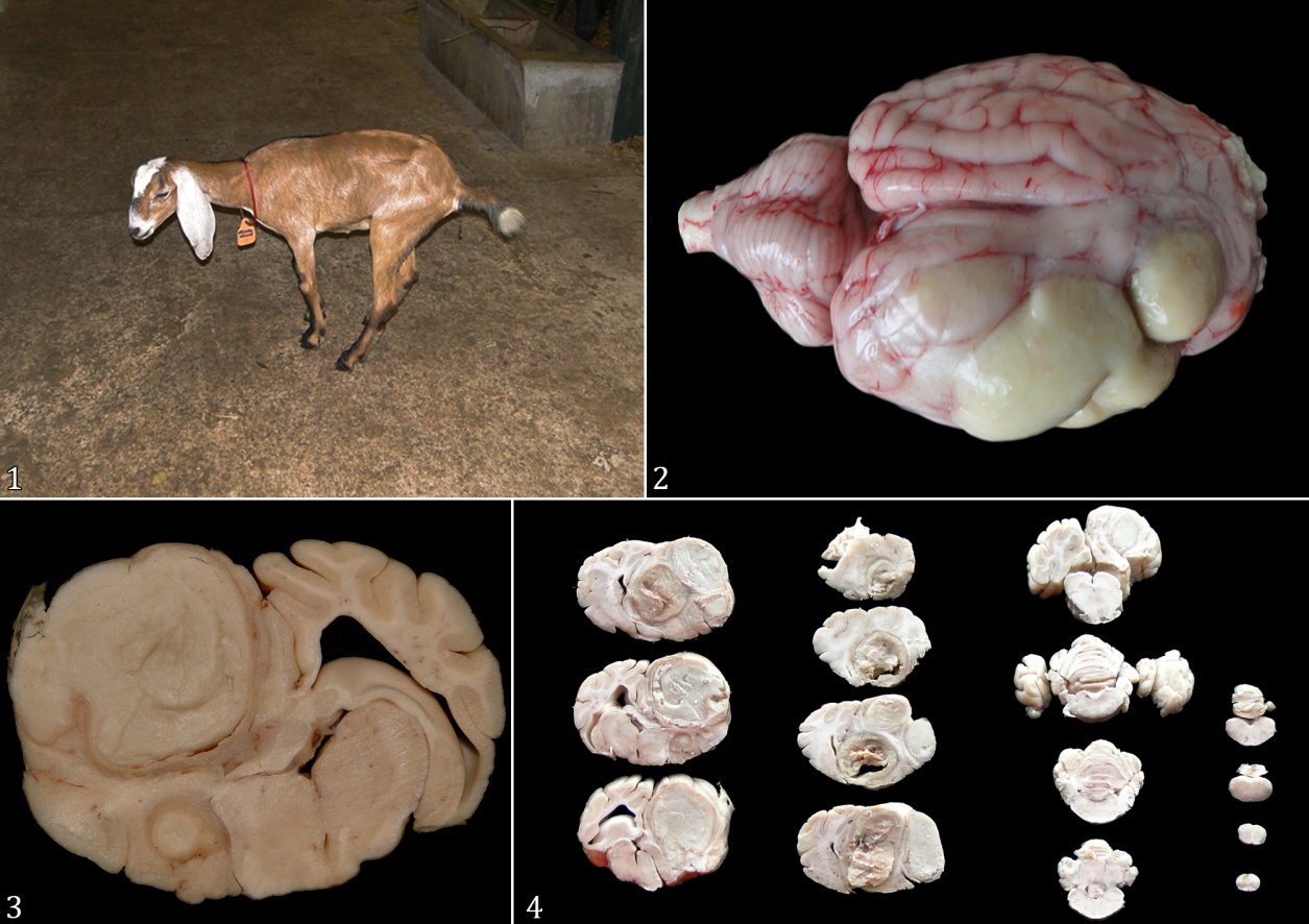
In the animals with brain damage (Sheep 1 and 2 and Goats 9 and 10), the clinical signs were decreased alertness, lateral decubitus with kicking movements, seizures, and spasticity (Fig.1). In Goat 9, impaired balance and rotational and vertical nystagmus were also noted.

Anglo-Nubian Goat 9 at two months of age. (1) Animal with the thoracic limbs facing backward and pelvic limbs forward, with a reduced support area. (2) Brain abscesses in the frontal, parietal, temporal, and occipital lobes. (3) Cross-section at the thalamus level, showing abscesses compressing the thalamus. (4) Figure 3 in cross sections after fixation in formalin.
In Sheep 3 with spinal cord injury in the C1-C5 segment, the initial clinical signs were ataxia, difficulty in remaining in a standing position, and spastic paresis of the extremities of the four limbs, mainly the pelvic limbs. However, it progressed to sternal decubitus and resulted in hyperreactive spinal pain reflexes in the extremities and loss of superficial sensitivity in the skin areas associated with the lesion.
Sheep 4 with lesions between C6 and T2 experienced flaccid paralysis of the thoracic limbs only, with no changes in the pelvic limbs.
Hyperreactive flexor reflexes, ataxia, and inability to stand on the pelvic limbs were observed in Sheep 5, 6, and 7 and Goat 11, which had an injury in the medullary segment T3-L3 (Fig.1-4).
Sheep 8 was diagnosed with a lesion in the L4-S2 segment, showing unchanged pain reflexes in the cranial segment to the lesion and decreased reflexes in the caudal segments to the lesion, ataxia, and flaccid pelvic limb paresis. The treated animals did not show a satisfactory response to the therapy.
CSF analysis and radiographic and dissection findings
Macroscopic examination of CSF from Sheep 8 with a compression lesion caused by an abscess in the medullary canal between L4 and S2 exhibited variations in the macroscopic appearance depending on the sampling site. The CSF was clear in the large cistern but yellowish in the lumbosacral space. In Sheep 3, radiographic examination revealed bony prominences directed toward the medullary canal, compressing the spinal cord, and in Goat 11, there was bone compression between segments T11 and L1.
In the dissected animals, abscesses were found in the bodies of the vertebrae projecting into the medullary canal in Sheep 4, 6, and 7; in the intervertebral disk between vertebrae L1 and L2 in Sheep 5; and in the ventral part of the medullary canal in segment L4-S2, approximately 6cm long, in Sheep 8. In the brain and the frontal, parietal, temporal, and occipital lobes, the abscesses caused an increase in the volume of the right hemisphere in Goat 9 (Fig.2-4) and parapituitary in Sheep 1. In Sheep 2 and Goat 10, there was subsidence of the frontal sinuses and a submeningeal hematoma in the brain with flattening of the frontal lobe coils. Sheep 3 displayed congenital stenosis of the medullary canal of the fifth cervical vertebra, and Goat 11 had spondylitis and ankylosis between segments T11 and L1. In addition, omphalitis occurred in Sheep 4 and 7, multiple abscesses in the lungs in Sheep 4, 5, 6, and 8, the spleen and liver in Sheep 8, and the liver in Goat 11. In addition, fibrinous pleurisy was observed in Sheep 6. The affected animals exhibited altered mental states and behavioral disturbances. Of the altered mental states, 6/11 were apathetic, 2/11 were hyperexcitable, and 2/11 were semicomatose. One animal was alert with no altered mental state (Table 1).
Discussion
This study found that of the animals treated for compressive lesions of the CNS, young sheep and adult goats were more frequently affected, although the number of cases studied was small. According to Radostits et al. (2002), abscesses in the spinal canal are common in neonates and are associated with chronic infections in other organs due to omphalitis, caudatectomy, pneumonia, and cheesy lymphadenitis lesions. These findings are consistent with those of the present study. In addition, males are at a higher risk of developing CNS lesions owing to trauma caused by aggressive behavior during fighting and head butting (Guedes et al. 2007). A similar finding was observed in Sheep 2.
The greater number of affected sheep can be explained by the predominance of sheep over goats in the demand for treatment at the Veterinary Hospital of the UFPA.
In sheep <12 months of age that were subjected to necropsy, the causes of CNS compression were abscesses that compressed the spinal cord (Sheep 4, 6, 7, and 8) and the brain (Sheep 1). In Sheep 4 and 7, in addition to CNS abscesses, inflammation of the umbilicus (omphalitis) was detected, which, according to Barros (2006), is one of the most common causes of abscesses compressing the spinal cord in newborn animals. Guedes et al. (2007) has also reported 12 cases of abscesses in the CNS of young and adult sheep and goats associated with Corynebacterium pseudotuberculosis and neonatal infection via the umbilical cord.
In Sheep 8, abscesses in the liver, lungs, and spleen, and in Sheep 5, those in the lungs indicated caseous lymphadenitis, suggesting the possibility of C. pseudotuberculosis involvement in the formation of abscesses in the medullary canal of these animals. These data are in agreement with studies performed by several other investigators in goats (Guedes et al. 2007, Rizzo et al. 2014, Santarosa et al. 2014) and sheep (Machado et al. 2003, Guedes et al. 2007, Souza et al. 2012), which also reported cases of CNS lesions due to infection with C. pseudotuberculosis.
In Sheep 8, the cause of the abscess in the medullary canal was a previous photosensitization. A deep dermatitis was found, which extended from the withers to the trunk, reached the spinous processes of the thoracic vertebrae and extended by continuity to the medullary canal, where the abscess originated, which also cannot be excluded as a cause of abscesses in the lungs, spleen, and liver. Furthermore, Ribas et al. (2013) has reported the occurrence of a vertebral abscess compressing the spinal cord of a bovine in association with dermatitis owing to heavy tick infestation, alluding the skin as the entry point for infection, similar to that noted in Sheep 8.
In 2-month-old Goat 9, the origin of the abscess that was compressing the brain could not be identified, but the animal’s caretaker indicated that it had undergone the hot iron mocha procedure four weeks before the onset of clinical signs, implying that this procedure was involved in the development of the abscess. Silva et al. (2009) has reported that hot iron mocha is used to remove a part of the corneal bud with knives and scalpels, exposing the frontal sinus and allowing pollutants to enter.
In Sheep 2 and Goat 10, which were >1 year old, compression injuries in the brain were caused by hematomas of traumatic origin. In Sheep 2, according to the animal’s tutor, the clinical signs occurred after a fight with another sheep. Necropsy revealed extensive hematomas in the frontal sinus and submeningeal space, which compressed the brain at a site corresponding to the frontal lobe. At necropsy, low bone strength of the cranial and rib bones was observed, which is typical of phosphorus-deficient animals, indicating that this deficiency could have contributed to the sinking of the cranial bones owing to the head impact. Studies conducted in buffaloes and cattle (Peixoto et al. 2005, Barbosa Neto et al. 2007, Barbosa et al 2014, 2016, 2021, Pinheiro et al. 2011, Malafaia et al. 2014, Bomjardim et al. 2015) have documented the occurrence of phosphorus deficiency in the state of Pará, reinforcing the suspicion of mineral deficiency in the animals of the present study.
In Goat 10, the hematoma might have originated from trauma sustained during handling. The fact that the animal was healthy the day before the appearance of the clinical signs associated with the head trauma noted at the necropsy supports this hypothesis.
In Goat 11, which was six years old, the cause of spinal cord compression was ankylosing spondylitis between segments T11 and L1; this consolidation was detected via radiographic examination. According to the necropsy findings of the animals in the aforementioned study, there were various causes for the formation of abscesses and hematoma, which compressed the CNS of goats and sheep. Barros (2006) and Mayhew (2008) have reported the presence of four main pathways for the penetration of an infectious agent into the CNS, namely, via the extension of an adjacent purulent lesion, via direct penetrating lesions, via centripetal lesions through peripheral nerves, and the hematogenous route.
In Sheep 3, bone deformity in the C1-C5 segment was responsible for spinal cord compression, which was evident on radiography and confirmed in necropsy. The occurrence of injury in this segment has also been reported by Barbosa et al. (2022a), Mackay & Van Metre (2015), and Borges et al. (2003).
The lesions diagnosed between the T3 and L3 vertebrae in this retrospective study were most common in sheep. These lesions were also observed by Mackay & Van Metre (2015) in calves in the thoracolumbar segment owing to abscesses, by Borges et al. (2003) in cattle with vertebral fractures, and by Barbosa et al. (2022a, 2022b) in retrospective studies of compression lesions in the CNS of cattle and buffalo in the Amazon. According to Hahn et al. (1999), the greater frequency of injuries between the T3 and L3 segments can be explained by the fact that this thoracic region exhibits a higher dorsoventral movement. Moreover, it is the main support point of body weight, demonstrating the predisposition of this anatomical region to the occurrence of CNS compression injuries.
The clinical signs exhibited by animals with lesions in the brain and brainstem are due to loss of function in the nuclei and pathways that regulate these activities in the corresponding regions (Riet-Correa et al. 2002, Borges et al. 2003). Spinal cord injuries vary depending on the segment affected and the extent of damage. In most cases, these lesions caused ataxia, paresis, or paralysis of the limbs; inability to stand up and remain standing; postural changes; and increased spinal reflexes. These lesions also caused loss of skin sensitivity in relation to their site of occurrence. These findings have also been reported by several authors (Riet-Correa et al. 2002, Borges et al. 2003, Rissi et al. 2010, Câmara et al. 2014, Barbosa et al. 2022a, 2022b) who mentioned the same anatomical and clinical correlates of CNS lesions. In Sheep 3 with spinal cord injury between C1 and C5, the observed changes in both fore and hind legs were due to damage to the upper limb motor neurons. Injury to these segments can result in paresis to paralysis, with loss of voluntary movements, normal to exaggerated reflexes, late muscle atrophy, normal to increased muscle tone, decreased proprioception, and decreased perception of superficial and deep pain. Furthermore, tetraparesis develops when the lesion is bilateral (Lahunta & Glass 2009).
Sheep 4 with a cervical swelling lesion (C6-T2) was expected to exhibit severe alterations in both thoracic and pelvic limbs. According to Riet-Correa et al. (2002) and Barbosa et al. (2022a), when the lesions are localized in the cervical swelling area and affect only the lower motor neurons, flaccid paralysis is observed exclusively in the forelimbs, which explains the clinical conditions in this animal.
The opacity with flocculent deposits in the CSF observed on macroscopic examination of Sheep 8 with a compression lesion caused by an abscess in the medullary canal is consistent with the findings of Barbosa et al. (2022a, 2022b), Pinheiro et al. (2018), Câmara et al. (2014), and Barros (2006) in ruminants. These researchers stated that a cloudy and viscous CSF can be observed in inflammatory processes such as spinal cord abscesses, meningitis, and brain abscesses, similar to those observed in the sheep studied. CSF analysis must consider the location of the lesion. In addition, according to Smith (2002), CSF may be normal if the abscess does not infiltrate the dura mater.
The treatment was performed in only two animals because of their poor prognosis, unlike the other animals in the study, which had an unfavorable prognosis. Furthermore, the unsatisfactory response to this treatment was due to the severity of the injuries that these two animals presented, which was evident during the necropsy. The diagnosis of CNS compression was confirmed via necropsy in all animals.
Conclusion
In this retrospective study in goats and sheep, compressive lesions of the central nervous system (CNS) affected the animals at different ages with varied etiologies and locations, demonstrating the importance of clinical examinations and necropsy findings in confirming the disease and the differential diagnosis.
References
-
Barbosa F.B., Bomjardim H.A., Helayel M.J.S.A., Faial K.C.F., Oliveira C.M.C., Malafaia P., Brito M.F. & Barbosa J.D. 2016. Avaliação econômica de três tipos de suplementação mineral para bovinos de corte no Estado do Pará. Pesq. Vet. Bras. 36(7):600-604. <https://dx.doi.org/10.1590/S0100-736X2016000700007>
» https://doi.org/https://dx.doi.org/10.1590/S0100-736X2016000700007 -
Barbosa J.D., Brito M.F., Bomjardim H.A., Duarte M.D., Barbosa C.C., Silveira N.S.S., Oliveira C.M.C. & Salvarani F.M. 2022a. Compressive lesions in the central nervous system of buffaloes. Pesq. Vet. Bras. 42:e07060. <https://dx.doi.org/10.1590/1678-5150-PVB-7060>
» https://doi.org/https://dx.doi.org/10.1590/1678-5150-PVB-7060 -
Barbosa J.D., Brito M.F., Duarte M.D., Albernaz T.T., Bomjardim H.A., Barbosa C.C., Oliveira C.M.C. & Salvarani F.M. 2022b. Compressive lesions in the central nervous system of cattle: a retrospective study of 50 cases in the Amazon biome. Pesq. Vet. Bras. 42:e07057. <https://dx.doi.org/10.1590/1678-5150-PVB-7057>
» https://doi.org/https://dx.doi.org/10.1590/1678-5150-PVB-7057 -
Barbosa J.D., Brito M.F., Oliveira C.M.C., Duarte M.D., Silveira N.S.S., Ferreira T.T.A., Silveira J.A.S., Bomjardim H.A., Barbosa C.C. & Malafaia P.A.M. 2021. Deficiências minerais em bovinos e bubalinos no Brasil: aspectos gerais, importância, diagnóstico, profilaxia e correção. Revta Bras. Buiatria 2(1):1-38. <https://dx.doi.org/10.4322/2763-955X.2021.010>
» https://doi.org/https://dx.doi.org/10.4322/2763-955X.2021.010 -
Barbosa J.D., Lima D.H.S., Belo-Reis A.S., Pinheiro C.P., Sousa M.G.S., Silva J.B., Salvarani F.M. & Oliveira C.M.C. 2014. Degenerative joint disease in cattle and buffaloes in the Amazon region: a retrospective study. Pesq. Vet. Bras. 34(9):845-850. <https://dx.doi.org/10.1590/S0100-736X2014000900007>
» https://doi.org/https://dx.doi.org/10.1590/S0100-736X2014000900007 -
Barbosa Neto J.D., Brito M.F., Duarte M.D., Albernaz T.T., Oliveira Júnior C.A., Riet-Correa G. & Riet -Correa F. 2007. Phosphorus deficiency in buffaloes in the state of Pará, northern Brazil. Ital. J. Anim. Sci. 6(2):971-973. <https://dx.doi.org/10.4081/ijas.2007.s2.971>
» https://doi.org/https://dx.doi.org/10.4081/ijas.2007.s2.971 - Barros C.S.L., Driemeier D., Dutra I.S. & Lemos R.A.A. 2006. Doenças do Sistema Nervoso de Bovinos no Brasil. Vallée, Montes Claros. 207p.
-
Bomjardim H.A., Oliveira C.M.C., Silveira J.A.S., Silva N.S., Duarte M.D., Faial K.C.F., Brito M.F. & Barbosa J.D. 2015. Deficiências minerais em vacas em lactação da bacia leiteira do município de Rondon do Pará, estado do Pará. Pesq. Vet. Bras. 35(5):409-416. <https://dx.doi.org/10.1590/S0100-736X2015000500004>
» https://doi.org/https://dx.doi.org/10.1590/S0100-736X2015000500004 -
Borges A.S., Silva D.P.G., Gonçalves R.C., Chiacchio S.B., Amorim R.M., Kuchembuck M.R.G., Vulcano L.C., Bandarra E.P. & Lopes R.S. 2003. Fraturas vertebrais em grandes animais: estudo retrospectivo de 39 casos (1987-2002). Arq. Bras. Med. Vet. Zootec. 55(2):127-132. <https://dx.doi.org/10.1590/S0102-09352003000200001>
» https://doi.org/https://dx.doi.org/10.1590/S0102-09352003000200001 -
Câmara A.C.L., Vale A.M., Batista J.S., Feijó F.M.C. & Soto-Blanco B. 2014. Suppurative intracranial processes in 15 domestic ruminants. Pesq. Vet. Bras. 34(5):421-426. <https://dx.doi.org/10.1590/S0100-736X2014000500006>
» https://doi.org/https://dx.doi.org/10.1590/S0100-736X2014000500006 -
Cardoso F.M., Pedroso P.M.O., Santos A.S., Vilanova M.S., Driemeier D. & Schmidt V. 2009. Lesão medular associada ao manejo incorreto de caprino. Acta Scient. Vet. 37(2):187-189. <https://dx.doi.org/10.22456/1679-9216.16249>
» https://doi.org/https://dx.doi.org/10.22456/1679-9216.16249 - Feitosa F.L.F. 2014. Semiologia Veterinária: a arte do diagnóstico. 3ª ed. Ed. Roca, São Paulo. 1406p.
-
Guedes K.M.R., Riet-Correa F., Dantas A.F.M., Simões S.V.D., Miranda Neto E.G., Nobre V.M.T. & Medeiros R.M.T. 2007. Doenças do sistema nervoso central em caprinos e ovinos no semi-árido. Pesq. Vet. Bras. 27(1):29-38. <https://dx.doi.org/10.1590/S0100-736X2007000100006>
» https://doi.org/https://dx.doi.org/10.1590/S0100-736X2007000100006 - Hahn C.N., Mayhew I.G. & Mackay R.J. 1999. Diseases of multiple or unknown sites, p.947-950. In: Colahan P.T., Mayhew I.G., Merritt A.M. & Moore J.N. (Eds), Equine Medicine and Surgery. Vol.1. 5th ed. Mosby, St. Louis.
-
Helmer J.F., Oliveira C.A., Cerqueira V.D., Bezerra P.S., Prado R.G.S., Pires C.R.S. & Riet-Correa G. 2020. Caracterização dos sistemas de produção de ovinos e caprinos na microrregião de Castanhal, Pará. Med. Vet., UFRPE, 14(3):202-209. <https://dx.doi.org/10.26605/medvet-v14n3-3862>
» https://doi.org/https://dx.doi.org/10.26605/medvet-v14n3-3862 -
IBGE 2022a. Rebanho de caprinos (bodes e cabras). Instituto Brasileiro de Geografia e Estatística, Rio de Janeiro. Available at <Available at https://www.ibge.gov.br/explica/producao-agropecuaria/caprinos/br > Accessed on Oct. 1, 2023.
» https://www.ibge.gov.br/explica/producao-agropecuaria/caprinos/br -
IBGE 2022b. Rebanho de ovinos (ovelhas e carneiros). Instituto Brasileiro de Geografia e Estatística, Rio de Janeiro. Available at <Available at https://www.ibge.gov.br/explica/producao-agropecuaria/ovino/br > Accessed on Oct. 1, 2023.
» https://www.ibge.gov.br/explica/producao-agropecuaria/ovino/br - Lahunta A. & Glass E. 2009. Veterinary Neuroanatomy and Clinical Neurology. 3rd ed. Elsevier, Amsterdam. 540p.
- Machado G.M., Moscardini A.R.C., Araújo G.R.S., Albuquerque P.I., Paludo G.R., Perecmanis S., Reis Jr. J.L. & Borges J.R.J. 2003. Compressão da medula espinhal por abscesso de Corynebacterium pseudotuberculosis em ovino: relato de caso. Anais do V Congresso Brasileiro de Buiatria, Salvador, BA. (Resumo)
- Mackay R.J. & Van Metre D.C. 2015. Diseases of the nervous system, p.917-1043. In: Smith B.P. (Ed.), Large Animal Internal Medicine. 5th ed. Mosby, St. Louis.
-
Malafaia P., Costa R.M., Brito M.F., Peixoto P.V., Barbosa J.D., Tokarnia C.H. & Döbereiner J. 2014. Equívocos arraigados no meio pecuário sobre deficiências e suplementação minerais em bovinos no Brasil. Pesq. Vet. Bras. 34(3):244-249. <https://dx.doi.org/10.1590/S0100-736X2014000300008>
» https://doi.org/https://dx.doi.org/10.1590/S0100-736X2014000300008 - Mayhew J. 2008. Infectious, inflammatory and immune diseases, p.225-293. In: Mayhew J. (Ed.), Large Animal Neurology. 2nd ed. Wiley-Blackwell, Singapore.
-
Nagy D.W. & Pugh D.G. 2012. Handling and examining sheep and goats, p.1-17. In: Pugh D.J. & Baird A.N. (Eds), Sheep and Goat Medicine. 2nd ed. Elsevier, St. Louis. <https://dx.doi.org/10.1016/B978-1-4377-2353-3.10001-0>
» https://doi.org/https://dx.doi.org/10.1016/B978-1-4377-2353-3.10001-0 -
Novais E.P.F. 2013. Doenças com sinais neurológicos em ovinos e caprinos no Distrito Federal e entorno. Dissertação de Mestrado, Universidade de Brasília, Brasília. 115p. Available at <Available at https://repositorio.unb.br/bitstream/10482/14331/1/2013_ErnanePaivaFerreiraNovais.pdf > Accessed on Dec. 29, 2023.
» https://repositorio.unb.br/bitstream/10482/14331/1/2013_ErnanePaivaFerreiraNovais.pdf -
Peixoto P.V., Malafaia P.A., Barbosa J.D. & Tokarnia C.H. 2005. Princípios de suplementação mineral em ruminantes. Pesq. Vet. Bras. 25(3):195-200. <https://dx.doi.org/10.1590/S0100-736X2005000300011>
» https://doi.org/https://dx.doi.org/10.1590/S0100-736X2005000300011 -
Pinheiro C.P., Bomjardim H.A., Andrade S.J.T., Faial K.C.F., Oliveira C.M.C. & Barbosa J.D. 2011. Níveis de fósforo, cobre, cobalto e zinco em bubalinos (Bubalus bubalis) na Ilha de Marajó, Estado do Pará. Pesq. Vet. Bras. 31(3):193-198. <https://dx.doi.org/10.1590/S0100-736X2011000300002>
» https://doi.org/https://dx.doi.org/10.1590/S0100-736X2011000300002 -
Pinheiro R.R., Brito R.R.L.L., Andrioli A., Alves F.S.F., Silva Neto J. & Gomes F.P. 2018. Meningite bacteriana em ovinos-relato de caso. Revta Bras. Saúde Prod. Anim. 12(1):92-100. <https://dx.doi.org/10.5935/1981-2965.20180010>
» https://doi.org/https://dx.doi.org/10.5935/1981-2965.20180010 - Radostits O.M., Gay C.C., Blood D.C. & Hinchcliff K.W. 2002. Clínica Veterinária: um tratado de doenças dos bovinos, ovinos, suínos, caprinos e eqüinos. 9ª ed. Guanabara Koogan, Rio de Janeiro. 1737p.
-
Ribas N.L.K.S., Carvalho R.I., Santos A.C., Valençoela R.A., Gouveia A.F., Castro M.B., Mori A.E. & Lemos R.A.A. 2013. Doenças do sistema nervoso de bovinos no Mato Grosso do Sul: 1082 casos. Pesq. Vet. Bras. 33(10):1183-1194. <https://dx.doi.org/10.1590/S0100-736X2013001000003>
» https://doi.org/https://dx.doi.org/10.1590/S0100-736X2013001000003 -
Riet-Correa F., Riet-Correa G. & Schild A.L. 2002. Importância do exame clínico para o diagnóstico das enfermidades do sistema nervoso em ruminantes e eqüídeos. Pesq. Vet. Bras. 22(4):161-168. <https://dx.doi.org/10.1590/S0100-736X2002000400006>
» https://doi.org/https://dx.doi.org/10.1590/S0100-736X2002000400006 -
Rissi D.R., Pierezan F., Oliveira-Filho J.C., Lucena R.B., Carmo P.M.S. & Barros C.S.L. 2010. Abordagem diagnóstica das principais doenças do sistema nervoso de ruminantes e equinos no Brasil. Pesq. Vet. Bras. 30(11):958-967. <https://dx.doi.org/10.1590/S0100-736X2010001100010>
» https://doi.org/https://dx.doi.org/10.1590/S0100-736X2010001100010 -
Rizzo H., Carvalho J.S., Fraga G.J.M., Lauria M.J.S. & Languidey P.H. 2014. Paralisia de membros pélvicos em reprodutor caprino causada por linfadenite caseosa no estado de Sergipe. Pubvet 8(22):2675-2805. <https://dx.doi.org/10.22256/pubvet.v8n22.1812>
» https://doi.org/https://dx.doi.org/10.22256/pubvet.v8n22.1812 - Santa Rosa J. & Santa Rosa M.D.G. 1999. Inflamações supuradas e granulomatosas no sistema nervoso de caprinos. Ciênc Vet. Trop. 2(2):108-114.
- Santarosa B.P., Dantas G.N., Amorim R.L., Chiacchio S.B., Oliveira Filho J.P., Amorim R.M., Ribeiro M.G. & Gonçalves R.C. 2014. Meningoencefalite supurativa por Corynebacterium pseudotuberculosis em cabra com linfadenite caseosa: relato de caso. Vet. Zootec. 21(4):537-542.
-
Silva L.A.F., Teixeira Neto A.R., Campos S.B.S., Brazil D.S., Helou J.B., Pucci R.L., Caetano L.B., Maranhão R.P.A. & Brandstetter L.R.G. 2009. Aspectos epidemiológicos da sinusite pós-descorna plástica ou mochação com ferro candente em bovinos. Acta Scient. Vet. 37(4):357-362. <https://dx.doi.org/10.22456/1679-9216.16400>
» https://doi.org/https://dx.doi.org/10.22456/1679-9216.16400 - Smith B.P. 2002. Large Animal Internal Medicine. Mosby, St. Louis, p.982-984.
- Souza S.O., Lemos L.A., Wouters A.T.B., Wouters F., Boabaid F.M., Casagrande R.A., Watanabe T.T.N., Guerra P. & Driemeier D. 2012. Linfadenite caseosa como causa de paralisia de membros pélvicos em ovinos no estado do Rio Grande do Sul. Acta Scient. Vet. 40(4):1085.