
 Mortality causes of Pitheciidae (order: Primates) species kept under human care
ABSTRACT:
Brazilian pitheciids include some of the world’s most endangered nonhuman primate species. Despite current efforts to conserve wild populations, no retrospective studies have described the causes of death in members of the Pitheciidae family kept under human care. Herein, we describe the spontaneous causes of mortality in pitheciids maintained under human care in Rio de Janeiro, Brazil. To this end, the post mortem reports of the Anatomy Pathology Sector of Federal University Rural of Rio de Janeiro (SAP/UFRuralRJ) from January 2019 to December 2024 were reviewed. During this period, we performed 497 necropsies and histopathological examinations in nonhuman primates, of which 26 were Pitheciidae. All individuals were adults; 61.5% (16/26) were females and 38.5% (10/26) were males. A conclusive diagnosis of the cause of death was made in 76.9% (20/26) of the cases. Regardless of the process category, the hepatobiliary system was the most affected in this study. The deaths were attributed to infectious causes in 55% (11/20) of the cases, noninfectious in 25% (5/20), and inflammatory with no specific etiology in 20% (4/20). Parasitic diseases were important causes of pitheciid illness in the captive scenario, mainly due to metazoan and protozoan infections. This translational study provides important information on the dynamics of diseases that affect pitheciids and may serve as a tool to improve these species’ prevention, management and conservation strategies.
INDEX TERMS:
Biodiversity conservation; conservation medicine; veterinary pathology; neotropical primates; primate diseases
RESUMO:
Os pitecídeos brasileiros incluem algumas das espécies de primatas não humanos mais ameaçadas do mundo. Apesar dos esforços atuais para a conservação de populações silvestre, nenhum estudo retrospectivo descreveu as causas de mortalidade de primatas não humanos da família Pitheciidae que são mantidos sob cuidados humanos. Aqui, descrevemos as causas de morte espontânea de pitecídeos mantidos sob cuidados humanos no Rio de Janeiro, Brasil. Os laudos post mortem do Setor de Anatomia Patológica da Universidade Federal Rural do Rio de Janeiro (SAP/UFRuralRJ) de janeiro de 2019 a dezembro de 2024 foram revisados. Neste período, foram realizadas 497 necropsias e exames histopatológicos em primatas não humanos e 26 espécimes de Pitheciidae foram incluídos neste estudo. Todos os indivíduos eram adultos, 61,5% (16/26) eram fêmeas e 38,5% (10/26) eram machos. Foi possível determinar a causa da morte em 76,9% (20/26) dos casos. Independentemente da categoria do processo, o sistema hepatobiliar foi o mais afetado neste estudo. As mortes foram atribuídas a causas infecciosas em 55% (11/20) dos casos, não infecciosas em 25% (5/20) e inflamatórias sem etiologia específica em 20% (4/20). Doenças parasitárias foram importantes causas de doenças de pitecídeos mantidos sob cuidados humanos, especialmente devido a infecções por metazoários e protozoários. Este estudo retrospectivo fornece informações importantes sobre a dinâmica de doenças que afetam os pitecídeos e servirá como uma ferramenta para melhorar as estratégias de manejo e conservação para essas espécies.
TERMOS DE INDEXAÇÃO:
Conservação da biodiversidade; medicina da conservação; patologia veterinária; primatas neotropicais; doenças de primatas
Introduction
Brazil remains the country with the greatest neotropical primate wealth in the world (Mittermeier et al. 2013). Despite the admirable magnitude of nonhuman primate diversity, threats impacting primate populations in Brazil are notable (Rylands & Mittermeier 2024). Government initiatives have been implemented in Brazil, considering the conservation of free-ranging species at risk of extinction (INEA 2015). However, the plans proposed at this time have a primary focus on anthropogenic implications, such as hunting, deforestation, habitat fragmentation, electrocution, and predation by domestic fauna (Peres 2001, Chiarello et al. 2008, Ribeiro et al. 2009, Canale et al. 2012).
The family Pitheciidae includes six genera in two subfamilies distributed over tropical South America (Fleagle & Seiffert 2020, Rylands & Mittermeier 2024). The subfamily Callicebinae comprises 34 species popularly known as titi monkeys, which are distributed in three genera: Callicebus, Cheracebus and Plecturocebus. The subfamily Pitheciinae has three genera: the saki monkeys (Pithecia), the bearded sakis (Chiropotes), and the uakaries (Cacajao). From the 2000s, neotropical primates’ surveys have resulted in Pitheciidae new species described as nine titi monkeys (eight of the genus Plecturocebus and one Cheracebus), five sakis (Pithecia) and three uacaris (Cacajao) (Byrne et al. 2016, Rylands & Mittermeier 2024).
Brazilian pitheciids include some of the world’s most endangered nonhuman primate species. According to the International Union for Conservation of Nature (IUCN) red list, 37.5% of the Pitheciidae taxa are potentially threatened. The pitheciids are all arboreal quadrupeds and leapers; many specialize as seed predators (Fleagle & Seiffert 2020). Habitat fragmentation is a key topic in the conservation of free-ranging pitheciidae species due to the high degree of arboreal specialization (Ferrari et al. 2013). Despite current efforts to conserve wild populations, few project strategies are focusing on conservation for Pitheciidae species kept under human care.
The only robust study that describes the mortality causes of Pitheciidae species was conducted on a population of Vieira’s titi monkey (Plecturocebus vieirai) kept at the Sorocaba Zoo, Brazil (Silva et al. 2024a). The currently available literature on fatal pitheciid diseases consists of case reports or case series of infectious diseases. Fatal herpes simplex virus-1 infection was reported in a family of white-faced saki monkeys (Pithecia pithecia) (Schrenzel et al. 2003). Yellow fever natural infection was described as the cause of titi monkeys (Callicebus spp.) deaths due to an epidemic wave in Brazil (Fernandes et al. 2021).
Understanding disease dynamics in the captive scenario is crucial for implementing future conservation, management and reintroduction strategies and actions for vulnerable populations. This retrospective study aims to describe the spontaneous causes of mortality of Pitheciidae nonhuman primates kept under human care in Rio de Janeiro, Brazil.
Materials and Methods
Ethical approval. All procedures were conducted in full compliance with the institutional and national ethical standards, including the approval by the Brazilian Ministry of the Environment (SISBIO 30939-12). The breeding colony of the “Centro de Primatologia do Rio de Janeiro” (22o29’14.51” S, 42o48’78” W) is authorized by “Instituto Brasileiro do Meio Ambiente e dos Recursos Naturais Renováveis” (IBAMA), under number 4989806, “Instituto Estadual do Ambiente do Rio de Janeiro” (INEA/RJ).
The post mortem reports of the “Setor de Anatomia Patológica” (Anatomy Pathology Sector - SAP) of “Universidade Federal Rural do Rio de Janeiro” (UFRuralRJ) from January 2019 to December 2024 were reviewed. All necropsy cases on specimens from Pitheciidae family (order: Primates) and maintained under human care were included in this study. The epidemiological data were acquired from the responsible technical veterinarian of the colonies. The cases that permitted the establishment of the cause of death were considered conclusive, and those without significant clinical history and morphological changes were deemed inconclusive. Based on the clinicopathological findings and ancillary tools, the cause of death of each case was categorized by body system and grouped into the following categories: infectious, noninfectious, and inflammatory with no specific etiology. The pathological findings of each case that were not directly related to the cause of death were considered comorbidities.
The infectious cases exhibited morphological evidence of metazoan structures, tachyzoites, bradyzoites, or alcohol-acid-resistant bacilli, whose etiology was confirmed by morphological, immunohistochemical or molecular methods or bacterial culture. The cases with morphological changes compatible with metabolic or toxin involvement, as well as neoplastic proliferations, were considered noninfectious. Inflammatory cases with no specific etiology were defined as cases in which it was not possible to associate the inflammatory reaction with a cause.
Necropsy and histopathology. In the cases of natural death or euthanasia in nonhuman primates, complete standardized necropsies were performed by two veterinary pathologists of the SAP/UFRuralRJ or by the colony technical responsible. During the procedure, personal protective equipment was used. A complete set of tissue samples including skin, skeletal muscle, tongue, esophagus, thyroid, trachea, lungs, heart, lymph nodes, liver, gallbladder, spleen, adrenal gland, kidneys, urinary bladder, stomach, small and large intestines, pancreas, and brain were collected from each primate and fixed in a 10% buffered formalin solution. The tissue samples stored in formalin were fixed for 24-48 hours and submitted to routine histological processing. Histological slides were stained with hematoxylin and eosin (HE) and then observed under optical microscopy. Histological sections with lesions morphologically compatible with tuberculosis were submitted to the Ziehl-Neelsen histochemical technique for evidence of acid-alcohol-resistant bacilli.
Immunohistochemistry. Tissue blocks of selected cases were cut into 3 μm slices and mounted onto silane-coated microscope slides. Infectious cases with morphological evidence of tachyzoites and bradyzoites, and in cases with granulomatous changes and with evidence of resistant alcohol-acid bacilli, were submitted to immunohistochemistry (IHC) using polyclonal antibodies, anti-Toxoplasma gondii (Dako®, Carpinteria, California, USA, dilution at 1:200) and anti-Mycobacterium tuberculosis (GeneTex, Inc. Cat. No. GTX 20905, dilution at 1:200), respectively, according to previous standardized protocols (Pereira et al. 2022, Schiffler et al. 2023). Positive controls for the reactions include a confirmed case of toxoplasmosis in Brachyteles arachnoides (Schiffler et al. 2023) and a confirmed case of tuberculosis in Chiropotes utahickae (Pereira et al. 2022). The IHC was also made by convenience in the neoplastic cases according to the manufacturers with minor modifications using anti-pan cytokeratin (Dako®, cloneAE1/AE3, ready to use), cytokeratin-7 (Dako®, Clone 7, dilution at 1:200), and anti-Hep-Par1 (Dako®, clone OCH1E5, dilution at 1:50). Normal skin tissue of a Callicebus personatus, a biliary adenocarcinoma of Leontopithecus chrysopygus, and a normal liver of a rhesus monkey were employed as a positive control for pan-cytokeratin, cytokeratin-7, and hep-par1, respectively. For negative controls, phosphate-saline buffer (PBS) replaced the primary antibody.
Molecular analysis. Selected cases with morphological evidence of hepatic trematode infection were employed in molecular analyses. DNA was extracted from duplicate 25 mg fragments of each liver tissue sample (n = 5) using the Qiagen Blood and Tissue Kit, following the manufacturer’s instructions. Extracted DNA was quantified using a Nanodrop 2000 spectrophotometer, and the concentration of all samples was adjusted to 50 ng/µL by dilution in AE buffer. The DNA (2 µL in a reaction volume of 25 µL) was examined using a nested polymerase chain reaction (nested PCR) approach. The first round of amplification was conducted using the primers 18SF (5’-CCT GGT AAG TGC AAG TCA GATGC-3’) and 5.8SR (5’-CAT GGC CGC AAT ATG CTT GCA-3’) in combination with the master mix and thermocycling conditions reported by Nguyen et al. (2017). This generated an amplicon of approximately 1100 base pairs (bp), comprising partial sequences of the genes encoding the 18S and 5.8S subunits of ribosomal RNA, as well as the full-length internal transcribed spacer region (ITS1) of Platynosomum illiciens. The nested PCR employed 2 µL of the first-round amplification products as a template. The master mix contained the primers nestF1 (5’-TCA GCT GGT GGC TGA CAT TA-3’) and nestR2 (5’-AGT GAT CCA CCG CTC AGA GT-3’), with MgCl2 at a final concentration of 2 mM. Cycling conditions were identical to those used in the first-round reaction, except that 25 cycles were used instead of 40. The presence of a product of 896 bp was determined via comparison to molecular weight standards on 1.5% agarose gels stained with ethidium bromide.
Amplicons were sequenced in both directions using the BigDye™ Terminator v3.1 Cycle Sequencing Kit in combination with the amplification primers and two internal primers (PlatSeq1: 5’-CCG CCT GGA TAT TTG AGT GT-3’ and PlatSeq2: 5’-ACA CTC AAA TAT CCA GGC GG-3’). Sequences were entered into the Basic Local Alignment Search Tool (BLAST) search algorithm and the National Center for Biotechnology Information (NCBI) nucleotide database to determine their identity.
Results
From routine pathological diagnosis at the SAP/UFRuralRJ, we performed 497 necropsies and histopathological examinations in nonhuman primates from January 2019 to December 2024. Twenty-six nonhuman primates were Pitheciidae specimens kept under human care and included in this research.
The pitheciids were from two Rio de Janeiro state colonies: 20 cases from the “Centro de Primatologia do Rio de Janeiro” (Rio de Janeiro Primatology Center), and six cases from the “Bioparque do Rio”, located in Guapimirim and Rio de Janeiro municipalities, respectively. All individuals were adults; 61.5% (16/26) were females and 38.5% (10/26) were males. A conclusive diagnosis with the determination of the death cause was made in 76.9% (20/26) of the cases.
Regardless of the process category in the conclusive cases, the hepatobiliary system was the most affected in this study, with 50% (10/20) of the deaths attributed to severe liver damage, followed by multisystemic infectious conditions in 20% (4/20). In 15% (3/20) of the cases, the cause of death was related to the respiratory system, 5% (1/20) to the digestive system, 5% (1/20) to the renal system, and 5% (1/20) to the female reproductive system.
The deaths were attributed to infectious causes in 55% (11/20) of the cases, noninfectious in 25% (5/20), and inflammatory without specific etiology in 20% (4/20). The parasitic diseases represented 81.8% (9/11) of the infectious cases, with 55.5% (5/9) due to metazoan and 44.5% (4/9) protozoan infections. The remaining 18.2% (2/9) infectious death causes were attributed to resistant alcohol-acid bacilli infection. The species, sex, age, cause of death by body system, category process and comorbidities of each case are shown in Tables 1-3.
Infectious death causes
Within the metazoan infectious cases, grossly, four Pithecia irrorata showed diffuse and severe hepatic size decrease, which were yellow to tan, firm and with multifocal to coalescing micro-nodulations (Fig. 1). There were no other significant macroscopic changes in these cases. Microscopically, severe chronic cholangiohepatitis with bridging fibrosis and micronodular hyperplasia was noted. In three cases, multiple intraductal elliptical structures were observed, ranging from 100 to 150 μm, with a thin eosinophilic cuticle, a body filled with parenchyma containing paired ceca, testis, vitellaria, and a uterus filled with light brown eggs with refringent shell, measuring on average 15 μm. These structures were morphologically compatible with adult trematodes, and the molecular examination confirmed the presence of Platynosomum fastosum (Fig. 2).

Platynosomiasis in a Pithecia irrorata. (1) Multifocal to coalescing yellow to tan, firm micro-nodulations on the hepatic surface. Inset: Hepatic cut surface with multifocal to coalescing and irregular nodules, and a transverse section of a thickened white biliary ductus. (2) Chronic cholangiohepatitis at hepatic portal space. Note intraductal trematodes with oral sucker (OS), pharynx (P), bilateral and paired cecum (C), uterus (U) and testis(T). HE, obj. 10x.
Upon molecular analysis, amplicons of the expected size (896 bp) were obtained from the hepatic samples. Sequence analysis demonstrated that these samples were identical, sharing 100% nucleotide similarity (896 bases/896 bases) with the sequences KU987972 and KU987674, deposited in the GenBank database as P. fastosum internal transcribed spacer 1 and 5.8S ribosomal RNA gene, derived from isolated parasites collected from feline liver in Vietnam. In addition, it novel sequences showed 100% nucleotide similarity (874 bases/874 bases) to the sequences MH156564 and MH156567 deposited in the Genbank as representing small subunit ribosomal RNA gene and internal transcribed spacer 1 of Platynosomum illiciens detected a House Geko (Hemidactylus mabouia), in the state of Ceará, Brazil (MH156564) or detected in a domestic cats (Felis catus) in the state of Minas Gerais, Brazil. The novel sequence obtained from the sample was deposited in GenBank under the accession number PV157520.
In one case of severe granulomatous hepatitis with bridging fibrosis, there is no microscopic evidence of adult parasites. There were, amid the granulomatous reaction of the hepatic parenchyma, a large number of barrel-shaped structures, with an average of 70 x 35 μm, with a thick, bi-operculated shell with radial striations and either an eosinophilic morula or small amounts of amorphous granular basophilic material (Fig. 3-4). Based on the morphological findings of the eggs, a diagnosis of Capillaria sp. was made.
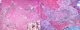
Hepatic capillariasis in a Pithecia irrorata. (3) Severe granulomatous hepatitis with bridging fibrosis and a large amount of intralesional eggs. HE, obj. 10x. Inset: bi-operculated egg with radial striations and eosinophilic morula. HE, obj. 40x (4) Marked evidentiation of collagen in the hepatic parenchyma. Masson’s Trichrome, obj. 10x.
A specimen of Callicebus personatus at necropsy showed lungs that failed to collapse and were heavy, wet, firm, consolidated, and red. The pulmonary parenchyma was filled with a large amount of foamy-white fluid on the cut surface. There was moderate hepatomegaly and splenomegaly, and a filiform structure of 7 cm in length was found free in the abdominal cavity. Microscopically, the alveolar septa are expanded by lymphocytes and histiocytes and are interspersed with many microfilariae. Those microfilariae were filiform structures without musculature, measuring 3-4 μm in width, with a thin amphophilic cuticle of 1 μm, and were filled with a myriad of basophilic nuclei of 1 x 2 μm. The microfilariae were also seen in the vascular lumen in the liver and spleen, causing moderate lymphohistiocytic hepatitis and splenitis, respectively. A filariasis diagnosis was made based on the microscopical morphological evidence of microfilariae.
During an outbreak of Toxoplasma gondii at the “Centro de Primatologia do Rio de Janeiro”, three pithecid specimens died, with the pathological data previously reported by Schiffler et al. (2023). An additional case was incorporated into the current retrospective study. A specimen of P. irrorata died without a clinical history. The liver was markedly enlarged upon necropsy with multifocal and irregular white areas surrounded by red halos at the capsular and cut surface. Microscopically, there was an acute, moderate, multifocal necrohemorrhagic hepatitis with intalesional protozoal tachyzoites (Fig. 5). Immunohistochemistry using anti-T. gondii antibodies revealed many free tachyzoites and bradyzoite cysts within the hepatocellular necrotic areas (Fig. 6).

Toxoplasmosis in a Plecturocebus caligatus. (5) Hepatic necrosis with intralesional tachyzoites. HE, obj. 40x. Inset: multiple intracytoplasmic tachyzoites in a macrophage. HE, obj. 40x. (6) Multifocal strong immunolabeling of bradyzoite cysts and tachyzoites within the hepatocellular necrosis area. Immunohistochemistry anti-Toxoplasma gondii, DAB chromogen, obj. 20x.
Two individuals with progressive weight loss died and were forwarded for necropsy. The clinicopathological, molecular, and immunohistochemical data regarding the Chiropotes utahickae case were previously described in a case series of tuberculosis in nonhuman primates (Pereira et al. 2022). A specimen of Plecturocebus vieirai presented multifocal, well-demarcated, elevated, firm, yellow-white nodules of different sizes in the liver. Microscopically, a severe multifocal granulomatous hepatitis with intralesional acid-fast bacilli was observed. Immunohistochemistry performed using anti-Mycobacterium tuberculosis antibodies demonstrated immunolabelling of intracytoplasmic and extracellular intact and degenerated bacilli within the granulomas in the liver. In this case, the tuberculosis was classified as extrapulmonary due to the absence of typical granulomatous pneumonia.
Noninfectious death causes
A P. irrorata was hospitalized for stabilization due to severe apathy and abdominal enlargement. Free abdominal fluid was drained, and medical care with fluid therapy, diuretic, analgesic and assisted feeding was established. Despite the medical efforts, P. irrorata died the following day and was submitted for necropsy. Grossly, the liver was small, firm, and pale yellow, with an irregular surface and multiple firm-elevated nodules varying between 0.1 and 0.3 cm. On the cut surface, there was marked evidence of the lobular pattern. Histologically, a severe hepatic bridging fibrosis with micronodular hyperplasia was observed. No other significant features were found in the gross and microscopic examination.
A Pithecia mittermeieri was found on the enclosure floor hypothermic and with severe apathy. Clinical management was performed, but the primate died the following day. The carcass was submitted to necropsy. The body condition score was 2 (on a 1-5 scale). The kidneys were bilaterally and severely retracted, with multiple, well-demarcated, sometimes elevated cystic areas throughout the capsular surface. Both kidneys were diffusely firm and small, with multifocal areas of adherence to the capsule in the renal parenchyma. On the cut section, there were multifocal to coalescent cystic areas in the remaining cortical region, which varied between 0.1 and 1.3 cm in diameter and were filled with translucent white fluid. A focal extensive dilation was observed in the ascending branch of the thoracic aorta, measuring 3.5 x 2 cm. On the cut section, the arterial wall was diffuse and severely irregular, yellow with multifocal white amorphous areas and with a large thrombus adhered to the vascular endothelium. No other significant macroscopic changes were observed. Microscopically, bilateral severe polycystic nephrosis and diffuse myxomatous degeneration and necrosis of the ascending aorta muscular tunica were observed. An aortic aneurysm diagnosis was made, but was considered a comorbidity in this case (Fig. 7-8). The cause of death was determined considering the severity of kidney changes associated with the clinical presentation.

Aortic aneurysm in a Pithecia mittermeieri with polycystic nephrosis. (7) Marked dilation of the ascending branch of the thoracic aorta. (8) Severe tubular dilatation in the renal cortex. HE, obj. 5x.
A specimen of C. personatus was found on the floor of its enclosure, suffering from severe distention and abdominal pain and was euthanized due to the poor prognosis. During necropsy, multiple irregular, non-demarcated, soft, dark-brown nodules were observed infiltrating the liver, mesentery, and uterus. Histologically, there was a poorly demarcated, infiltrative, and densely cellular neoplasm composed of moderately differentiated hepatocytes disposed in small nests and interspersed with multinucleated giant cells. The IHC anti-Hep-Par1 revealed multifocal and weak immunolabeling in the neoplastic cells (Fig. 9-10). There is no immunolabeling against anti-pan cytokeratin and anti-cytokeratin 7. Based on those findings, we made a diagnosis of metastatic giant cell hepatocellular carcinoma.

Multinucleated giant cell hepatocellular carcinoma in a Callicebus personatus. (9) High pleomorphic neoplastic hepatocytes with marked karyomegaly. Note a neoplastic multinucleated giant cell (black arrow). HE, obj. 40x. (10) Multifocal intracytoplasmic immunolabeling anti-Hep-Par1 in the neoplastic cells. IHC, DAB chromogen, obj. 40x.
A female specimen of C. personatus was found dead without a previous clinical history. Upon necropsy, the abdominal cavity was distended by an irregular and enlarged uterus with multiple irregular, non-demarcated, non-encapsulated, firm, red-dark friable nodules. Histological examination revealed that the uterine architecture was replaced by an infiltrative, densely cellular neoplastic proliferation composed of poorly differentiated spindle-to-polyhedral epithelial cells arranged in small nests. The IHC revealed diffuse and strong immunolabeling against anti-pan cytokeratin and anti-cytokeratin 7. A poorly differentiated uterine carcinoma diagnosis was made based on the pathological and immunohistochemical features.
A single specimen of C. personatus was found dead without a clinical history. Grossly, all hepatic lobes were diffuse and severely reduced in size, with irregular multifocal soft areas. No other significant changes were observed during the necropsy. Microscopically, there was a diffuse hepatocellular atrophy with multifocal microvacuolar lipidic changes.
Inflammatory with no specific etiology death causes
All four pitheciids in this group were found dead in the enclosure without a clinical history. A P. irrorata showed multifocal irregular depressed areas surrounded by a red halo in the duodenal mucosa. A free, red-viscous content was noted in the intestinal lumen. Microscopically, moderate multifocal chronic lymphoplasmacytic necrotic duodenitis with villous fusion was noted. A Plecturocebus moloch presented diffuse and severe subcutaneous edema and a large amount of free and translucent fluid in the abdominal cavity. In the liver, there was diffuse and marked evidence of the lobular pattern with multifocal red-dotted areas on the capsular and cut surfaces. Microscopically, there was a severe acute multifocal fibrinohemorrhagic hepatitis.
In two cases, the death was related to pulmonary changes. A Callicebus melanochir presented non-collapsed lungs with multifocal to coalescent red-dark areas on the pleural surface. The pulmonary parenchyma was filled with a large amount of foamy red fluid on the cut surface. The histology revealed a severe acute multifocal to coalescent fibrinonecrotic interstitial pneumonia. A Callicebus nigrifrons with non-collapsed and edematous lungs showed a severe subacute multifocal to coalescent lymphohistiocytic interstitial pneumonia.
Discussion
This 6-year translational research highlights hepatobiliary disturbances as the leading cause of spontaneous death of pitheciids kept under human care. The liver was the only organ affected in all infectious conditions, either by parasitic or bacterial agents. The main helminth found to cause severe hepatobiliary damage in this study was Platynosomum illiciens (=Platynosomum fastosum), a trematode with low host specificity (Pinto et al. 2022). The species names illiciens and fastosum are included because they are considered synonyms (Assis et al. 2021). Our research group diagnosed a Pithecia irrorata at the BioParque do Rio, a case of Platynosomum sp. causing icterus, hepatic insufficiency and death (Pereira et al. 2019). In the captivity scenario, platynosomosis is often considered asymptomatic, and fatal outcomes have been rarely reported in both captive and free-ranging nonhuman primates, mainly in callitrichids (Assis et al. 2021, Pereira et al. 2021, Pinto et al. 2022, Macêdo et al. 2025). A previous case series describes portal fibrosis in Callicebus moloch and Chiropotes satanas with platynosomosis (Pereira et al. 2021), as seen in our P. irrorata cases. The reason for the severity of hepatic bridging fibrosis due to platynosomosis in pitheciid species remains uncertain. Still, it seems to be triggered by the chronic host response against P. illiciens at the bile ducts. A survey focusing on alimentary and hepatobiliary system lesions caused by parasites found that out of 24 nonhuman primates, two cases (8.3%) of P. illiciens infection in the Callithrix jacchus host (Silva et al. 2024b).
Capillariasis has been previously reported to cause severe granulomatous hepatitis with bridging fibrosis in different species of nonhuman primates worldwide (Pizzi et al. 2008, Pereira et al. 2016, Rondón et al. 2024, Marchiori et al. 2024). Adult nematodes do not always appear in histopathological sections. However, the morphology of the eggs distributed throughout the liver parenchyma, as observed in our case, is strong diagnostic evidence (Miller et al. 2020). Some other nematode eggs share morphological similarities with Capillaria hepatica but differ in size and tissue location (Li et al. 2010). Based on the egg morphology and location, we made a putative diagnosis of Capillaria sp. infection in a P. irrorata individual.
Unfortunately, we did not perform parasitological identification of the adult nematode found free in the abdominal cavity of the Callicebus personatus, and there is no evidence of adults in any of the organs examined histologically. Based on the morphology of the microfilariae, a nonspecific filariasis diagnosis was made. Filarioids of the genus Dipetalonema (Spirurida: Onchocercidae) are parasites of a large group of New World nonhuman primates. In a study conducted in southern Brazil with Dipetalonema spp. infection in 35 Alouatta guariba clamitans and two Sapajus nigritus, the authors describe that at histology, 27% of positive cases presented microfilariae inside blood vessels of the lung, spleen, liver, and brain (Ehlers et al. 2023). The main pathological features reported include polyserositis in the thoracic and abdominal cavities, with lymphohistiocytic and eosinophilic inflammatory responses.
Toxoplasmosis is an important zoonotic disease caused by the parasite Toxoplasma gondii and is especially fatal for neotropical primates (Schiffler et al. 2023). Due to the evolutionary arboreal habits (Catão-Dias et al. 2013, Fleagle & Seiffert 2020), the pitheciids are sensitive to T. gondii infection and present an acute disease with necrohemorrhagic changes in many organs, mainly in the liver, which was affected in all cases in this study. In the captive environment, we hypothesized that the most probable source of infection would be inadequate sanitization of leaves, fruits and foliage contamination with infectious oocysts. Therefore, special attention should be provided to preparing and providing food for these primates in captivity.
Tuberculosis is an ancient problem in nonhuman primate colonies worldwide. Our group has previously described a study with the clinicopathological aspects of tuberculosis in New and Old World monkeys (Pereira et al. 2022). Extrapulmonary tuberculosis was not common in nonhuman primates, and a Saimiri ustus (Primates: Cebidae) presented only typical tuberculosis granulomatous inflammation in the liver, as seen in the Plecturocebus vierai from this study. For humans, risk factors involved in the development of extrapulmonary tuberculosis include an increase in the patient’s age, female gender, concurrent HIV infection and comorbidities such as chronic renal disease, diabetes mellitus or immunosuppression (Ramírez-Lapausa et al. 2015). In our P. vierai case, there is no evidence of immunosuppression, but it shares some of these factors, including female gender and chronic renal disease presentation. Due to the scarcity of extrapulmonary tuberculosis cases in nonhuman primates, it is still impossible to make a correlation between these findings and risk factors for extrapulmonary tuberculosis in nonhuman primates.
The cause of hepatic bridging fibrosis with micronodular hyperplasia in a P. irrorata is obscure. We cannot determine the factor that leads to marked fibrotic proliferation. Infectious agents or toxin involvement cannot be ruled out, regardless of the apparent absence of these evidences. A Pithecia mittermeieri presented concomitant polycystic nephrosis and an aneurysm on the aortic thoracic branch. Interestingly, there are descriptions of a possible association between renal cystic disease and aortic aneurysms in humans (Bailey et al. 2013, Brownstein et al. 2019). Despite the controversy, the proposed mechanism is related to the direct action of matrix metalloproteinases, which are detected in the fluid of human kidney cystic lesions and lead to arterial wall degeneration (Harada et al. 2002, Bailey et al. 2013). However, there is no description of matrix metalloproteinases in the polycystic nephrosis fluids of P. mittermeieri.
The interest in neoplasms in neotropical primates is increasingly evident in the scientific community. To the best of our knowledge, no reports of spontaneous neoplasms in pitheciids are available in the literature. The prevalence of neoplasia in captive neotropical primates is considered low. In a retrospective study conducted in southern Brazil, which analyzed 146 necropsies of neotropical primates from 2000 to 2018, only 1.8% of cases presented neoplasms as the cause of death (Ehlers et al. 2022). Another study evaluated 86 necropsied neotropical primates and found that 3.5% of deaths were attributed to neoplasias (Casagrande et al. 2013). Our research group reported a six-year study on reproductive neoplasia from Platyrrhini parvorder female primates, which identified 2.6% cases (6/228). From these, three were ovarian neoplasms without clinical significance, and three were uterine adenocarcinomas, which were considered the cause of death (Pereira et al. 2025). Due to the scarcity of specific reports on neoplasms in primates of the Pitheciidae family, tumors in these animals may be rare or underreported. Herein, the neoplastic cases accounted for 10% of the pitheciid deaths. Spontaneous neoplasias in adult primates can be age-related and directly associated with the life expectancy increase provided by adequate management conditions in captivity.
Gastrointestinal diseases are a significant cause of death in primates, primarily those associated with bacteria (Silva et al. 2024a) and nematode infection (Silva et al. 2024b). However, we didn’t see any evidence of bacterial infections causing gastrointestinal damage in our study. Disorders of the respiratory system were seen causing death both in systemic infectious conditions and in cases without a specific etiology. Toxoplasmosis, tuberculosis, and filariasis should be included as causes of respiratory illness in captive pitheciids. The data presented here represent the routine of a veterinary pathology laboratory. Veterinary Diagnostic Laboratories’ efforts have contributed to animal disease diagnoses and research in Brazil (Riet-Correa et al. 2025). However, there are still broad country areas with limited data on wildlife diseases.
Conclusions
This translational research highlights infectious diseases as the leading cause of spontaneous deaths in pitheciids kept under human care conditions. The main metazoan agent found in this study was Platynosomum illiciens and should be included as an important cause of cholangiohepatitis with severe bridging fibrosis in pitheciid species, mainly in the Pithecia irrorata. Pitheciids are sensitive to the Toxoplasma gondii infection, and severe necrotizing hepatitis was observed in all cases in this study. Tuberculosis should be included as a cause of granulomatous hepatitis in pitheciids, even in cases without pulmonary involvement. Herein, we report that spontaneous neoplasms can lead to the death of adult Pitheciidae specimens from captivity. The causes of noninfectious diseases in pitheciids remain unclear and must be elucidated.
This retrospective study provides essential information on the dynamics of diseases that affect pitheciids and will serve as a tool to improve prevention, management and conservation strategies for those species in captivity.
Acknowledgments
We thank the “Coordenação de Aperfeiçoamento de Pessoal de Nível Superior” (CAPES) for granting scholarships, financing code 001. We thank the “Conselho Nacional de Desenvolvimento Científico e Tecnológico - (CNPq) for supporting this study by the “Chamada Pública MCTI/CNPq 14/2023”. This research was also supported by resources from the approved Project related to the Basic Research Support Notice (APQ1) 2023 (SEI-260003/003363/2023) from “Fundação de Amparo à Pesquisa do Estado do Rio de Janeiro” (FAPERJ).
References
-
Assis RCP, Campos DR, Borges DA, Avelar BR, Pereira JASM, Matias CAR, Lopes CWG, McIntosh D, Scott FB. Platynosomum illiciens (Trematoda: Dicrocoeliidae) in a hybrid marmoset (Callithrix sp.) in the Municipality of Seropédica, RJ, Brazil - case report. Rev Bras Parasitol Vet 2021; https://doi.org/10.1590/S1984-29612021012
» https://doi.org/10.1590/S1984-29612021012 -
Bailey MA, Griffin KJ, Windle AL, Lines SW, Scott DJA. Cysts and swellings: a systematic review of the association between polycystic kidney disease and abdominal aortic aneurysm. Ann Vasc Surg 2013; https://doi.org/10.1016/j.avsg.2012.05.013
» https://doi.org/10.1016/j.avsg.2012.05.013 -
Brownstein AJ, Mahmood SUB, Saeyeldin A, Mejia CV, Zafar MA, Li Y, Rizzo JA, Dahl NK, Erben Y, Ziganshin BA, Elefteriades JA. Simple renal cysts and bovine aortic arch: markers for aortic disease. Open Heart 2019; https://doi.org/10.1136/openhrt-2018-000862
» https://doi.org/10.1136/openhrt-2018-000862 -
Byrne H, Rylands AB, Carneiro JC, Alfaro JWL, Bertuol F, Silva MNF, Messias M, Groves CP, Mittermeier RA, Farias I, Hrbek T, Schneider H, Sampaio I, Boubli JP. Phylogenetic relationships of the New World titi monkeys (Callicebus): first appraisal of taxonomy based on molecular evidence. Front Zool 2016; https://doi.org/10.1186/s12983-016-0142-4
» https://doi.org/10.1186/s12983-016-0142-4 -
Canale GR, Peres CA, Guidorizzi CE, Gatto CAF, Kierulff MCM. Pervasive defaunation of forest remnants in a tropical biodiversity hotspot. PLoS One 2012; https://doi.org/10.1371/journal.pone.0041671
» https://doi.org/10.1371/journal.pone.0041671 -
Casagrande RA, Silva TCE, Pescador CA, Borelli V, Souza Jr JC, Souza ER, Traverso SD. Toxoplasmose em primatas neotropicais: estudo retrospectivo de sete casos. Pesq Vet Bras 2013; https://doi.org/10.1590/S0100-736X2013000100017
» https://doi.org/10.1590/S0100-736X2013000100017 -
Catão-Dias JL, Epiphanio S, Kierulff MCM. Neotropical primates and their susceptibility to Toxoplasma gondii: New insights for an old problem, p.253-289. In: Brinkworth JF, Pechenkina K. Primates, Pathogens, and Evolution. 2013; https://doi.org/10.1007/978-1-4614-7181-3_9
» https://doi.org/10.1007/978-1-4614-7181-3_9 - Chiarello AG, Aguiar LMS, Cerqueira R, Melo FR, Rodrigues FHG, Silva VMF. Mamíferos ameaçados de extinção no Brasil, p.681-880. In: Machado ABM, Drummond GM, Paglia AP. Livro Vermelho da Fauna Brasileira Ameaçada de Extinção. 1ª ed. Vol. 2. Brasília, DF: MMA; Belo Horizonte: Fundação Biodiversitas; 2008. (Biodiversidade; 19).
-
Ehlers LP, Slaviero M, Bianchi MV, Mello LS, De Lorenzo C, Surita LE, Alievi MM, Driemeier D, Pavarini SP, Sonne L. Causes of death in neotropical primates in Rio Grande do Sul State, Southern Brazil. J Med Primatol 2022; https://doi.org/10.1111/jmp.12557
» https://doi.org/10.1111/jmp.12557 -
Ehlers LP, Slaviero M, De Lorenzo C, Fagundes-Moreira R, Souza VK, Perles L, Baggio-Souza V, Bezerra-Santos MA, Modrý D, Benovics M, Panziera W, Driemeier D, Pavarini SP, Soares JF, Otranto D, Sonne L. Pathological findings associated with Dipetalonema spp. (Spirurida, Onchocercidae) infection in two species of Neotropical monkeys from Brazil. Parasitol Res 2023; https://doi.org/10.1007/s00436-023-07895-3
» https://doi.org/10.1007/s00436-023-07895-3 -
Fernandes NCCA, Cunha MS, Guerra JM, Diaz-Delgado J, Ressio RA, Cirqueira CS, Kanamura CT, Fuentes-Castillo D, Catão-Dias JL. Yellow fever as cause of death of titi monkeys (Callicebus spp.). Vet Pathol 2021; https://doi.org/10.1177/03009858211009781
» https://doi.org/10.1177/03009858211009781 -
Ferrari SF, Silva Júnior JS, Souza MA, Albernaz ALK, Oliveira MM, Jerusalinsky L. Conservation of Pithecidiis: Brazil. In: Veiga LM, Barnett AA, Ferrari SF, Norconk MA. Evolutionary Biology and Conservation of Titis, Sakis and Uacaris. 2013; https://doi.org/10.1017/CBO9781139034210.040
» https://doi.org/10.1017/CBO9781139034210.040 -
Fleagle JG, Seiffert ER. The phylogeny of primates. In: Kaas JH. Evolutionary Neuroscience. 2020; https://doi.org/10.1016/B978-0-12-820584-6.00020-9
» https://doi.org/10.1016/B978-0-12-820584-6.00020-9 -
Harada H, Furuya M, Ishikura H, Shindo J, Koyanagi T, Yoshiki T. Expression of matrix metalloproteinase in the fluids of renal cystic lesions. J Urol 2002; https://doi.org/10.1016/S0022-5347(05)64822-7
» https://doi.org/10.1016/S0022-5347(05)64822-7 - INEA. Centro de Primatologia do Rio de Janeiro. Rio de Janeiro: Instituto Estadual do Ambiente; 2015.
-
Li C-D, Yang H-L, Wang Y. Capillaria hepatica in China. World J Gastroenterol 2010; https://doi.org/10.3748/wjg.v16.i6.698
» https://doi.org/10.3748/wjg.v16.i6.698 -
Macêdo IL, Sousa DER, Souza AR, Toledo Costa GRT, Scalon MC, Duarte MA, Paludo GR, Hoppe EGL, Oliveira WJ, Passos PHO, Romano APM, Lima EMM, Melo CB, Castro MB. Hepatobiliary platynosomosis in black-tufted marmosets (Callithrix penicillata): a lethal threat for wildlife and captive populations. Am J Primatol 2025; https://doi.org/10.1002/ajp.23701
» https://doi.org/10.1002/ajp.23701 -
Marchiori E, Bono L, Voltan L, Dotto G, Tessarin C, Marcer F. Gastrointestinal parasites in non-human primates in zoological gardens in northern Italy. Animals 2024; https://doi.org/10.3390/ani14172607
» https://doi.org/10.3390/ani14172607 -
Miller MA, Duignan PJ, Dodd E, Batac F, Staedler M, Tomoleoni JA, Murray M, Harris H, Gardiner C. Emergence of a zoonotic pathogen in a coastal marine sentinel: Capillaria hepatica (syn. Calodium hepaticum)-associated hepatitis in southern sea otters (Enhydra lutris nereis). Front Mar Sci 2020; https://doi.org/10.3389/fmars.2020.00335
» https://doi.org/10.3389/fmars.2020.00335 - Mittermeier RA, Rylands AB, Wilson DE. Handbook of the Mammals of the World. Vol. 3. Primates. Barcelona: Lynx Edicions; 2013.
-
Pereira AHB, Carvalho TMS, Pereira GO, Roberto LRO, Troccoli F, Verona C, Ubiali DG. Colangite proliferativa eosinofílica por Platynosomum sp. em parauacu (Pithecia irrorata). Pesq Vet Bras 2019;39(Supl.)32-33. https://pvb.com.br/portal/visualiza_fasciculo/198
» https://pvb.com.br/portal/visualiza_fasciculo/198 -
Pereira AHB, Lopes CAA, Pissinatti TA, Pinto ACA, Oliveira DRA, Leal GM, Oliveira LC, Redner P, Barbosa BEP, Moreira SB, Pissinatti A, Maruyama FH, Nakazato L, Dutra V, Ubiali DG. Pulmonary granuloma is not always the tuberculosis Hallmark: pathology of tuberculosis stages in New World and Old World monkeys naturally infected with the Mycobacterium tuberculosis complex. J Comp Pathol 2022; https://doi.org/10.1016/j.jcpa.2022.09.011
» https://doi.org/10.1016/j.jcpa.2022.09.011 -
Pereira WLA, Benigno RNM, Silva KSM, Bernal MKM, Aguirra LRVM. Hepatic capillariasis in two captive Callithrix penicillata Braz J Vet Res Anim Sci 2016; https://doi.org/10.11606/issn.1678-4456.bjvras.2016.110446
» https://doi.org/10.11606/issn.1678-4456.bjvras.2016.110446 -
Pereira WLA, Conga DMF, Silva KSM, Silva RJ, Imbeloni AA. Anatomopathological lesions of infection caused by Platynosomum Illiciens (Braun, 1901) in neotropical primates kept in captivity. J Med Primatol 2021; https://doi.org/10.1111/jmp.12504
» https://doi.org/10.1111/jmp.12504 -
Pereira AHB, Rocha FC, Carvalho MPS, Barbosa BEP, Balthazar DA, Moreira SB, Pissinatti A, Ubiali DG. Female reproductive system neoplasms in neotropical primates. J Med Primatol 2025; https://doi.org/10.1111/jmp.70027
» https://doi.org/10.1111/jmp.70027 -
Peres CA. Synergistic effects of subsistence hunting and habitat fragmentation on Amazonian forest vertebrates. Conserv Biol 2001; https://doi.org/10.1046/j.1523-1739.2001.01089.x
» https://doi.org/10.1046/j.1523-1739.2001.01089.x -
Pinto HA, Melo AL, Mati VLT. Platynosomum Illiciens Trends Parasitol 2022; https://doi.org/10.1016/j.pt.2021.08.004
» https://doi.org/10.1016/j.pt.2021.08.004 - Pizzi R, Gordon JC, Flach EJ, Routh AD, Clark B, Boardman WSJ. Capillaria hepatica (syn Calodium hepaticum) in primates in a zoological collection in the UK. Vet Rec 2008;163:690-691. PMid:19060319
- Ramírez-Lapausa M, Menendez-Saldana A, Noguerado-Asensio A. Extrapulmonary tuberculosis: an overview. Rev Esp Sanid Penit 2015;17:3-11.
-
Ribeiro MC, Metzger JP, Martensen AC, Ponzoni FJ, Hirota MM. The Brazilian Atlantic Forest: How much is left, and how is the remaining forest distributed? Implications for conservation. Biol Conserv 2009; https://doi.org/10.1016/j.biocon.2009.02.021
» https://doi.org/10.1016/j.biocon.2009.02.021 -
Riet-Correa F, Lemos RAA, Guizelini CC. The importance of veterinary diagnostic laboratories for disease surveillance, research, and postgraduate studies in animal health in Brazil. Pesq Vet Bras 2025; https://doi.org/10.1590/1678-5150-PVB-7633
» https://doi.org/10.1590/1678-5150-PVB-7633 -
Rondón S, Cavallero S, Di Filippo MM, De Liberato C, Berrilli F, Capitani N, D’Amelio S. Intestinal parasites infecting captive non-human primates in Italy. Front Vet Sci 2024; https://doi.org/10.3389/fvets.2023.1270202
» https://doi.org/10.3389/fvets.2023.1270202 -
Rylands AB, Mittermeier RA. Taxonomy and systematics of the Neotropical primates: a review and update. Front Conserv Sci 2024; https://doi.org/10.3389/fcosc.2024.1391303
» https://doi.org/10.3389/fcosc.2024.1391303 -
Schiffler FB, Pereira AHB, Moreira SB, Arruda IF, Moreira FRR, D’arc M, Claro IM, Pissinatti TA, Cavalcante LTF, Miranda TS, Cosentino MAC, Oliveira RC, Fernandes J, Assis MRS, Oliveira JG, Silva TAC, Galliez RM, Faffe DS, Jesus JG, Silva MSB, Bezerra MF, Ferreira Junior OC, Tanuri A, Castiñeiras TM, Aguiar RS, Faria NR, Almeida AP, Pissinatti A, Sabino EC, Amendoeira MRR, Lemos ERS, Ubiali DG, Santos AFA. Lessons from a multilaboratorial task force for diagnosis of a fatal toxoplasmosis outbreak in captive primates in Brazil. Microorganisms 2023; https://doi.org/10.3390/microorganisms11122888
» https://doi.org/10.3390/microorganisms11122888 -
Schrenzel MD, Osborn KG, Shima A, Klieforth RB, Maalouf GA. Naturally occurring fatal herpes simplex virus 1 infection in a family of white-faced saki monkeys (Pithecia pithecia pithecia). J Med Primatol 2003; https://doi.org/10.1034/j.1600-0684.2003.01040.x
» https://doi.org/10.1034/j.1600-0684.2003.01040.x -
Silva ML, Caiaffa MG, Costa ALM, Ferreira-Machado E, Ervedosa TB, Navas-Suárez PE, Guerra JM, Matos FN, Gonzaga CRR, Fernandes NCCA, Teixeira RHF. Retrospective study of the mortality of the Vieira’s titi monkey (Plecturocebus vieirai) at the Sorocaba Zoo, Brazil. J Med Primatol 2024a; https://doi.org/10.1111/jmp.12685
» https://doi.org/10.1111/jmp.12685 -
Silva RAF, Lima TS, Dias RFF, Santos NTA, Oliveira RL, Oliveira JB, Araújo JL, Lucena RB. Pathological features of gastrointestinal and hepatobiliary parasitosis in neotropical primates in Northeast Brazil. Pesq Vet Bras 2024b; https://doi.org/10.1590/1678-5150-PVB-7301
» https://doi.org/10.1590/1678-5150-PVB-7301
-
Data Availability Statement
The data supporting this research’s findings are deposited in the archives of the SAP/UFRuralRJ and will be made available upon request to the corresponding author.
The data supporting this research’s findings are deposited in the archives of the SAP/UFRuralRJ and will be made available upon request to the corresponding author.


