Metastatic simple tubular mammary carcinoma in an ocelot (Leopardus pardalis)
ABSTRACT:
Mammary neoplasms are common in domestic felines but are rarely reported in wild felids. On the other hand, understanding the diseases that affect these animals is crucial, considering the importance of wild felines for the environmental balance. Therefore, we aim to describe a case of mammary neoplasia in an ocelot (Leopardus pardalis). A 12-year-old female ocelot housed in a breeding facility located in the state of Paraíba, Brazil, presented anorexia and dehydration for 48 hours, followed by death. The necropsy revealed a firm mass in the abdominal region and a single irregular, ulcerated mass between the caudal abdominal and inguinal mammary glands. All lesions were located on the right side. The masses were multilobulated, irregular, soft, and white on sectioning, with friable and dark (necrotic) areas. Histopathological examination revealed multiple multilobulated, partially encapsulated, non-delimited masses without free borders, composed of ducts, sometimes forming densely solid areas. Metastases were also observed in the lung and the internal muscle of the pelvic limb in the femoral region. Immunohistochemistry (IHC) was performed using anti-cytokeratin and anti-vimentin antibodies. IHC revealed cytokeratin positivity in the mammary gland. Vimentin was negative in the neoplastic cells and positive in the adjacent connective tissue. Thus, histopathological examination led to what seems to be the first diagnosis of metastatic simple tubular mammary carcinoma in an ocelot.
INDEX TERMS:
Wild animals; mammary tumor; felines; neoplasia
RESUMO:
Neoplasia mamárias são comuns em felinos domésticos, porém pouco relatadas em felídeos selvagens. Por outro lado, considerando a importância do felinos silvestres para o equilíbrio ambiental, torna-se fundamental o conhecimento das enfermidades que acometem esses animais. Dessa forma, objetivamos descrever um caso de neoplasia mamária em uma jaguatirica (Leopardus pardalis). Uma Jaguatirica fêmea, de 12 anos, mantida em um criatório situado no estado da Paraíba, Brasil, apresentou quadro de inapetência e desidratação por 48 horas, seguido de morte. Após a morte foi realizada a necropsia, na qual foi possível observar uma massa firme na região abdominal e uma massa única irregular e ulcerada entre a mama abdominal caudal e inguinal. Todas as lesões situavam-se no lado direito. Ao corte essas massas eram multilobuladas, irregulares, macias e brancas, com áreas friáveis e escuras (necróticas). No exame histopatológico foram visualizadas múltiplas massas multilobuladas, parcialmente encapsuladas, não delimitadas, sem bordas livres, constituídas por ductos, por vezes, formando áreas densamente sólidas. Metástases também foram observadas no pulmão e no músculo interno do membro pélvico na região do fêmur. A imuno-histoquímica (IHQ) foi com anticorpos anti-citoceratina e anti-vimentina. A IHQ revelou positividade para a citoceratina na glândula mamária. A vimentina foi negativa nas células neoplásicas e positivas no tecido conjuntivo adjacente. Assim, o exame histopatológico conduziu ao que parece ser o primeiro diagnóstico de carcinoma mamário tubular simples metastático em uma jaguatirica.
TERMOS DE INDEXAÇÃO:
Animais selvagens; tumor mamário; felinos; neoplasia
Introduction
Neotropical felines have a wide geographic distribution in Brazil, especially in tropical and subtropical forests (Oliveira et al. 2013). The ocelot is considered the third-largest neotropical felid, weighing between six and 18 kg. This species plays a crucial role in maintaining ecological biodiversity by controlling prey populations, as its diet mainly consists of small and medium-sized vertebrates, including large rodents, such as agoutis, pacas, monkeys and sloths (Noss et al. 1996, Soulé & Terborgh 1999, Terborgh et al. 2001, Estes et al. 2011). Regarding the risk of extinction, this felid is currently classified as Least Concern (LC) by the IUCN (Oliveira et al. 2013). Although it is not an endangered felid, measures must be taken to ensure the species’ survival, such as controlling the loss and fragmentation of its natural habitat and killing and trafficking of these animals (Sunquist & Sunquist 2004).
The knowledge of diseases affecting wild animals is essential for conserving these species. The increase in cases of malignant neoplasms reported in the literature in animals under human care highlights the importance of studying morbidity indicators in these species (Norton et al. 2018). In these conditions, animals are subject to greater exposure to carcinogens, such as pollutants, through air, water, and food, as well as modifications in species behavior (Bollo et al. 2011, Gombač et al. 2015). In ocelots, the literature describes cases of auricular squamous cell carcinoma (Leme et al. 2003), squamous cell carcinoma of the ear canal (Leme et al. 2003), pulmonary adenocarcinoma (Oliveira et al. 2021), uterine leiomyoma (Gonçalves & Oliveira 2000), and hepatocellular carcinoma (Miranda et al. 2015).
Malignant mammary neoplasia is a disorder characterized by the development of cancerous cells with a high ability to invade adjacent tissues and organs, with an incidence of 80% in domestic felines (Cassali et al. 2020). It is typically characterized as firm, nodular proliferation with variable dimensions and invasiveness, which may be adhered to the skin and muscle tissue. In large wild felids, there are reports of mammary tumors in tigers (Panthera tigris), jaguars (Panthera onca), leopards (Panthera pardus), pumas (Puma concolor), and jungle cats (Felis chaus) (Sadler et al. 2016, Cruz-Ochoa et al. 2017). According to a literature review conducted by the authors, no description of mammary carcinoma in ocelots was found. In this sense, the objective is to describe a case of metastatic simple tubular mammary carcinoma in an ocelot (Leopardus pardalis) in Brazil.
Case Report
Ethical approval. The authors of this article declare that the current study data were not submitted for evaluation by the Ethics and Research Committee (CEUA) of the “Universidade Federal da Paraíba” (UFPB). However, we are aware of the content of the Brazilian resolutions of the National Council for the Control of Animal Experimentation (CONCEA) when studies involve animals.
This report describes cases of animals with natural disease and spontaneous death brought by their owners to the Veterinary Pathology Laboratory of the “Universidade Federal da Paraíba” (UFPB). Therefore, the procedures were necessary and part of routine diagnostics. Hence, the authors take full responsibility for the data presented and are available for any inquiries should they be requested by the competent authorities.
An unspayed 12-year-old ocelot (Leopardus pardalis), housed in a breeding facility since a young age in the state of Paraíba, developed mammary nodules. The animal did not reproduce during its time in the enclosure, remaining without contact with a male of the species and not receiving estrus-inhibiting drugs. The duration of the nodules’ progression was not reported. However, it was observed that the animal presented signs of anorexia and dehydration for 48 hours, followed by death. According to the breeder, the feline had masses in the abdominal region. During the necropsy, a firm mass measuring approximately 4 x 3 cm in diameter, covered by skin, was observed at the level of the right abdominal mammary gland, along with an ulcerated mass (ulceration of 2 cm in diameter) of the same size between the right caudal abdominal and inguinal mammary glands. These masses were multilobulated, irregular, soft, and white on sectioning, with friable and dark (necrotic) areas. The lobes were interspersed with firm, white areas (fibrous tissue). No masses were found in the other mammary glands. Samples from all organs were collected and sent to the “Laboratório de Patologia” of the “Universidade Federal da Paraíba” (UFPB), fixed in 10% buffered formalin. After 48 hours of fixation, the tissue fragments were cleaved, routinely processed, embedded in paraffin, sectioned in series (4 μm), and stained with hematoxylin and eosin (HE).
In the histopathological examination of the mammary gland, multiple multilobulated masses were observed, partially encapsulated, non-delimited, without free borders, and composed of ducts, sometimes forming densely solid areas (Fig.1). These tubules were composed of multiple layers (two to five layers) of cells with scant cytoplasm, indistinct borders, large oval nuclei with loose chromatin and an evident nucleolus. In some areas, the tubules were markedly ectatic (Fig.2). In this study, the mitotic count was 120 figures in an area of 2.37 mm², indicating the rapid progression and, consequently, the occurrence of metastases. Adjacent to the neoplastic lobules was the proliferation of well-differentiated fibrous connective tissue.

Simple tubular carcinoma in the mammary gland of an ocelot (Leopardus pardalis). Multilobulated mammary mass, non-delimited without free borders, composed of ducts. HE, obj.4x.

Simple tubular carcinoma in the mammary gland of an ocelot (Leopardus pardalis). Areas with solid formation featuring neoplastic cells with scant cytoplasm and indistinct borders. Ectatic tubules are noted in some areas. HE, obj.20x.
The presence of metastatic neoplastic cell aggregates was observed in the lung, diaphragm, and internal muscle of the pelvic limb near the femur, along with lymphatic invasion in the analyzed areas. The diagnosis of simple tubular mammary carcinoma, with metastases in distant locations from the primary neoplasm, was confirmed and classified as Grade III, according to the classification system for mammary carcinoma in cats (Mills et al. 2015).
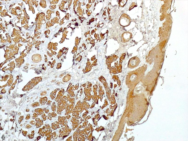
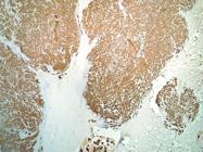
Immunohistochemical (IHC) investigation was performed using anti-cytokeratin antibodies (clone AE1/AE3, dilution 1:500, Dako, Carpinteria, California, USA) and anti-vimentin antibodies (clone V9, dilution 1:100, Dako, Carpinteria, California, USA). The immunolabeling was visualized using 3-3’ diaminobenzidine (DAB). The sections were counterstained with Harris hematoxylin. The IHC revealed cytokeratin AE1/AE3 positivity in the mammary gland (Fig.3 and 4). Vimentin was negative in the neoplastic cells and positive in the adjacent connective tissue.
Simple tubular carcinoma in the mammary gland of an ocelot (Leopardus pardalis). Positive immunolabeling for AE1/AE3 in the neoplastic cells, epidermis, and adjacent skin follicles. Counterstaining with Harris hematoxylin, obj.4x.

Simple tubular carcinoma in the mammary gland of an ocelot (Leopardus pardalis). Cytoplasmic immunolabeling for AE1/AE3 in neoplastic cells. Counterstaining with Harris hematoxylin, obj.40x.
Discussion
The diagnosis of simple tubular mammary carcinoma was established through histopathological examination and confirmed by IHC, which characterizes what seems to be the first description of mammary neoplasia in an ocelot (Leopardus pardalis). The investigative methods for the definitive diagnosis of this condition should primarily include histopathological evaluation of a biopsy or necropsy (Sadler et al. 2016). In the present case, a biopsy was not performed since the nodule was only noticed after the ocelot showed signs of anorexia. The aggressive behavior of these animals, both in the wild and under human care, complicates handling, making early identification of neoplastic masses challenging. Therefore, the behavioral characteristics of wild animals present challenges for monitoring their health.
Macroscopically, mammary tumors in wild felines can occur as an infiltrative, firm, and multilobulated mass (Šoštaric-Zuckermann et al. 2018), as observed in the animal of this investigation. A histopathological study in a tiger (Panthera tigris) (Finotello et al. 2011) also revealed simple tubular mammary carcinoma, with the mammary gland stroma exhibiting a mass organized into ductular structures and solid areas. However, the tumor exhibited a low mitotic count in the tigress, with 2-3 mitoses per high-power field (HPF). In the present study, however, the mitotic count was considered high, with an average of 120 aberrant figures in a 2.37 mm² field, indicating the rapid progression and, consequently, the occurrence of metastases. The same observation was made in domestic animals with rapid progression of mammary tumors (Prates et al. 2023). It is important to emphasize that the histological classification used for large felines is the same as for domestic felines, given the limited descriptions available for wild animals. In this classification, distinct histopathological features are scored and summed to produce groups that correlate with the degree of malignancy (Mills et al. 2015).
In general, metastases from mammary carcinomas can be expected in wild felids, with the liver, lungs, and lymph nodes being the most commonly affected sites (Frazier et al. 1994). In the ocelot, the lymph nodes were not submitted for histopathological evaluation, so we cannot rule out the involvement of this organ. On the other hand, muscles distant from the mammary gland, such as the diaphragm, as well as the pelvic limb (internal region) at the level of the femur, were affected by metastases.
Immunohistochemical techniques are used to facilitate cancer subclassifications, such as differentiation of lesions and assessment of prognosis. Pan-cytokeratin is a marker for epithelial cells, carcinomas, and some sarcomas (Kang et al. 2006). The use of pan-cytokeratin has proven effective for identifying neoplastic cells in other wild felids affected by different types of neoplasms (Bollo et al. 2011). The use of vimentin, in this case, allowed the marking of mesenchymal tissue, which was non-neoplastic, helping rule out the occurrence of mixed-type mammary neoplasia (Prates et al. 2023).
The animal in question was 12 years old, highlighting that advanced age is an important predisposing factor for the development of mammary carcinoma in felines kept under human care (Sorenmo et al. 2009, Šoštaric-Zuckermann et al. 2018). Therefore, it is important for breeding facilities to develop methods for periodic preventive assessments and early diagnosis of these middle-aged or elderly animals. As in the present case, decreased appetite is a clinical sign considered a consequence of tumor progression and can progress to death (Sadler et al. 2016). However, the onset of this sign may indicate that the condition is already advanced, with an unfavorable prognosis.
Conclusion
The ocelot (Leopardus pardalis) can develop mammary tumors with metastatic potential to the lungs and muscles. This neoplasia demonstrated clinicopathological features similar to those seen in domestic felines. However, due to the lack of data on the clinical progression and prognosis of tumors in this species, the importance of periodic mammary gland evaluation in species under human care is emphasized, as well as the necessity of performing a necropsy to obtain information on tumor behavior in this group of felines.
Acknowledgements
To the “Coordenação de Aperfeiçoamento de Pessoal de Nível Superior” (CAPES), Finance Code 001, and “Conselho Nacional de Desenvolvimento Científico e Tecnológico” (CNPq), Process 304804/2018-5, for granting the necessary financial support for the development of this study.
References
-
Bollo E, Scaglione FE, Tursi M, Tursi M, Schröder C, Degiorgi G, Belluso E, Capella S, Bellis D. Malignant pleural mesothelioma in a female lion (Panthera leo). Res Vet Scien 2011; https://doi.org/10.1016/j.rvsc.2010.08.005, PMid:20846704
» https://doi.org/https://doi.org/10.1016/j.rvsc.2010.08.005 -
Cassali GD, Jark PC, Gamba C, Damasceno KA, Lima AE, De Nardi AB, Ferreira E, Horta RS, Firmo BF, Sueiro FAR, Rodrigues LCS, Nakagaki KYR. Consensus regarding the diagnosis, prognosis and treatment of canine and feline mammary tumors - 2019. Braz J Vet Pat 2020; https://doi.org/10.24070/bjvp.1983-0246.v13i3p555-574
» https://doi.org/https://doi.org/10.24070/bjvp.1983-0246.v13i3p555-574 -
Cruz-Ochoa PF, Ochoa-Amaya JE, Cruz-Casallas PE. Patología comparada de neoplasias en carnívoros salvajes. Orinoquia 2017; https://doi.org/10.22579/20112629.393
» https://doi.org/https://doi.org/10.22579/20112629.393 -
Estes JA, Terborgh J, Brashares JS, Power ME, Berger J, Bond WJ, Carpenter SR, Essington TE, Holt RD, Jackson JBC, Marquis RJ, Oksanen L, Oksanen T, Paine RT, Pikitch EK, Ripple WJ, Sandin SA, Scheffer M, Schoener TW, Shurin JB, Sinclair ARE, Soulé ME, Virtanen R, Wardle DA. Trophic downgrading of planet Earth. Science 2011; https://doi.org/10.1126/science.1205106, PMid:21764740
» https://doi.org/https://doi.org/10.1126/science.1205106 -
Finotello R, Ressel L, Verin R, Di Lollo S, Baroni G, Piccinini R, Poli A. Mammary carcinoma in a tiger (Panthera tigris): morphological and immunohistochemical study. J Zoo Wildl Med 2011; https://doi.org/10.1638/2010-0044.1, PMid:22946385
» https://doi.org/https://doi.org/10.1638/2010-0044.1 - Frazier KS, Hines ME, Ruiz C, Herron AJ, Altman NH. Immunohistochemical differentiation of multiple metastatic neoplasia in a jaguar (Panthera onca). J Zoo Wildl Med 1994;25(2):286-293.
-
Gombač M, Dolenšek T, Jaušovec D, Kvapil P, Švara T, Pogačnik, M. Simultaneous occurrence of pancreatic adenocarcinoma and Brunner’s gland adenoma in a Siberian tiger (Panthera tigris altaica). J Comp Pathol 2015; https://doi.org/10.1016/j.jcpa.2015.08.008, PMid:26422412
» https://doi.org/https://doi.org/10.1016/j.jcpa.2015.08.008 - Gonçalves GF, Oliveira ST. Leiomioma uterino em jaguatirica (Leopardus pardalis) relato de caso. Arq Ciênc Vet Zoo Unipar 2000;3(2):185-188.
-
Kang M-S, Park M-S, Kwon S-W, Ma S-A, Cho D-Y, Kim D-Y, Kim Y. Amyloid-producing odontogenic tumour (calcifying epithelial odontogenic 26 tumour) in the mandible of a Bengal tiger (Panthera tigris tigris). J Comp Pathol 2006; https://doi.org/10.1016/j.jcpa.2005.09.004, PMid:16540113
» https://doi.org/https://doi.org/10.1016/j.jcpa.2005.09.004 -
Leme MCM, Martins AMCRPF, Bodini MES, Carvalho PR, Portugal MASC. Carcinoma de células escamosas em uma jaguatirica (Leopardus pardalis). Arq Inst Biol 2003; https://doi.org/10.1590/1808-1657v70p2172003
» https://doi.org/https://doi.org/10.1590/1808-1657v70p2172003 -
Mills SW, Musil KM, Davies JL, Hendrick S, Duncan C, Jackson ML, Kidney B, Philibert H, Wobeser BK, Simko E. Prognostic value of histologic grading for feline mammary carcinoma: a retrospective survival analysis. Vet Pathol 2015; https://doi.org/10.1177/0300985814543198, PMid:25060990
» https://doi.org/https://doi.org/10.1177/0300985814543198 -
Miranda DFH, Souza FAL, Fonseca LS, Almeida HM, Braga JFV, Costa FAL, Silva SMMS. Carcinoma hepatocelular metastático em Jaguatirica (Leopardus pardalis). Pesq Vet Bras 2015; https://doi.org/10.1590/S0100-736X2015001100007
» https://doi.org/https://doi.org/10.1590/S0100-736X2015001100007 -
Norton BB, Tunseth D, Holder K, Briggs M, Hayek L-AC, Murray S. Causes of morbidity in captive African lions (Panthera leo) in North America, 2001-2016. Zoo Biol 2018; https://doi.org/10.1002/zoo.21435, PMid:30070397
» https://doi.org/https://doi.org/10.1002/zoo.21435 -
Noss RF, Quigley HB, Hornocker MG, Merrill T, Paquet PC. Conservation biology and conservation of carnivores in the Rocky Mountains. Conser Bio 1996; https://doi.org/10.1046/j.1523-1739.1996.10040949.x
» https://doi.org/https://doi.org/10.1046/j.1523-1739.1996.10040949.x -
Oliveira AR, Pereira FMAM, Santos DO, Carvalho TP, Soares-Neto LL, Santos RL. Pulmonary adenocarcinoma in a captive ocelot (Leopardus pardalis): morphologic and immunophenotypic characterization - case report. Arq Bras Med Vet Zootec 2021; https://doi.org/10.1590/1678-4162-12324
» https://doi.org/https://doi.org/10.1590/1678-4162-12324 - Oliveira TG, Almeida LB, Campos CB. Avaliação do risco de extinção da jaguatirica Leopardus pardalis (Linnaeus, 1758) no Brasil. Bio Bras 2013;3(1):66-75.
-
Prates KS, Oliveira PL, Bueno TS, Damasceno KA, Driemeier D, Sonne L, Pavarini SP, Bertagnolli AC. A comparative analysis of anatomopathological features and COX-2 expression of mammary neoplasms with malignant mesenchymal components in female dogs. Pesq Vet Bras 2023; https://doi.org/10.1590/1678-5150-PVB-7186
» https://doi.org/https://doi.org/10.1590/1678-5150-PVB-7186 -
Sadler RA, Craig LE, Ramsay EC, Helmick K, Collins D, Garner MM. Clinicopathologic features of mammary masses in captive lions (Panthera leo). J Zoo Anim Med 2016; https://doi.org/10.1638/2015-0087.1, PMid:27010273
» https://doi.org/https://doi.org/10.1638/2015-0087.1 -
Sorenmo KU, Kristiansen VM, Cofone MA, Shofer FS, Breen A-M, Langeland M, Mongil CM, Grondahl AM, Teige J, Goldschmidt MH. Canine mammary gland tumours; a histological continuum from benign to malignant; clinical and histopathological evidence. Vet Comp Oncol 2009; https://doi.org/10.1111/j.1476-5829.2009.00184.x, PMid:19691645
» https://doi.org/https://doi.org/10.1111/j.1476-5829.2009.00184.x -
Šoštaric-Zuckermann IC, Artuković B, Smolecy O, Gudan Kurilj A, Huber D, Grabarević Ž, Hohšteter M. Metastatic tubulopapillary mammary carcinoma in a leopard (Panthera pardus). J Comp Path 2018; https://doi.org/10.1016/j.jcpa.2017.10.163
» https://doi.org/https://doi.org/10.1016/j.jcpa.2017.10.163 -
Soulé ME, Terborgh J. Conserving nature at regional and continental scales - a scientific program for North America. BioScience 1999; https://doi.org/10.2307/1313572
» https://doi.org/https://doi.org/10.2307/1313572 -
Sunquist M, Sunquist F. Wild cats of the world. J Mam 2004; https://doi.org/10.1644/1545-1542(2004)085<0365:WCOTW>2.0.CO;2
» https://doi.org/https://doi.org/10.1644/1545-1542(2004)085<0365:WCOTW>2.0.CO;2 -
Terborgh J, Lopez L, Nuñez P, Rao M, Shahabuddin G, Orihuela G, Riveros M, Ascanio R, Adler GH, Lambert TD, Balbas L. Ecological meltdown in predator-free forest fragments. Science 2001; https://doi.org/10.1126/science.1064397, PMid:11729317
» https://doi.org/https://doi.org/10.1126/science.1064397
-
1
Ethics committee.- The authors of this article declare that the current study data were not submitted for evaluation by the Ethics and Research Committee (CEUA) of the Universidade Federal da Paraíba. However, we are aware of the content of the Brazilian resolutions of the National Council for the Control of Animal Experimentation (CONCEA) [https://www.in.gov.br/web/dou/-/resolucao-n-55-de-5-de-outubro-de-2022-434869177] when studies involve animals.
-
Data Availability Statement
The raw data supporting the conclusions of this article will be made available by the authors on request.
The raw data supporting the conclusions of this article will be made available by the authors on request.